. 2014 Jul;60(1):278-89.
doi: 10.1002/hep.27084. Epub 2014 May 28.

Clonal tracing of Sox9+ liver progenitors in mouse oval cell injury

Affiliations

Clonal tracing of Sox9+ liver progenitors in mouse oval cell injury

Branden D Tarlow et al. Hepatology. 2014 Jul.

Abstract

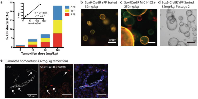
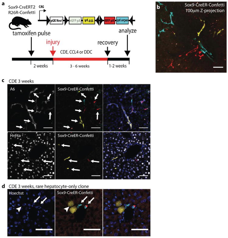
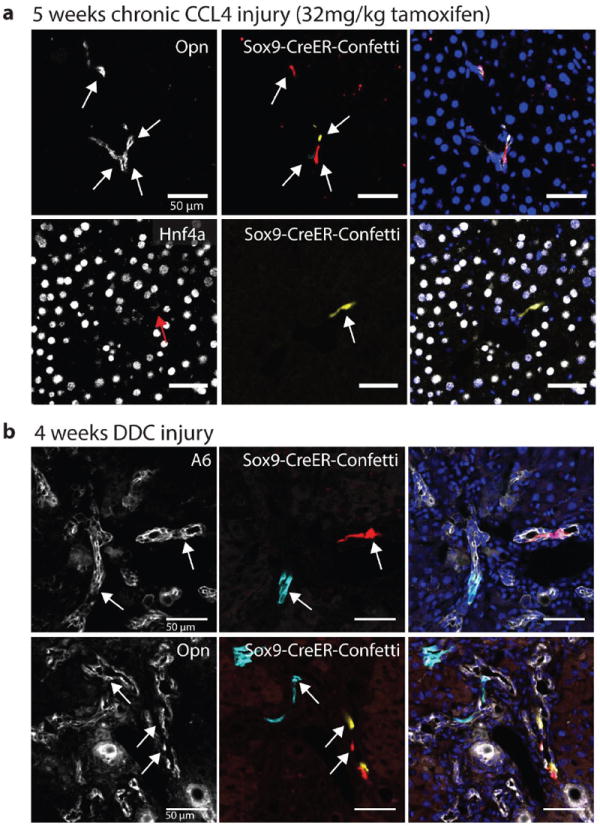
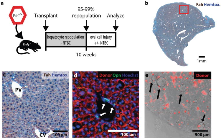
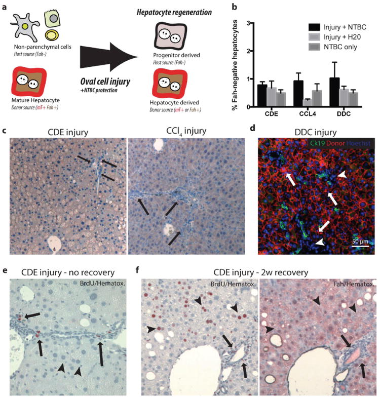
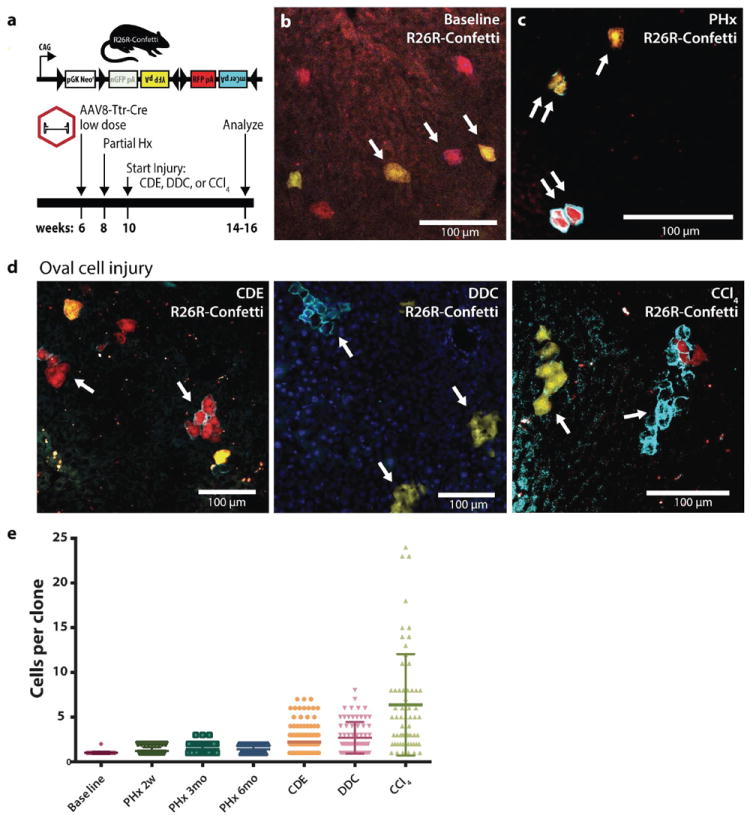
Proliferating ducts, termed "oval cells," have long been thought to be bipotential, that is, produce both biliary ducts and hepatocytes during chronic liver injury. The precursor to oval cells is considered to be a facultative liver stem cell (LSC). Recent lineage tracing experiments indicated that the LSC is SRY-related HMG box transcription factor 9 positive (Sox9(+) ) and can replace the bulk of hepatocyte mass in several settings. However, no clonal relationship between Sox9(+) cells and the two epithelial liver lineages was established. We labeled Sox9(+) mouse liver cells at low density with a multicolor fluorescent confetti reporter. Organoid formation validated the progenitor activity of the labeled population. Sox9(+) cells were traced in multiple oval cell injury models using both histology and fluorescence-activated cell sorting. Surprisingly, only rare clones containing both hepatocytes and oval cells were found in any experiment. Quantitative analysis showed that Sox9(+) cells contributed only minimally (<1%) to the hepatocyte pool, even in classic oval cell injury models. In contrast, clonally marked mature hepatocytes demonstrated the ability to self-renew in all classic mouse oval cell activation injuries. A hepatocyte chimera model to trace hepatocytes and nonparenchymal cells also demonstrated the prevalence of hepatocyte-driven regeneration in mouse oval cell injury models.

Conclusion: Sox9(+) ductal progenitor cells give rise to clonal oval cell proliferation and bipotential organoids, but rarely produce hepatocytes in vivo. Hepatocytes themselves are the predominant source of new parenchyma cells in prototypical mouse models of oval cell activation.


Figures

Figure 1
Figure 1. Sox9 clonal lineage tracing identifies ductal progenitors but not hepatocytes in homeostasis
(a) Sox9-CreERT2 R26R-Confetti+/- mice dosed with tamoxifen at limiting dilutions produced recombination in ductal progenitor cells (CD45- CD31- MIC1-1C3+) (b) FACS sorted MIC1-1C3+, Sox9-CreERT2 Confetti+ marked single cells initiated self-renewing organoids (YFP sorted) after 10 days culture or (c) 14 days (YFP+, mCerulean+, or RFP+ sorter) (d) Clonally labeled organoids self-renewed after passage. (e) Sox9-CreERT2 clonally marked cells continued to express ductal marker Opn after 3 months of homeostasis (scale bars = 50μm).
Figure 2
Figure 2. Sox9+ ducts rarely give rise to hepatocytes in CDE injury
(a) Experimental scheme: 4-6 weeks old Sox9-CreERT2 R26R-Confetti+/- mice were given a single dose of tamoxifen followed by oval cell injury two weeks later (b) CDE diet produced ductal proliferation after low density Sox9-labeling. A Z-projection generated from confocal analysis of 100 μ m thick liver section (32mg/kg tamoxifen). Ductal proliferation was not associated with hepatocyte-differentiation (c) Immunostaining for ductal markers A6 (top), Hnf4a (bottom) confirmed Sox9-CreERT2 marked cells did not differentiate into hepatocytes after recovery from CDE injury. (d) 2 YFP+ cells with distinct hepatocyte morphology (arrowhead) were adjacent to clonally unrelated mCerulean+ and RFP+ cholangiocytes (arrows). Scale bars = 50μm.
Figure 3
Figure 3. Sox9+ ducts do not differentiate into hepatocytes in DDC or CCl4 injury
(a) Immunofluorescence for ductal marker Opn (top) and hepatocyte marker Hnf4a (bottom) showed Sox9-CreERT2 marked cells retained ductal fate after regeneration from 5 weeks CCl4 injury (arrows indicate a unique clone). (b) Immunofluorescence for ductal markers A6 (top) and Opn (bottom) continued to co-localize with Sox9-CreERT2 marked clones after 4 weeks of DDC injury. Scale bars = 50μm.
Figure 4
Figure 4. Robust and specific replacement of adult hepatocytes but not other cells in Fah-chimeric mice
(a) Experimental scheme: gravity enriched wildtype or mTomatoFah+/+ hepatocytes were transplanted into the spleen of Fah-/- mice and allowed to repopulate (95-99%). (b) Fah-immunostaining (brown) shows >99% of hepatocytes were donor derived after 10 weeks repopulation (bar = 1mm) (c) Complete central vein (CV) to portal vein (PV) repopulation with donor Fah+ hepatocytes (brown). (d) Donor cell marker (mTomato red) did not colocalize with Osteopontin+ (green) host duct cells. (e) Organoids (arrows) formed from dissociated chimeric liver were host derived (mTomato negative). mTomato+ hepatocytes did not form organoid spheres after 12 days culture.
Figure 5
Figure 5. Progenitor derived hepatocytes are not required for adaptation to oval cell injury in chimeric mice
(a) Schematic: Hepatocyte transplantation into Fah-/- mice generated chimeric mice, host nonparenchymal cells were Fah- and unmarked. Donor hepatocytes were Fah+ and mTomato-marked. If nonparenchymal cells gave rise to hepatocytes after oval cell injury, we hypothesized that progenitor-derived Fah- hepatocytes would displace marked Fah+ hepatocytes. (b) Image quantification of Fah-negative hepatocytes demonstrates approximately 1% of hepatocytes remain host derived after injury. (c) No Fah-negative progenitor derived hepatocytes were observed at the majority portal regions (arrow = ductal proliferation) after 3 weeks CDE injury plus NTBC rescue or 10 weeks CCl4 injury plus NTBC (arrow = portal inflammation, ductal proliferation but no Fah- hepatocytes). (d) After 4 weeks DDC injury plus NTBC treatment CK19+ (green) ductal proliferations were adjacent to donor mTomato (red) hepatocytes (arrows) and inflammatory nonparenchmal cell (arrowhead, mTomato-negative) (bar=50μm). (e) BrdU uptake (2-hour pulse) was observed in proliferating ductal and nonparenchymal cells (arrow) but not hepatocytes at the peak of CDE injury. Hepatocytes show microvesicularsteatosis. (f) BrdU uptake (2 weeks BrdU water) during recovery from CDE injury was observed in hepatocytes (arrowheads), inflammatory cells, and cholangiocytes (arrows). Serial sectioning demonstrates that regenerating hepatocytes are lineage-marked mature hepatocytes (Fah+) and not derived from a nonparenchymal progenitor (Fah-).
Figure 6
Figure 6. Mature hepatocytes self-renew in oval cell injuries
(a) Experimental scheme: Hepatocytes were marked at low density in heterozygous R26R-Confetti+/- mice using low dose rAAV8-Ttr-Cre. Partial hepatectomy was performed and injuries were initiated after complete regeneration. (b) Prior to hepatectomy single hepatocytes were marked. (c) Partial hepatectomy induced hepatocyte replication producing clones of 1 (single arrow) or two identically marked cells (double arrow). (d) Oval cell injury with CDE diet, DDC diet, or chronic carbon tetrachloride induced mature hepatocyte replication and self-renewal or clonal expansion. (e) Dot plot histogram of mature hepatocyte colony size at baseline, partial hepatectomy followed by 2 weeks to 6 months homeostasis, and after oval cell injury (line = mean ± SD). Scale bars = 100μm.

References

    1. Farber E. Similarities in the sequence of early histological changes induced in the liver of the rat by ethionine, 2-acetylamino-fluorene, and 3’-methyl-4-dimethylaminoazobenzene. Cancer Res. 1956;16:142–148. - PubMed
    1. Sell S. Comparison of liver progenitor cells in human atypical ductular reactions with those seen in experimental models of liver injury. Hepatology. 1998;27:317–331. - PubMed
    1. Roskams T, De Vos R, Van Eyken P, Myazaki H, Van Damme B, Desmet V. Hepatic OV-6 expression in human liver disease and rat experiments: evidence for hepatic progenitor cells in man. Journal of Hepatology. 1998;29:455–463. - PubMed
    1. Itoh T, Miyajima A. Liver regeneration by stem/progenitor cells. Hepatology. 2013 - PubMed
    1. Grisham JW, Porta E. Origin and fate of proliferated hepatic ductal cells in the rate: electron microscopic and autoradiographic studies. Exp Mol Pathol. 1964;86:242–261. - PubMed

Publication types

MeSH terms