. 2014 May 30;289(22):15166-78.
doi: 10.1074/jbc.M113.531152. Epub 2014 Apr 3.

Centrobin-centrosomal protein 4.1-associated protein (CPAP) interaction promotes CPAP localization to the centrioles during centriole duplication

Affiliations

Centrobin-centrosomal protein 4.1-associated protein (CPAP) interaction promotes CPAP localization to the centrioles during centriole duplication

Radhika Gudi et al. J Biol Chem. .

Abstract

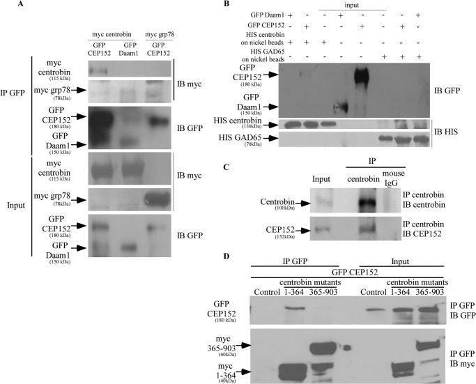
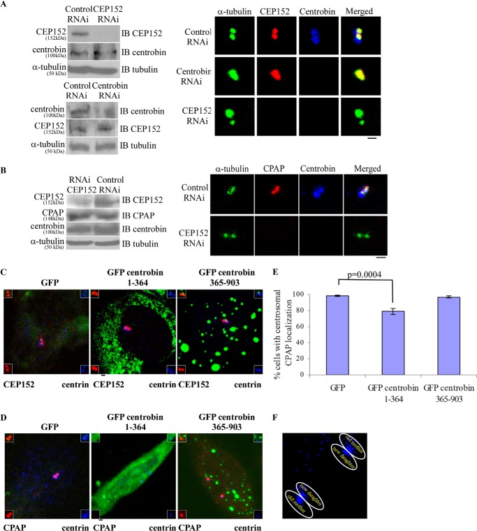
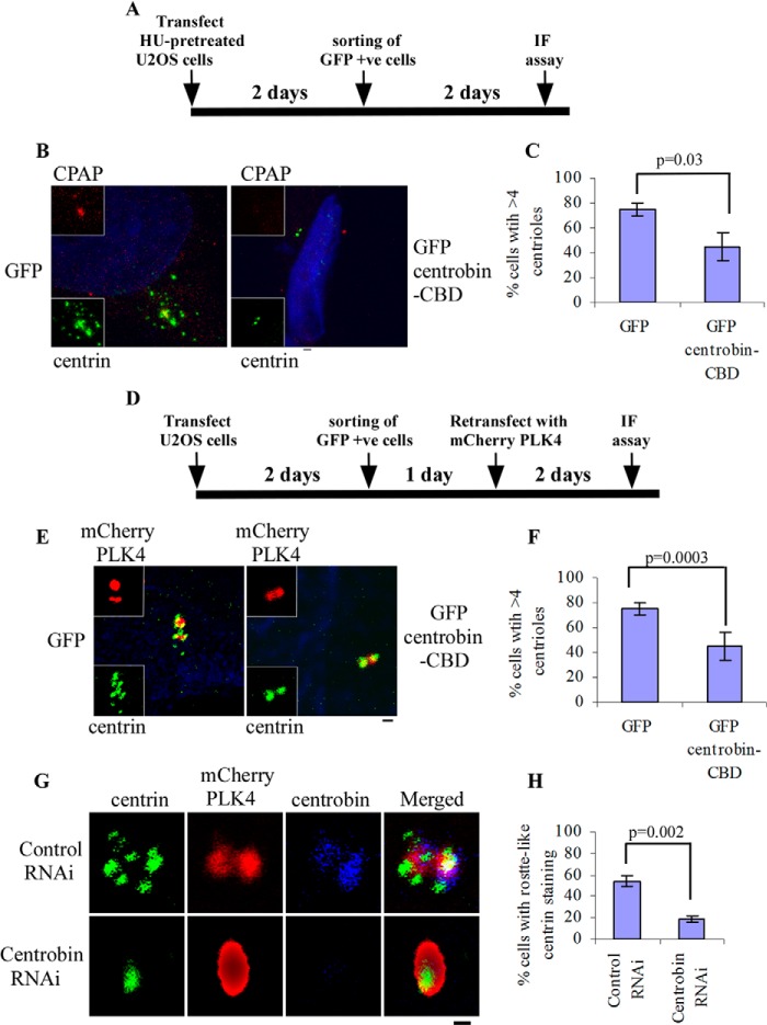
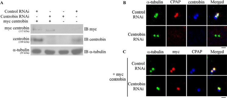
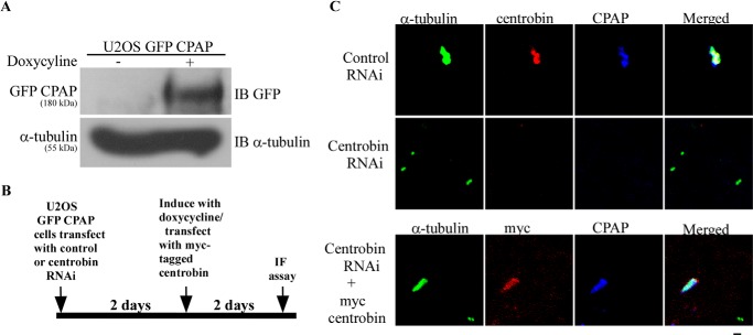
Centriole duplication is the process by which two new daughter centrioles are generated from the proximal end of preexisting mother centrioles. Accurate centriole duplication is important for many cellular and physiological events, including cell division and ciliogenesis. Centrosomal protein 4.1-associated protein (CPAP), centrosomal protein of 152 kDa (CEP152), and centrobin are known to be essential for centriole duplication. However, the precise mechanism by which they contribute to centriole duplication is not known. In this study, we show that centrobin interacts with CEP152 and CPAP, and the centrobin-CPAP interaction is critical for centriole duplication. Although depletion of centrobin from cells did not have an effect on the centriolar levels of CEP152, it caused the disappearance of CPAP from both the preexisting and newly formed centrioles. Moreover, exogenous expression of the CPAP-binding fragment of centrobin also caused the disappearance of CPAP from both the preexisting and newly synthesized centrioles, possibly in a dominant negative manner, thereby inhibiting centriole duplication and the PLK4 overexpression-mediated centrosome amplification. Interestingly, exogenous overexpression of CPAP in the centrobin-depleted cells did not restore CPAP localization to the centrioles. However, restoration of centrobin expression in the centrobin-depleted cells led to the reappearance of centriolar CPAP. Hence, we conclude that centrobin-CPAP interaction is critical for the recruitment of CPAP to procentrioles to promote the elongation of daughter centrioles and for the persistence of CPAP on preexisting mother centrioles. Our study indicates that regulation of CPAP levels on the centrioles by centrobin is critical for preserving the normal size, shape, and number of centrioles in the cell.

Keywords: CEP152; CPAP; Centriole; Centrobin; Centrosome; Cloning; Confocal Microscopy; Gene Expression.


Figures

FIGURE 1.
FIGURE 1.
Centrobin binds to CEP152 in vivo and in vitro via its C-terminal 364 aa. A, lysates of 293T cells transfected with Myc-centrobin or Myc-GRP78 and GFP-CEP152 or GFP-Daam1 expression vectors were subjected to immunoprecipitation with an anti-GFP antibody. Immunoblotting was done with anti-Myc and -GFP antibodies. B, His-centrobin and His-GAD65 was purified from IPTG-induced E. coli BL21 bacteria using nickel beads. These nickel beads were then incubated with lysates of 293T cells expressing GFP-CEP152 or GFP-Daam1. Immunoblotting was performed using anti-GFP and anti-His antibodies. C, endogenous centrobin was immunoprecipitated from lysates of 293T cells using the anti-centrobin monoclonal antibody, followed by immunoblotting with the indicated antibodies. D, 293T cells were co-transfected with control or GFP-CEP152 and Myc-centrobin fragment 1–364 or 365–903 expression vectors. Immunoprecipitation was performed with anti-GFP antibody, and immunoblotting was done using anti-GFP and anti-Myc antibodies.
FIGURE 2.
FIGURE 2.
Overexpression of the N-terminal centrobin fragment (aa 1–364) results in loss of CPAP, but not CEP152, from the centrioles. A, HeLa cells transfected with control, centrobin, or CEP152-siRNAs were treated with HU and stained with the anti-centrobin (blue), anti-CEP152 (red), and anti-α-tubulin (green) antibodies. Left, cellular lysate levels of centrobin or CEP152. B, HeLa cells were transfected with control or CEP152 siRNA, treated with HU, and stained with anti-centrobin (blue), anti-CPAP (red), and anti-α-tubulin (green) antibodies. Left, lysates of HeLa cells expressing control or CEP152 siRNA or centrobin siRNA that were tested for cellular levels of centrobin, CEP152, and CPAP. C and D, HeLa cells, pretreated with HU for 8 h, were transfected with GFP or GFP centrobin fragment 1–364 or 365–903. After 4 days, cells were stained with anti-CEP152 (C) or anti-CPAP (D) and anti-centrin antibodies to mark the centrioles. Insets, marked by white boxes, show enlarged centrosomes. Insets at the top left show enlarged centrioles with merged red and green channels, whereas insets at the top right show merged far red and green channels. E, the frequency of cells with CPAP in the centriole in GFP, GFP-centrobin(1–364), and GFP-centrobin(365–903) fragment-expressing cells was examined. 100 cells were counted per group. Each bar represents mean ± S.D. (error bars) of values obtained from three independent experiments, and statistical significance was calculated by Student's t test. Images are maximum intensity projections of Z stacks obtained using an Olympus FV10i confocal microscope. Scale bar, 2 μm. F, enlarged centrioles from the middle image of C are shown to pinpoint different centrioles.
FIGURE 3.
FIGURE 3.
Centrobin interacts with CPAP, and centrobin-CBD expression results in disappearance of CPAP from the centrioles. A, lysates of 293T cells expressing Myc-centrobin or Myc-GRP78 and GFP-CPAP or GFP-Daam1 were subjected to immunoprecipitation with anti-GFP antibody, and immunoblotting was performed using anti-Myc and anti-GFP antibodies. B, His-centrobin and His-GAD65 were purified from IPTG-induced E. coli BL21 bacteria using nickel beads. These nickel beads were incubated with lysates of 293T cells expressing GFP-CPAP or GFP-Daam1. IB was performed using anti-GFP and anti-His antibodies. C, endogenous centrobin was immunoprecipitated from lysates of 293T cells using the anti-centrobin monoclonal antibody, followed by immunoblotting with the indicated antibodies. D, His-centrobin, GST, and GST-CPAP fragments spanning residues 1–996 and 970–1338 were purified from IPTG-induced E. coli BL21 bacteria. His-centrobin that was imidazole-eluted off of the beads was incubated with GST or GST-CPAP fragments on the glutathione beads at 4 °C. GST beads were washed and subjected to SDS-PAGE and Western blot. IB was performed with anti-GST and anti-His antibodies. input, 5% imidazole eluted His-centrobin that was used in the binding experiment. E, schematic representation of centrobin fragments generated by deletion mutagenesis to decipher the CPAP binding region. F and G, CPAP binding fragment was identified by cotransfection of 293T cells with Myc-CPAP and control or GFP-centrobin fragment-expressing vectors followed by IP using anti-GFP antibody and IB using anti-GFP and anti-Myc antibodies. H, HU-pretreated HeLa cells were transfected with GFP or GFP(183–364) (centrobin-CBD) vectors as depicted in the left panel, stained with anti-CPAP (red) and anti-centrin (green) antibodies to visualize the centrioles. DAPI (blue) shows nucleus (middle). The bar diagram shows the percentage of cells with detectable levels of CPAP in centrioles (right). Error bars, S.D. Asterisks (B and D), nonspecific bands. IF, immunofluorescence.
FIGURE 4.
FIGURE 4.
Centrobin-CBD inhibits centriole duplication and PLK4 overexpression-associated centrosome amplification. A, experimental scheme depicting the time line and details for B and C. B, U2OS cells, pretreated with HU for 8 h, were transfected with GFP or GFP centrobin-CBD expression vectors. 96 h after transfection, cells were stained with anti-centrin (green) and anti-CPAP (red) antibodies and DAPI (blue). C, bar diagram shows the percentage of cells with amplified centrioles from B. D, experimental scheme depicting the time line and details for E and F. E, U2OS cells were transfected with GFP or GFP centrobin-CBD expression vectors, followed by retransfection with mCherry PLK4 (red) after 2 days. Cells were stained with anti-centrin (green) antibody and DAPI (blue). F, bar diagram shows the percentage of cells with amplified centrioles from E. G, U2OS cells transfected with control or centrobin siRNA were retransfected with mCherry PLK4 (red) expression vector and stained with anti-centrin (green) and anti-centrobin (blue) antibodies. H, the bar diagram shows the percentage of cells with amplified centrioles from G. Scale bar of the microscopy images, 2 μm. Insets marked by white boxes show enlarged centrosomes. Images in B and C are maximum intensity projections of Z stacks obtained using the Olympus FV10i microscope, whereas E–G show images acquired using the Zeiss 510 Meta microscope. Images were deconvoluted using the Metamorph software. For B, F, and H, 100 cells were counted per group; each bar represents the mean ± S.D. (error bars) of values obtained from three independent experiments; and statistical significance was calculated by Student's t test or two-way analysis of variance.
FIGURE 5.
FIGURE 5.
Centrobin depletion results in loss of CPAP from the centrosomes. A, HeLa cells were transfected with control or centrobin siRNAs for 48 h, followed by retransfection with control or Myc-centrobin-expressing vectors. Cell lysates were subjected to immunoblotting with anti-Myc, anti-centrobin, and anti-tubulin antibodies to demonstrate the knockdown of centrobin and expression of Myc centrobin. B, HeLa cells were transfected with control or centrobin siRNA for 72 h and stained with anti-centrobin (blue), anti-CPAP (red), and anti-α-tubulin (green) antibodies. C, HeLa cells were treated as described in A, followed by retransfection with Myc-centrobin vector and staining with anti-CPAP (blue), anti-Myc (red), and α-tubulin (green) antibodies. Scale bar for microscopy images, 2 μm. Images in A and B were acquired using the Olympus FV10i microscope and are maximum intensity projections of Z stacks.
FIGURE 6.
FIGURE 6.
Centrobin-depleted cells do not show CPAP recruitment to the centrioles and centriole elongation. A, U2OS cells expressing inducible GFP-CPAP were transfected with control or centrobin siRNA for 2 days and treated with doxycycline for another 2 days. Cells were then subjected to immunoblotting with anti-GFP and anti-tubulin antibodies. B, experimental scheme of C. C, cells described in A were stained with anti-α-tubulin (green), anti-centrobin (red), and anti-CPAP (blue) antibodies to mark the centrioles (top and middle rows). Cells described in A were retransfected with Myc-tagged centrobin after 2 days of RNAi treatment and treated with doxycycline, followed by staining with anti-α-tubulin (green), anti-Myc (red), and anti-CPAP (blue) antibodies (bottom row). Scale bar, 2 μm. Images were acquired using the Olympus FV10i microscope and are maximum intensity projections of Z stacks. IF, immunofluorescence.
FIGURE 7.
FIGURE 7.
Model depicting the order in which proteins are recruited to the procentriole. Centriole duplication is initiated by CEP152-mediated localization of PLK4 to the proximal end of the mother centriole. CEP152 binds to centrobin directly (dotted green arrow) or indirectly through other interacting partners (solid green arrow), which facilitates recruitment of centrobin to the procentrioles. Direct interaction between centrobin and CPAP mediates elongation of the daughter centriole.

Similar articles

Cited by

References

    1. Lüders J., Stearns T. (2007) Microtubule-organizing centres: a re-evaluation. Nat. Rev. Mol. Cell Biol. 8, 161–167 - PubMed
    1. Bornens M. (2002) Centrosome composition and microtubule anchoring mechanisms. Curr. Opin. Cell Biol. 14, 25–34 - PubMed
    1. Job D., Valiron O., Oakley B. (2003) Microtubule nucleation. Curr. Opin. Cell Biol. 15, 111–117 - PubMed
    1. Nigg E. A., Raff J. W. (2009) Centrioles, centrosomes, and cilia in health and disease. Cell 139, 663–678 - PubMed
    1. Kitagawa D., Kohlmaier G., Keller D., Strnad P., Balestra F. R., Flückiger I., Gönczy P. (2011) Spindle positioning in human cells relies on proper centriole formation and on the microcephaly proteins CPAP and STIL. J. Cell Sci. 124, 3884–3893 - PubMed

Publication types

MeSH terms

LinkOut - more resources