Overlapping demyelinating syndromes and anti–N-methyl-D-aspartate receptor encephalitis

Maarten J Titulaer et al. Ann Neurol. 2014 Mar.

Abstract

Objective: To report the clinical, radiological, and immunological association of demyelinating disorders with anti–Nmethyl- D-aspartate receptor (NMDAR) encephalitis.

Methods: Clinical and radiological analysis was done of a cohort of 691 patients with anti-NMDAR encephalitis. Determination of antibodies to NMDAR, aquaporin-4 (AQP4), and myelin oligodendrocyte glycoprotein (MOG) was performed using brain immunohistochemistry and cell-based assays.

Results: Twenty-three of 691 patients with anti-NMDAR encephalitis had prominent magnetic resonance imaging (MRI) and/or clinical features of demyelination. Group 1 included 12 patients in whom anti-NMDAR encephalitis was preceded or followed by independent episodes of neuromyelitis optica (NMO) spectrum disorder (5 cases, 4 anti-AQP4 positive) or brainstem or multifocal demyelinating syndromes (7 cases, all anti-MOG positive). Group 2 included 11 patients in whom anti-NMDAR encephalitis occurred simultaneously with MRI and symptoms compatible with demyelination (5 AQ4 positive, 2 MOG positive). Group 3 (136 controls) included 50 randomly selected patients with typical anti-NMDAR encephalitis, 56 with NMO, and 30 with multiple sclerosis; NMDAR antibodies were detected only in the 50 anti-NMDAR patients, MOG antibodies in 3 of 50 anti-NMDAR and 1 of 56 NMO patients, and AQP4 antibodies in 48 of 56 NMO and 1 of 50 anti-NMDAR patients (p<0.0001 for all comparisons with Groups 1 and 2). Most patients improved with immunotherapy, but compared with anti-NMDAR encephalitis the demyelinating episodes required more intensive therapy and resulted in more residual deficits. Only 1 of 23 NMDAR patients with signs of demyelination had ovarian teratoma compared with 18 of 50 anti-NMDAR controls (p50.011).

Interpretation: Patients with anti-NMDAR encephalitis may develop concurrent or separate episodes of demyelinating disorders, and conversely patients with NMO or demyelinating disorders with atypical symptoms (eg, dyskinesias, psychosis) may have anti-NMDAR encephalitis.


Figures

Figure 1
Figure 1
Flow chart summarizing the patients included in the study “NMDAR-DSE” or group 1 includes 5 patients with sequential episodes of anti-NMDAR encephalitis and NMO or NMO spectrum disorder (NMOSD), and 7 patients with sequential episodes of anti-NMDAR encephalitis and unusual symptoms and MRI findings suggesting demyelination, but without criteria for NMO or NMOSD. The arrows indicate which of the episodes occurred first, and the profile of antibodies associated with demyelination (AQP4, MOG). “NMDAR + clinical/MRI demyelination” or group 2, includes 11 patients in whom anti-NMDAR encephalitis developed simultaneously with MRI and/or clinical features of demyelination (8 of them with unusual symptoms for anti-NMDAR encephalitis). Controls include three groups of patients with classical features of anti-NMDAR encephalitis, NMO, or multiple sclerosis (relapsing-remitting [RRMS], secondary progressive [SPMS], primary progressive [PPMS], clinically isolated syndrome [CIS]). All 136 patients were tested for NMDAR, NMO, and AQP4 antibodies and results are shown in the chart.
Figure 2
Figure 2
Demonstration of co-occurrence of NMDAR antibodies with aquaporin 4 (AQP4), or myelin oligodendrocyte glycoprotein (MOG) HEK293 cells transfected with the AQP4-M23 isoform (A), EGFP-N1-hMOG (B), and GluN1/GluN2B subunits of the NMDAR (C); (A) serum from patient #1 (red) shows reactivity with AQP4-M23 transfected HEK293 cells (green, commercial antibody against AQP4; blue: DAPI; yellow: merged reactivities), in contrast, a control serum is negative. (B) CSF from patient #6 (red) shows reactivity with MOG-EGFP transfected HEK293 cells (green), in contrast, a control CSF is negative. (C) serum from patient #1 and CSF from patient #6, also show reactivity with NR1/NR2B-transfected HEK293 cells (green, commercial antibody against NR1 subunit), in contrast, a control CSF is negative. A–C: magnification ×400, all bars = 10 micrometer.
Figure 3
Figure 3
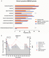
Clinical symptoms of the anti-NMDAR encephalitis episodes for the three groups of patients. (A) Comparison of the indicated groups of patients. §p = 0.031; p = 0.002. The p-values are uncorrected values. (B) Clinical course and follow-up of antibodies in a patient with anti-NMDAR encephalitis and DSE with MOG-antibodies. MP methylprednisolone; PE plasma exchange; CTX cyclophosphamide
Figure 4
Figure 4
Brain and spinal cord MRI of patients with anti-NMDAR encephalitis separated in time from demyelinating episodes (A–D) Patient 3 of table 1: Increased T2/FLAIR signal abnormalities involving the right temporal lobe (A), caudate nucleus and frontal lobes (B), deterioration over time (C), and near-resolution after treatment (D). (E–J) patient 7: T2/FLAIR increased signal in the left parietal region during Sydenham's chorea (E), subtle increase of the area of abnormal signal two weeks into the anti-NMDAR encephalitis (F); new T2 lesions in the brainstem and Th9 and Th11–12 during the first demyelinating episode (G–H); new T2/FLAIR abnormalities in the left facial collicular region and cerebellum during the second demyelinating episode (I), and near-resolution after treatment (J). (K–P) patient 9: T2/FLAIR sequences showing no lesions during anti-NMDAR encephalitis (K); several new lesions during the first demyelinating episode (L–M) and second demyelinating episode (N). In the first demyelinating episode there was also involvement of the spinal cord at Th6 (O–P). (Q–T) patient 10: increased T2 signal (Q) and areas of contrast enhancement (R) during an episode of demyelination. Different abnormalities, including increased T2 signal in cortical frontal regions (S) without contrast enhancement (T) occurred during an episode of anti-NMDAR encephalitis. (U–X) patient 11: increased FLAIR signal in the cortex of the right hemisphere during anti-NMDAR encephalitis (U); during a demyelinating episode with symptoms of optic neuritis, the right optic nerve shows increased T2 signal (V) with contrast enhancement (W), along with enhancement in pre-chiasmal right and left optic nerves (X).
Figure 5
Figure 5
Brain and spinal cord MRI of patients with anti-NMDAR encephalitis occurring simultaneously with extensive, atypical FLAIR-T2 MRI-abnormalities (A–B) Patient 17 of table 3, increased T2/FLAIR signal abnormalities involving the periventricular, subcortical white matter (A), with hypointense T1 signals (B). (C–D) Patient 13, increased FLAIR signal abnormalities in the pons, periventricular region of the fourth ventricle (C), and the third ventricle (D). (E–H) Patient 22, increased FLAIR signal abnormality in the left parietal region (E), with ring-enhancement with Gadolinium at T1 (F); the abnormality improved after immunotherapy (G), showing resolution of the gadolinium enhancement (H). (I–L) Patient 19, increased FLAIR signal abnormalities, involving the brainstem (I), subcortical white matter (J), and increased T2 signal in the spinal cord C3–C6 and Th3–Th4 (K), with gadolinium enhancement (L).

Comment in

Similar articles

Cited by

References

    1. Dalmau J, Gleichman AJ, Hughes EG, et al. Anti-NMDA-receptor encephalitis: case series and analysis of the effects of antibodies. Lancet Neurol. 2008;7:1091–1098. - PMC - PubMed
    1. Hughes EG, Peng X, Gleichman AJ, et al. Cellular and synaptic mechanisms of anti-NMDA receptor encephalitis. J Neurosci. 2010;30:5866–5875. - PMC - PubMed
    1. Mikasova L, De RP, Bouchet D, et al. Disrupted surface cross-talk between NMDA and Ephrin-B2 receptors in anti-NMDA encephalitis. Brain. 2012;135:1606–1621. - PubMed
    1. Titulaer MJ, McCracken L, Gabilondo I, et al. Treatment and prognostic factors for long-term outcome in patients with anti-NMDA receptor encephalitis: an observational cohort study. Lancet Neurol. 2013;12:157–165. - PMC - PubMed
    1. Kruer MC, Koch TK, Bourdette DN, et al. NMDA receptor encephalitis mimicking seronegative neuromyelitis optica. Neurology. 2010;74:1473–1475. - PMC - PubMed

MeSH terms