

Comment
doi: 10.1002/mrm.25214.
Epub 2014 Apr 3.
Biophysical mechanisms of myelin-induced water frequency shifts
Affiliations
- PMID: 24700617
- PMCID: PMC6366856
- DOI: 10.1002/mrm.25214
Item in Clipboard
Comment
Biophysical mechanisms of myelin-induced water frequency shifts
Magn Reson Med.
2014 Jun.
No abstract available
Figures
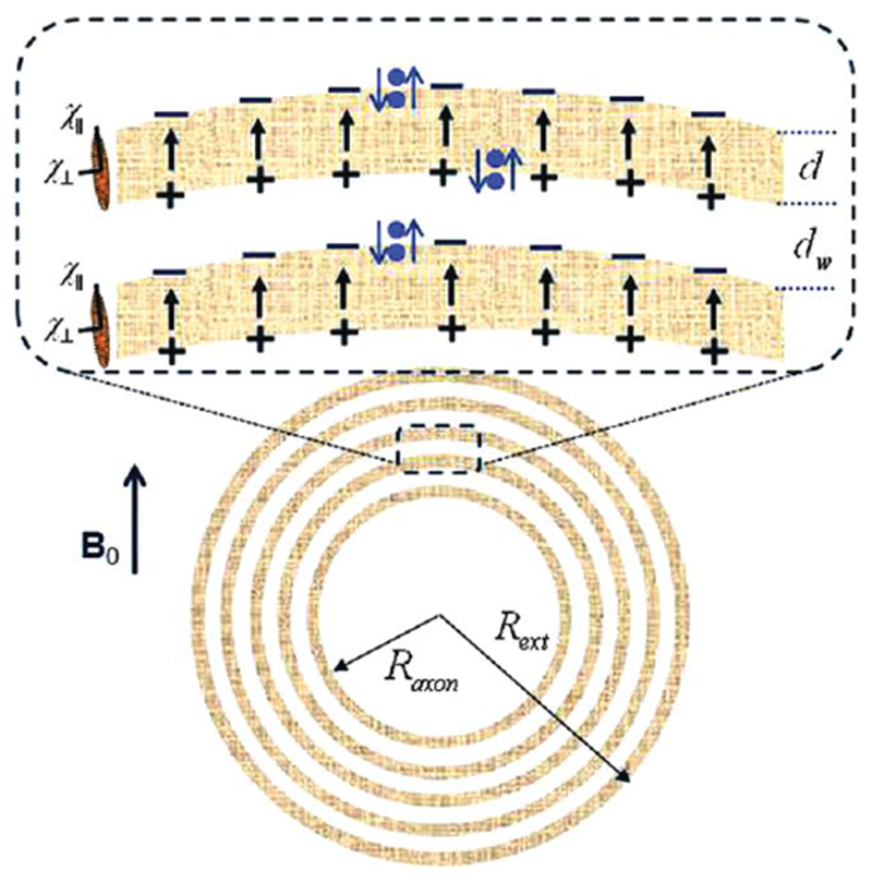
Schematic structure of an axon with radius Raxon surrounded by a myelin sheath of the external radius Rext, consisting of lipid layers of thickness d (marked in gray) separated by aqueous phases (layers) of thickness dw. Each lipid layer is formed by highly organized, radially oriented long molecules (shown as an ellipsoids at left) with anisotropic magnetic susceptibility. In the presence of magnetic field B0, the lipid layers become magnetized and create an additional magnetic field that can be described as a result of magnetostatic charges ρ = −div M formed on the layers’ surfaces (surface charges) and inside the lipid layers (volume charges). The surface magnetostatic charges (which are of interest for the proposed “hop in, hop out” mechanism and are shown as + and − signs) are equal to ρS = ±H0 sin α ⋅ cosφ ⋅ χ∥, where α and φ are the polar and azimuthal angles (Z-direction is along the axon). The signs of the surface charges and the direction of the field H within the lipid layers correspond to χ∥ < 0. Blue dots represent water molecules performing a “Hokey Pokey”–like dance from aqueous to lipid layers. When a water molecule jumps from water layer to lipid layer, it experiences an additional field H (shown as arrows) induced by the surface charges. The projection of this field on B0 is equal to H = −H0 ⋅ χ∥ ⋅ sin2 α ⋅ cos2
φ.
Comment on
-
On the role of neuronal magnetic susceptibility and structure symmetry on gradient echo MR signal formation.Magn Reson Med. 2014 Jan;71(1):345-53. doi: 10.1002/mrm.24629. Epub 2013 Feb 4. Magn Reson Med. 2014. PMID: 23382087 Free PMC article.
-
Frequency shifts in the myelin water compartment.Magn Reson Med. 2014 Jun;71(6):1953-5. doi: 10.1002/mrm.24983. Epub 2014 Apr 3. Magn Reson Med. 2014. PMID: 24700549 Free PMC article. No abstract available.
Similar articles
-
Frequency shifts in the myelin water compartment.Magn Reson Med. 2014 Jun;71(6):1953-5. doi: 10.1002/mrm.24983. Epub 2014 Apr 3. Magn Reson Med. 2014. PMID: 24700549 Free PMC article. No abstract available.
-
On the role of neuronal magnetic susceptibility and structure symmetry on gradient echo MR signal formation.Magn Reson Med. 2014 Jan;71(1):345-53. doi: 10.1002/mrm.24629. Epub 2013 Feb 4. Magn Reson Med. 2014. PMID: 23382087 Free PMC article.
-
[Propagation of excitation in a model of nerve fiber].Biofizika. 1967 Sep-Oct;12(5):900-7. Biofizika. 1967. PMID: 5623583 Russian. No abstract available.
-
The role of tissue microstructure and water exchange in biophysical modelling of diffusion in white matter.MAGMA. 2013 Aug;26(4):345-70. doi: 10.1007/s10334-013-0371-x. Epub 2013 Feb 27. MAGMA. 2013. PMID: 23443883 Free PMC article. Review.
-
Problems concerning the measurements of electrical events in myelinated nerve fibers.Rev Roum Physiol (1990). 1990 Apr-Jun;27(2):99-108. Rev Roum Physiol (1990). 1990. PMID: 2088514 Review.
Cited by
-
Evaluation of the Blood-Brain Barrier, Demyelination, and Neurodegeneration in Paramagnetic Rim Lesions in Multiple Sclerosis on 7 Tesla MRI.J Magn Reson Imaging. 2024 Mar;59(3):941-951. doi: 10.1002/jmri.28847. Epub 2023 Jun 5. J Magn Reson Imaging. 2024. PMID: 37276054 Free PMC article.
-
Ultra-short T2 components imaging of the whole brain using 3D dual-echo UTE MRI with rosette k-space pattern.Magn Reson Med. 2023 Feb;89(2):508-521. doi: 10.1002/mrm.29451. Epub 2022 Sep 25. Magn Reson Med. 2023. PMID: 36161728 Free PMC article.
-
Improved magnetic resonance myelin water imaging using multi-channel denoising convolutional neural networks (MCDnCNN).Quant Imaging Med Surg. 2022 Mar;12(3):1716-1737. doi: 10.21037/qims-21-404. Quant Imaging Med Surg. 2022. PMID: 35284287 Free PMC article.
-
Effects of biological tissue structural anisotropy and anisotropy of magnetic susceptibility on the gradient echo MRI signal phase: theoretical background.NMR Biomed. 2017 Apr;30(4):10.1002/nbm.3655. doi: 10.1002/nbm.3655. Epub 2016 Nov 11. NMR Biomed. 2017. PMID: 27862452 Free PMC article. Review.
-
The impact of fibre orientation on T1-relaxation and apparent tissue water content in white matter.MAGMA. 2018 Aug;31(4):501-510. doi: 10.1007/s10334-018-0678-8. Epub 2018 Feb 20. MAGMA. 2018. PMID: 29464463
References
Publication types
MeSH terms
Grants and funding
LinkOut - more resources
Full Text Sources
Other Literature Sources
Medical
