Review
. 2014 Jul;93(7 Suppl):7S-19S.
doi: 10.1177/0022034514529150. Epub 2014 Apr 3.

Rare bone diseases and their dental, oral, and craniofacial manifestations

Affiliations
Review

Rare bone diseases and their dental, oral, and craniofacial manifestations

B L Foster et al. J Dent Res. 2014 Jul.

Abstract

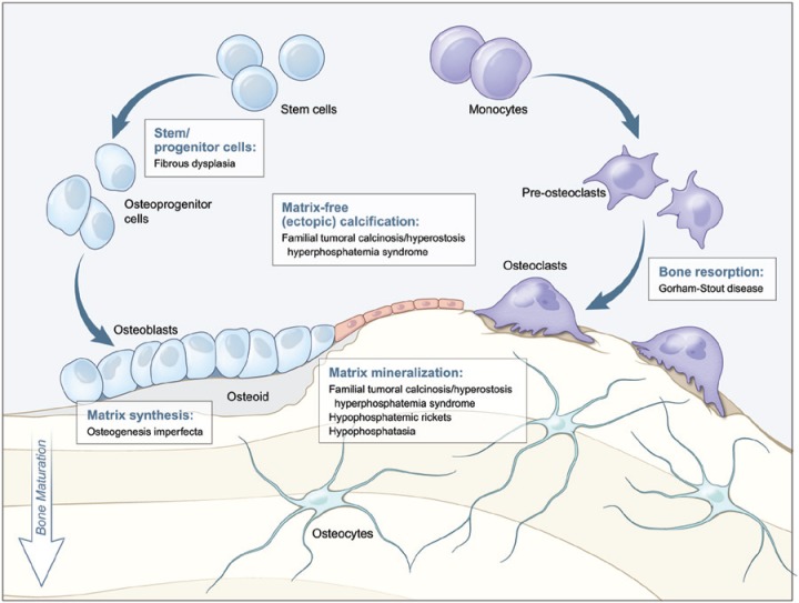
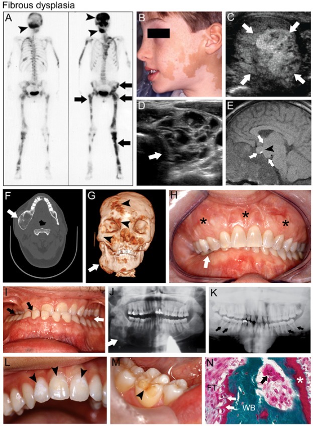
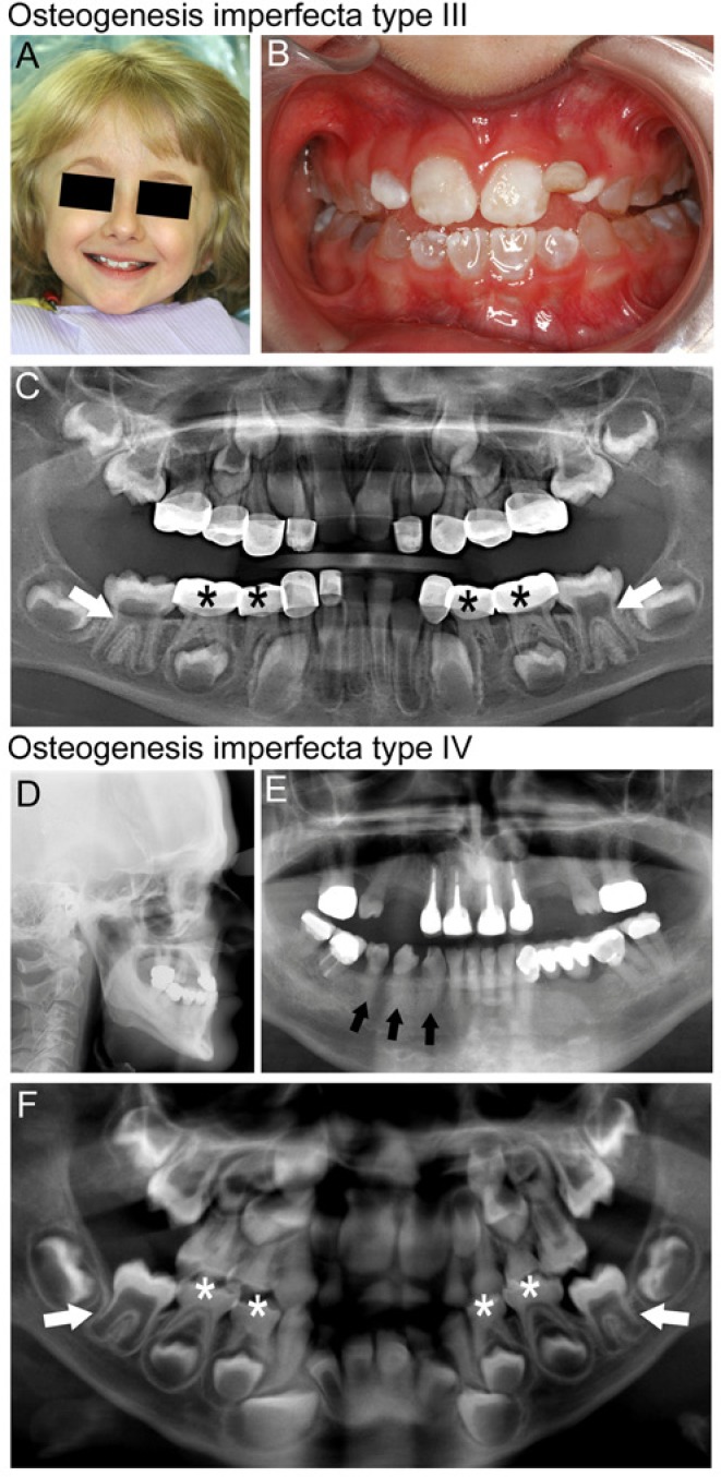
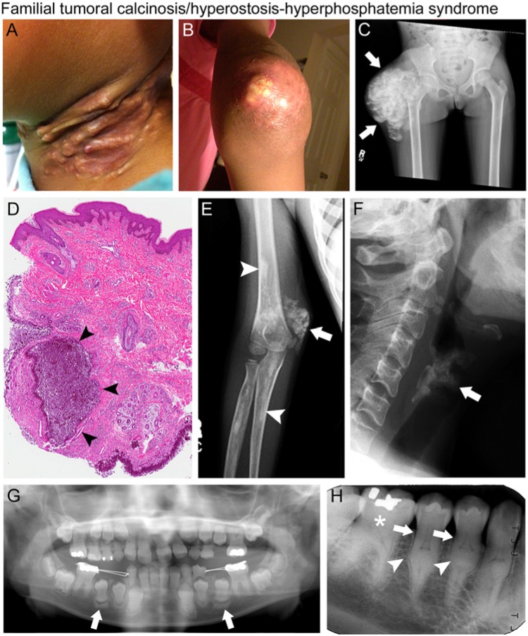
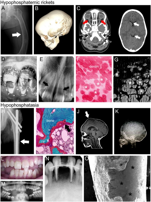
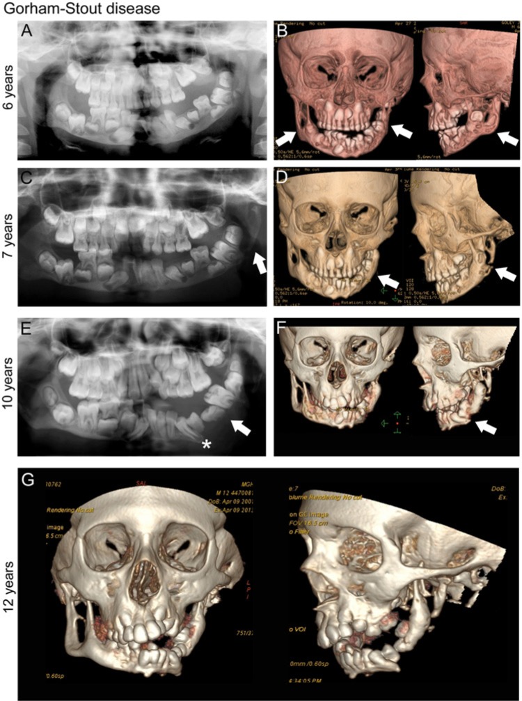
Hereditary diseases affecting the skeleton are heterogeneous in etiology and severity. Though many of these conditions are individually rare, the total number of people affected is great. These disorders often include dental-oral-craniofacial (DOC) manifestations, but the combination of the rarity and lack of in-depth reporting often limit our understanding and ability to diagnose and treat affected individuals. In this review, we focus on dental, oral, and craniofacial manifestations of rare bone diseases. Discussed are defects in 4 key physiologic processes in bone/tooth formation that serve as models for the understanding of other diseases in the skeleton and DOC complex: progenitor cell differentiation (fibrous dysplasia), extracellular matrix production (osteogenesis imperfecta), mineralization (familial tumoral calcinosis/hyperostosis hyperphosphatemia syndrome, hypophosphatemic rickets, and hypophosphatasia), and bone resorption (Gorham-Stout disease). For each condition, we highlight causative mutations (when known), etiopathology in the skeleton and DOC complex, and treatments. By understanding how these 4 foci are subverted to cause disease, we aim to improve the identification of genetic, molecular, and/or biologic causes, diagnoses, and treatment of these and other rare bone conditions that may share underlying mechanisms of disease.

Keywords: Gorham-Stout disease; familial hypophosphatemic rickets; fibrous dysplasia of bone; hyperphosphatemic familial tumoral calcinosis; hypophosphatasia; osteogenesis imperfecta.


Conflict of interest statement

The authors declare no potential conflicts of interest with respect to the authorship and/or publication of this article.

Figures

Figure 1.
Figure 1.
Diseases affecting specific stages of bone formation and remodeling. This model outlines key stages of bone development (differentiation of stem/progenitor cells, matrix production, mineralization, and resorption) affected by the genetic disorders highlighted in this review. For each developmental process (indicated by text boxes), associated diseases caused by defects in that process are identified. Tooth development depends on parallel processes outlined for bone formation, though notably does not undergo physiological resorption as part of a remodeling process.
Figure 2.
Figure 2.
Fibrous dysplasia (FD). (A) Technetium-99 bone scintigraphy of a female patient with FD/McCune-Albright syndrome (MAS) shows patchy areas of increased radiotracer uptake by FD in the craniofacial bones (black arrowheads), pelvis, and long bones (black arrows). (B) Café-au-lait macules on the face of a boy with MAS macules display the typical jagged “coast of Maine” borders. (C) Testicular ultrasound of a patient with MAS displays heterogeneous mixed cystic and solid lesions characteristic of Leydig cell hyperplasia (white arrows). (D) Thyroid ultrasound of a patient with MAS and hyperthyroidism demonstrates characteristic heterogeneity with a cystic, “Swiss cheese”-like appearance (white arrow). (E) Magnetic resonance imaging of the brain of a patient with MAS-associated growth hormone excess displays a large, dumbbell-shaped growth-hormone- and prolactin-secreting pituitary macroadenoma (white arrows); the bright spot (black arrowhead) indicates the posterior pituitary. (F) Computed tomography (CT) of the head in this MAS patient shows right mandibular expansion by FD (white arrow). (G) Three-dimensional volume rendering of CT images of the same patient seen in panel F shows expansion of FD in the right mandible (white arrow), as well as smaller potential areas of FD in the right fronto-orbital region and midface (black arrowheads). (H) Severe expansion of the maxilla by FD (black stars) is associated with malocclusion and right-sided overbite (white arrow). (I) In another FD patient, malocclusion shows a right lingual crossbite (black arrows) and a left posterior open bite (white arrow), possibly from mandibular enlargement due to growth hormone excess. (J) Panoramic radiograph of mandibular FD shows radiolucent ground glass trabeculation (white arrow). (K) Another MAS patient with growth hormone excess displays generalized FD radiopacity, particularly in the maxilla, and taurodontic pulp chambers (black arrows). (L) Enamel hypomineralization (black arrowheads) and (M) dentin dysplasia (black arrowheads) are also dental features often seen in patients with FD/MAS. (N) Histologic features of FD shown in Goldner’s trichrome-stained section include immature irregular woven bone (WB) with Sharpey’s fibers (white arrows) in a fibrous tissue (FT) matrix of variable cellularity. Note the unmineralized osteoid (white star) indicating osteomalacia, and an osteoclast resorbing bone in Howship’s lacuna (black arrow). Images in C, D, F, E, and J reproduced with permission from Akintoye et al., 2013. Image in K reproduced with permission from Akintoye et al., 2003.
Figure 3.
Figure 3.
Osteogenesis imperfecta (OI). Clinical and radiographic findings in individuals with OI types III (A-C) and IV (D-F). (A) Facial appearance of child with OI type III shows a triangular face with prominent forehead and blue sclera. (B) Dentition of this child shows blue-gray discoloration characteristic of dentinogenesis imperfecta (DI). Discoloration is less pronounced in the maxillary permanent incisors than in the primary teeth. (C) A panoramic radiograph of the dentition of this child with OI type III shows cervical constriction and large pulp chambers in the six-year molars (white arrows), and diminished pulp space in the primary dentition (black stars) that has been restored with stainless steel crowns. (D) A cephalometric radiograph of an adult individual with OI type IV shows midface deficiency and a steep mandibular plane angle. (E) A panoramic radiograph of the dentition of the same individual displays obliteration of the pulp space and poor contrast of the roots to the bone due to the hypomineralized dentin (for example, mandibular teeth indicated by black arrows). This individual has required multiple root canals as a result of pulpal necrosis associated with DI. (F) A panoramic radiograph of an individual with OI type IV showing delayed dentin formation and large pulp chambers in the early-developing permanent dentition (see white arrows for examples), while the primary dentition is undergoing pulpal obliteration due to increased dentin formation (see white stars for examples).
Figure 4.
Figure 4.
Familial tumoral calcinosis/hyperostosis-hyperphosphatemia syndrome (FTC/HHS). Clinical, radiographic, and histologic findings of patients with FTC/HHS. (A) Erythematous, inflamed subcutaneous calcifications of the neck and (B) periarticular ectopic calcification of the elbow in a 9-year-old girl. (C) Radiograph with extensive periarticular ectopic calcification of the right hip (white arrows). (D) Histology of ectopic calcification with amorphous calcium deposits (arrowheads) surrounded by a thick infiltration of mono- and poly-nucleated macrophages (“foreign body macrophages”), associated with a chronic inflammatory reaction. (E) Radiograph showing periarticular ectopic calcification of the right elbow (white arrow) and diaphyseal hyperostosis of the radius and humerus (white arrowheads). (F) Lateral neck radiograph showing age-inappropriate calcification of the thyroid cartilage in a 36-year-old woman (white arrow). (G) Panoramic dental radiograph of a 9-year-old girl with blunted, bulbous roots and obliteration of the pulp chamber in the maxillary and mandibular permanent central and lateral incisors. The erupting permanent premolars exhibit calcification at the root apices. Sclerotic bony changes are also present around the erupting teeth (white arrows). (H) Periapical radiograph of mandibular right sextant showing thistle-shaped roots (white arrows), midroot bulges (white arrowheads), and mineralized molar pulps and root canals (white star) in a 36-year-old woman. Images in F and H reproduced with permission from Dumitrescu et al., 2009.
Figure 5.
Figure 5.
X-linked hypophosphatemic rickets (XLH) and hypophosphatasia (HPP). Representative images of individuals with hypophosphatemic rickets (A-G) and hypophosphatasia (H-O). (A) Skeletal pathology resulting from osteomalacia includes bowing of the femur (arrow) due to softening of the bone. (B) The computed tomography (CT) scan of a 3-year-old female with XLH shows scaphocephaly (elongation of the skull), suggestive of premature fusion of the sagittal suture. (C) Brain CT (left image) from the same patient shows enlargement of optic nerve sheaths (red arrows) due to papilledema (optic disc swelling), while orbital CT shows ventricular narrowing and decreased size of ventricles (white arrows) due to synostoses, contributing to elevated intracranial pressure. (D) Enamel discoloration indicating hypoplasia in a juvenile individual diagnosed with XLH, and (E) dental radiograph from the same individual features thin dentin and wide pulp chambers (white arrows). (F) Histologic staining reveals interglobular dentin hypomineralization, reflecting inhibition of mineralization foci to fuse into a unified mineralization front, a pattern replicated in (G) scanning electron microscopy from an adult individual with XLH. (H) Radiograph of pseudofracture of the proximal femur (white arrow) in adult diagnosed with HPP. (I) Goldner’s trichrome stain of iliac crest biopsy from adult diagnosed with HPP, exhibiting excess osteoid (red layer indicated by black arrow). (J) Magnetic resonance imaging (MRI) of the skull of a six-year-old individual diagnosed with childhood HPP exhibiting craniosynostosis and resulting bregmatic eminence (white arrow). (K) Radiograph of a four-year-old HPP individual with hypomineralization of cranial vault exhibiting severe “copper beaten” skull appearance, as well as deformations of cranial vault shape. Alterations in cranial shape in J and K are indicative of increased cranial pressure. (L) Clinical photograph of a fourteen-year-old individual with HPP who experienced spontaneous exfoliation of the lower mandibular incisor during toothbrushing. (M) Panoramic radiograph of the same individual revealing delayed eruption of several permanent teeth (e.g., lower premolars), enlarged pulp chambers, and thin dentin. (N) Periapical radiograph of the same patient at 20 yr old, showing loss of lower incisor, endodontic treatment, and splinting of remaining incisors, with generalized loss of alveolar bone. (O) Scanning electron microscopy of incisor root from HPP patient indicates lack of cementum and exposure of dentin (*), as well as extensive resorption over the root surface. Images in B and C adapted from Glass et al. (2011); reproduced under open access policy. Images in D-F are adapted from Pereira et al. (2004) and are reproduced with permission. Image in G was adapted from Foster et al. (2013) and is reproduced by permission. Images in H and I are adapted from Berkseth et al. (2013) and are reproduced with permission. Images in J and K are adapted from Collmann et al. (2009) and are reproduced with permission. Images in L, M, and O are adapted from Rodrigues et al. (2012) and are reproduced with permission. Image in N is adapted from Rodrigues et al. (2012), and is reproduced by permission.
Figure 6.
Figure 6.
Gorham-Stout disease (GSD). Progressive destruction of the mandible in an individual with GSD, initially presenting at age 4 yr with mobility of left mandibular primary teeth, and premature exfoliation of the left mandibular primary first molar. (A) Panoramic radiograph, age 6 yr. Note normal complement of permanent teeth (left mandibular primary first molar #L missing) and abnormal left mandibular condyle. (B) Computed tomography (CT), age 6 yr. Destruction of normal cortical architecture of mandibular condyle, ramus, and body, bilaterally (white arrows). (C) Panoramic radiograph, age 7 yr (post-treatment with Interferon-α2b and zoledronic acid). Further bony changes with complete loss of left mandibular angle (white arrow). (D) CT, age 7 yr. Remodeling changes of the left mandibular condyle and ramus (white arrows). (E) Panoramic radiograph, age 10 yr. Loss of bony continuity in the left mandible (white arrow); teeth appear to be “floating.” Permanent tooth formation has progressed (left mandibular canine, white star). (F) CT, age 10 yr. Tooth buds appear to hold remaining bone together in left mandible. (G) CT, age 12 yr. Interval disease progression and bone loss of the bilateral mandible. Radiographs courtesy of Dr. Leonard B. Kaban, Massachusetts General Hospital, Department of Oral and Maxillofacial Surgery, Boston, MA, USA.

Similar articles

Cited by

References

    1. Akintoye SO, Lee JS, Feimster T, Booher S, Brahim J, Kingman A, et al. (2003). Dental characteristics of fibrous dysplasia and McCune-Albright syndrome. Oral Surg Oral Med Oral Pathol Oral Radiol Endod 96:275-282. - PubMed
    1. Akintoye SO, Boyce AM, Collins MT. (2013). Dental perspectives in fibrous dysplasia and McCune-Albright syndrome. Oral Surg Oral Med Oral Pathol Oral Radiol 116:e149-e155. - PMC - PubMed
    1. Al-Jamali J, Glaum R, Kassem A, Voss PJ, Schmelzeisen R, Schon R. (2012). Gorham-Stout syndrome of the facial bones: a review of pathogenesis and treatment modalities and report of a case with a rare cutaneous manifestations [sic]. Oral Surg Oral Med Oral Pathol Oral Radiol 114:e23-e29. - PubMed
    1. Benet-Pages A, Orlik P, Strom TM, Lorenz-Depiereux B. (2005). An FGF23 missense mutation causes familial tumoral calcinosis with hyperphosphatemia. Hum Mol Genet 14:385-390. - PubMed
    1. Berdal A, Molla M, Descroix V. (2011). Vitamin D and oral health. In: Vitamin D. Feldman D, Pike JW, Adams JS, editors. San Diego, CA: Academic Press, pp. 521-532.

Publication types

MeSH terms

Supplementary concepts