Characterization of Leukocyte Formin FMNL1 Expression in Human Tissues
- PMID: 24700756
- PMCID: PMC4174630
- DOI: 10.1369/0022155414532293
Characterization of Leukocyte Formin FMNL1 Expression in Human Tissues
Abstract
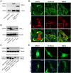
Formins are cytoskeleton regulating proteins characterized by a common FH2 structural domain. As key players in the assembly of actin filaments, formins direct dynamic cytoskeletal processes that influence cell shape, movement and adhesion. The large number of formin genes, fifteen in the human, suggests distinct tasks and expression patterns for individual family members, in addition to overlapping functions. Several formins have been associated with invasive cell properties in experimental models, linking them to cancer biology. One example is FMNL1, which is considered to be a leukocyte formin and is known to be overexpressed in lymphomas. Studies on FMNL1 and many other formins have been hampered by a lack of research tools, especially antibodies suitable for staining paraffin-embedded formalin-fixed tissues. Here we characterize, using bioinformatics tools and a validated antibody, the expression pattern of FMNL1 in human tissues and study its subcellular distribution. Our results indicate that FMNL1 expression is not restricted to hematopoietic tissues and that neoexpression of FMNL1 can be seen in epithelial cancer.
Keywords: FMNL1; FRL1; actin cytoskeleton; formin; immunohistochemistry.
© The Author(s) 2014.
Conflict of interest statement
Figures


Similar articles
-
The carboxy-terminus of the formin FMNL1ɣ bundles actin to potentiate adenocarcinoma migration.J Cell Biochem. 2019 Sep;120(9):14383-14404. doi: 10.1002/jcb.28694. Epub 2019 Apr 11. J Cell Biochem. 2019. PMID: 30977161
-
Human Macrophages Utilize the Podosome Formin FMNL1 for Adhesion and Migration.Cellbio (Irvine, Calif). 2015 Mar;4(1):1-11. doi: 10.4236/cellbio.2015.41001. Epub 2015 Mar 4. Cellbio (Irvine, Calif). 2015. PMID: 26942206 Free PMC article.
-
The formin FRL1 (FMNL1) is an essential component of macrophage podosomes.Cytoskeleton (Hoboken). 2010 Sep;67(9):573-85. doi: 10.1002/cm.20468. Cytoskeleton (Hoboken). 2010. PMID: 20617518 Free PMC article.
-
Formins: emerging players in the dynamic plant cell cortex.Scientifica (Cairo). 2012;2012:712605. doi: 10.6064/2012/712605. Epub 2012 Sep 26. Scientifica (Cairo). 2012. PMID: 24278734 Free PMC article. Review.
-
Fifteen formins for an actin filament: a molecular view on the regulation of human formins.Biochim Biophys Acta. 2010 Feb;1803(2):152-63. doi: 10.1016/j.bbamcr.2010.01.014. Epub 2010 Jan 25. Biochim Biophys Acta. 2010. PMID: 20102729 Review.
Cited by
-
Formin protein FMNL1 is a biomarker for tumor-infiltrating immune cells and associated with well immunotherapeutic response.J Cancer. 2023 Sep 11;14(16):2978-2989. doi: 10.7150/jca.86965. eCollection 2023. J Cancer. 2023. PMID: 37859818 Free PMC article.
-
Discovery of anti-Formin-like 1 protein (FMNL1) antibodies in membranous nephropathy and other glomerular diseases.Sci Rep. 2022 Aug 11;12(1):13659. doi: 10.1038/s41598-022-17696-w. Sci Rep. 2022. PMID: 35953506 Free PMC article.
-
Overexpression of Drosophila NUAK or Constitutively-Active Formin-Like Promotes the Formation of Aberrant Myofibrils.Cytoskeleton (Hoboken). 2025 Jan 29:10.1002/cm.21999. doi: 10.1002/cm.21999. Online ahead of print. Cytoskeleton (Hoboken). 2025. PMID: 39876757
-
Emerging functions of FMNL1 in myeloid neoplasms: insights from bioinformatics to biological and pharmacological landscapes.Transl Cancer Res. 2024 Nov 30;13(11):6105-6116. doi: 10.21037/tcr-24-1091. Epub 2024 Nov 27. Transl Cancer Res. 2024. PMID: 39697714 Free PMC article.
-
Formin Proteins FHOD1 and INF2 in Triple-Negative Breast Cancer: Association With Basal Markers and Functional Activities.Breast Cancer (Auckl). 2018 Aug 24;12:1178223418792247. doi: 10.1177/1178223418792247. eCollection 2018. Breast Cancer (Auckl). 2018. PMID: 30158824 Free PMC article.
References
-
- Faix J, Grosse R. (2006). Staying in shape with formins. Dev Cell 10:693-706 - PubMed
-
- Favaro P, Traina F, Machado-Neto JA, Lazarini M, Lopes MR, Pereira JK, Costa FF, Infante E, Ridley AJ, Saad ST. (2013). FMNL1 promotes proliferation and migration of leukemia cells. J Leukoc Biol 94:503-512 - PubMed
LinkOut - more resources
Full Text Sources
Other Literature Sources
Research Materials

