. 2014 Apr 4;344(6179):94-7.
doi: 10.1126/science.1248523.

Optical control of muscle function by transplantation of stem cell-derived motor neurons in mice

Affiliations

Optical control of muscle function by transplantation of stem cell-derived motor neurons in mice

J Barney Bryson et al. Science. .

Abstract

Damage to the central nervous system caused by traumatic injury or neurological disorders can lead to permanent loss of voluntary motor function and muscle paralysis. Here, we describe an approach that circumvents central motor circuit pathology to restore specific skeletal muscle function. We generated murine embryonic stem cell-derived motor neurons that express the light-sensitive ion channel channelrhodopsin-2, which we then engrafted into partially denervated branches of the sciatic nerve of adult mice. These engrafted motor neurons not only reinnervated lower hind-limb muscles but also enabled their function to be restored in a controllable manner using optogenetic stimulation. This synthesis of regenerative medicine and optogenetics may be a successful strategy to restore muscle function after traumatic injury or disease.


Figures

Fig. 1
Fig. 1. Expression of Gdnf in ChR2 motor neurons enhances survival and enables them to mature electrically in vitro.
(A) Embryoid bodies derived from CAG::ChR2-YFP/Gdnf transgenic ESCs and parental controls stained for the pan-motor neuron marker Isl1/2. GFP and YFP signals were detected by direct fluorescence. (B) Confocal images of MACS-sorted ESC motor neurons derived from CAG::ChR2-YFP (MACS, magnetic-activated cell sorting) and CAG::ChR2-YFP/Gdnf ESCs immunostained for Gdnf and GFP/YFP. (C) Survival analysis of sorted CAG:: ChR2-YFP and CAG::ChR2-YFP/Gdnf ESC motor neurons (MNs) (200 cells per well) plated on ESC astrocytes at indicated time points. CAG::ChR2-YFP motor neurons were cultured with (10 ng/ml) or without recombinant Gdnf (two replicates, analysis of variance with Bonferroni correction, *P < 0.25). Error bars indicate SEM. One representative of three separate experiments is shown. Wt, wild type. (D) Optogenetic stimulation (blue bars) of CAG::ChR2-YFP/Gdnf ESC motor neurons cultured on ESC astrocytes. Scale bars in (A) and (B), 50 µm.
Fig. 2
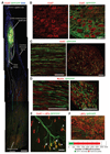
Fig. 2. Robust axonal growth and reinnervation of distal muscles after engraftment of ChR2 motor neurons.
(A) Image montage of a whole nerve and muscle section showing ChR2 motor neuron cell bodies at the graft site and axon projection (dashed lines indicate approximate trajectory). Scale bar, 500 µm. (B) Confocal image of engrafted ChR2 motor neurons immunolabeled for choline acetyl-transferase (ChAT; left image) and GFP and/or YFP (merged image at right). Scale bar, 50 µm. (C) Confocal image of longitudinal and transverse common peroneal nerve sections showing both ChR2 motor neurons and endogenous axons. Scale bar, 50 µm. (D) Confocal images of engrafted ChR2 motor neuron axons showing myelination. Scale bar, 50 µm. (E) Confocal z-stack of ChR2 motor neuron axon terminals innervating multiple neuromuscular junctions within the TS muscle. Arrows indicate preterminal collateral sprouting, arrowheads denote terminal sprouting, and the asterisk indicates an endogenous motor axon. Scale bar, 50 µm. (F) Two-dimensional projection image of a TS muscle showing proportion of neuromuscular junctions (NMJs) innervated by engrafted ChR2 motor neurons, relative to the total number of end plates present [labeled with α-bungarotoxin (αBTx)]. Quantification is shown below. Representative images shown here are compiled from n = 4 engrafted nerves from three separate experiments.
Fig. 3
Fig. 3. Restoration of muscle function, in a controlled manner, using optical stimulation of engrafted ChR2 motor neurons in vivo.
(A) Schematic showing optical stimulation and isometric muscle tension recordings. EB, embryoid body. Representative twitch (B), tetanic (C), and repetitive tetanic (D) contraction traces obtained from the TA muscle, induced by optical stimulation. Blue line, muscle force; red line, electrical trigger signals sent to the LED unit. (E) Quantification of twitch and tetanic contraction of TA and EDL muscles. Time to peak contractile force, from initiation of the electrical trigger to the LED unit (F) or from the initiation of muscle contraction (G), is shown alongside direct electrical nerve stimulation (n values represent optical/electrical stimulation, respectively, compiled from four separate experiments). (H) Representative fatigue traces from TA muscles (different animals) produced by optical (top) or electrical (bottom) stimulation for 180 s. (I) Representative TA muscle optical stimulation motor-unit number estimate trace. The asterisk indicates square-wave trigger voltage to the LED unit and oscilloscope trigger. (J) Motor-unit number quantification of TA and EDL muscles after optical versus electrical stimulation. (K) Analysis of average motor-unit force. The dashed line indicates the normal EDL value. All error bars indicate SEM.

Comment in

References

    1. Glenn WW, Phelps ML. Neurosurgery. 1985;17:974–984. - PubMed
    1. Onders RP, et al. Surg Endosc. 2009;23:1433–1440. - PubMed
    1. Gernandt B. Acta Physiol Scand. 1946;12:255–260.
    1. Zhang SC. J Hematother Stem Cell Res. 2003;12:625–634. - PubMed
    1. Filbin MT. Nat Rev Neurosci. 2003;4:703–713. - PubMed

Publication types

MeSH terms

Substances