. 2014 Oct;25(10):2303-15.
doi: 10.1681/ASN.2013070690. Epub 2014 Apr 3.

AKI after conditional and kidney-specific knockdown of stanniocalcin-1

Affiliations

AKI after conditional and kidney-specific knockdown of stanniocalcin-1

Luping Huang et al. J Am Soc Nephrol. 2014 Oct.

Abstract

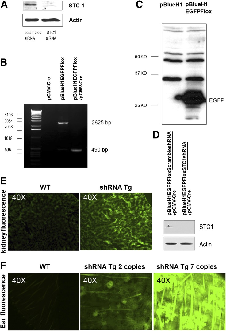
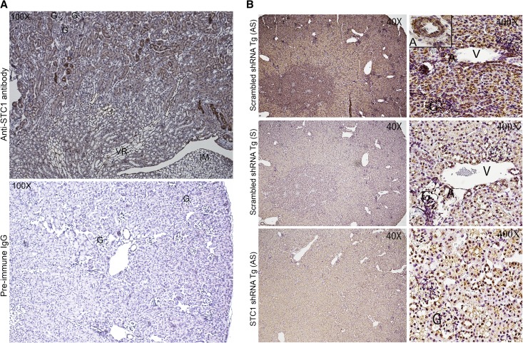
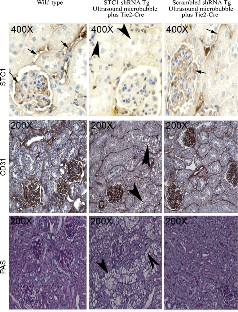
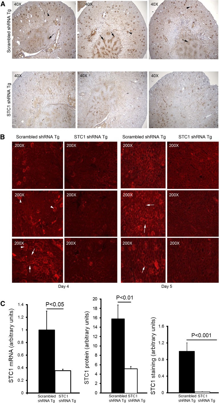
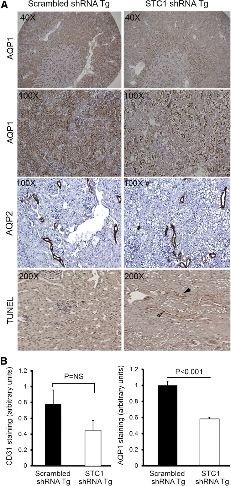
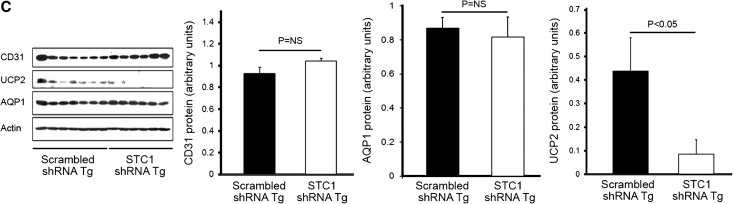
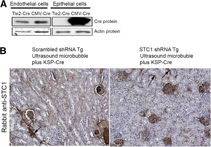
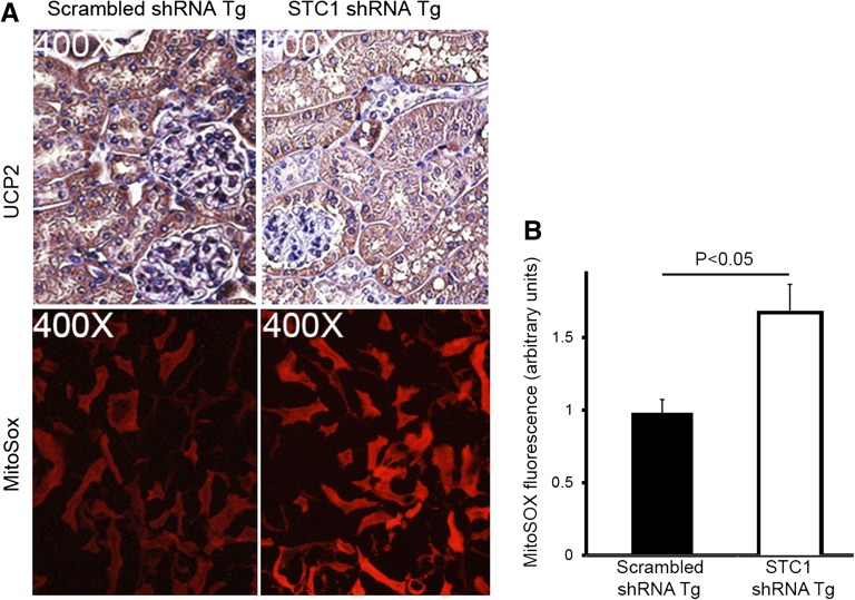
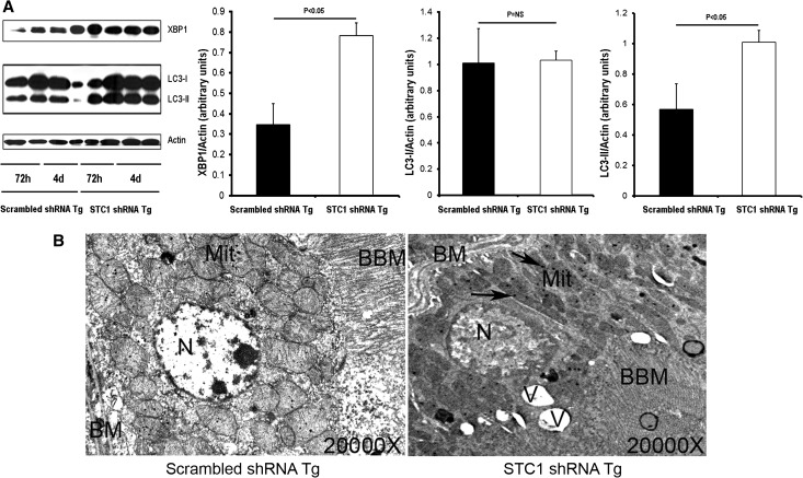
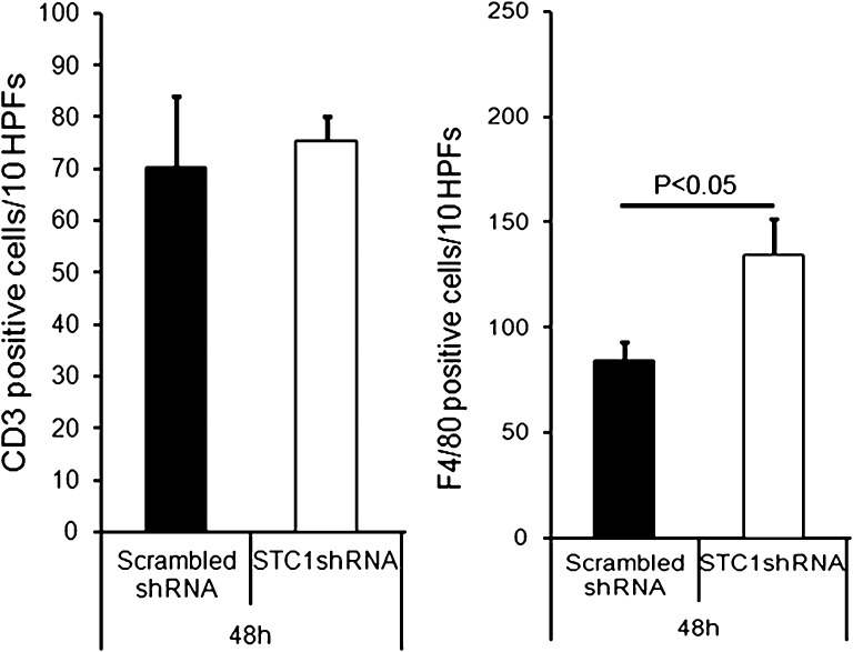
Stanniocalcin-1 is an intracrine protein; it binds to the cell surface, is internalized to the mitochondria, and diminishes superoxide generation through induction of uncoupling proteins. In vitro, stanniocalcin-1 inhibits macrophages and preserves endothelial barrier function, and transgenic overexpression of stanniocalcin-1 in mice protects against ischemia-reperfusion kidney injury. We sought to determine the kidney phenotype after kidney endothelium-specific expression of stanniocalcin-1 small hairpin RNA (shRNA). We generated transgenic mice that express stanniocalcin-1 shRNA or scrambled shRNA upon removal of a floxed reporter (phosphoglycerate kinase-driven enhanced green fluorescent protein) and used ultrasound microbubbles to deliver tyrosine kinase receptor-2 promoter-driven Cre to the kidney to permit kidney endothelium-specific shRNA expression. Stanniocalcin-1 mRNA and protein were expressed throughout the kidney in wild-type mice. Delivery of tyrosine kinase receptor-2 promoter-driven Cre to stanniocalcin-1 shRNA transgenic kidneys diminished the expression of stanniocalcin-1 mRNA and protein throughout the kidneys. Stanniocalcin-1 mRNA and protein expression did not change in similarly treated scrambled shRNA transgenic kidneys, and we observed no Cre protein expression in cultured and tyrosine kinase receptor-2 promoter-driven Cre-transfected proximal tubule cells, suggesting that knockdown of stanniocalcin-1 in epithelial cells in vivo may result from stanniocalcin-1 shRNA transfer from endothelial cells to epithelial cells. Kidney-specific knockdown of stanniocalcin-1 led to severe proximal tubule injury characterized by vacuolization, decreased uncoupling of protein-2 expression, greater generation of superoxide, activation of the unfolded protein response, initiation of autophagy, cell apoptosis, and kidney failure. Our observations suggest that stanniocalcin-1 is critical for tubular epithelial survival under physiologic conditions.


Figures

Figure 1.
Figure 1.
Validation of shRNA transgenic construct and genotyping of shRNA Tg mice. (A) siRNA knockdown of STC1 protein in C2C12 cells: Mouse C2C12 cells were transfected with 19-bp STC1 siRNA or 19-bp scrambled siRNA; 48 hours after transfection, cells were lysed and equal amounts of protein were run on SDS-PAGE; Western blot was probed with rabbit anti-STC1 antibody. (B) Cre recombinase excision of floxed PGK-EGFP reporter in pBlueH1EGFPFlox-transfected HEK293 cells: HEK293 cells were transfected with pCMV-Cre, pBlueH1EGFPFlox, or pCMV-Cre plus pBlueH1EGFPFlox; 48 hours after transfection, cells were scraped and collected by centrifugation; DNA was isolated and subjected to PCR amplification using primers that flank the PGK-EGFP sequences in pBlueH1EGFPFlox. DNA isolated from cells transfected with pCMV-Cre alone, yielded no product; DNA isolated from cells transfected with pBlueH1EGFPFlox alone yielded an expected 2625-bp product; DNA isolated from cells cotransfected with pCMV-Cre and pBlueH1EGFPFlox yielded a truncated PCR product of expected size (490 bp), confirming functional Flox P sites in pBlueH1EGFPFlox. (C) Expression of EGFP in pBlueH1EGFPFlox-transfected HEK293 cells: HEK293 cells were transfected with empty vector (pBlueH1), or pBlueH1EGFPFlox (PGK-driven EGFP); 48 hours after transfection, cells were lysed and equal amounts of protein were resolved on SDS-PAGE; Western blot was probed with anti-GFP; EGFP expression is observed in cells transfected with pBlueH1EGFPFlox but not in cells transfected with pBlueH1, confirming active EGFP reporter. (D) Knockdown of native STC1 expression in Att20/D16v-F2 cells transfected with pCMV-Cre and pBlueH1FloxEGFPSTC1shRNA. Att20/D16v-F2 cells were co-transfected with pCMV-Cre and pBlueH1FloxEGFPSTC1shRNA, or pCMV-Cre and pBlueH1FloxEGFPScrambledshRNA; 48 hours later, cells were lysed and equal amounts of protein were resolved on SDS-PAGE; Western blots were reacted with anti-STC1 and anti-actin; knockdown of STC1 protein is observed in cells cotransfected with pCMV-Cre and pBlueH1FloxEGFPSTC1shRNA but not in cells co-transfected with pCMV-Cre and pBlueH1FloxEGFPScrambledshRNA; these findings confirm H1-driven shRNA expression upon excision of floxed reporter and efficient knockdown of native STC1 expression in cells transfected with pBlueH1FloxEGFPSTC1shRNA, but not in cells transfected with pBlueH1FloxEGFPScrambledshRNA. Mouse genotyping: In addition to genotyping of shRNA Tg mice by PCR using EGFP-specific primers, transgenic mouse genotype and an estimate of copy number of the transgene may be discerned by green fluorescence intensity of kidney or ear skin fresh sections under fluorescence microscope: (E) kidney; (F) skin.
Figure 2.
Figure 2.
(A) Wide distribution of STC1 protein in the kidney and strong staining in blood vessels and some cortical tubules. Formalin-fixed and EDTA-treated WT kidney sections were stained with goat anti-STC1. Upper panel shows wide distribution of STC1 immunoreactivity (albeit with variable staining intensity); note strong staining in some cortical tubules, possibly representing CD and blood vessels including medullary rays. No staining is observed in preimmune serum-treated sections (lower panel). G, glomerulus; IM, inner medulla; VR, vasa recta. (B) Wide distribution of STC1 mRNA in the kidney. Upper panels: scrambled shRNA Tg kidneys harvested 4 days after the delivery of pTie2-Cre and probed with antisense STC1 probe reveal expression of STC1 mRNA (brown color) in the entire kidney; left upper corner inset in the cortical panel shows expression of STC1 mRNA in the endothelium and adventitia of a midsize artery. Middle panels: consecutive sections from scrambled shRNA Tg kidneys, shown in the upper panels, were probed with sense STC1 probe and show minimal background signal. Lower panel: sections from STC1 shRNA Tg kidneys harvested 4 days after the delivery of pTie2-Cre and probed with anti-sense STC1 probe; note diminished expression of STC1 mRNA in the entire kidney. A, artery; G, glomerulus; V, vein.
Figure 3.
Figure 3.
Expression of STC1 protein in proximal tubules and glomerular and peritubular capillaries: Loss of STC1 immunoreactivity in shRNA Tg kidneys after delivery of pTie2-Cre is accompanied by tubular vacuolization. pTie2-Cre was delivered to the right kidney of STC1 shRNA and scrambled shRNA Tg mice; kidneys were harvested 4 days later. Methacarn-fixed kidney sections were stained with rabbit anti-STC1 antibody (top panels); anti-CD31 (middle panels); periodic acid–Schiff (bottom panels). Sections from WT kidneys were included for comparison. Strong immunoreactivity for STC1 in peritubular and glomerular capillaries (arrows) and positive staining in epithelial cells (brown hue). Similar distribution of STC1 immunoreactivity is observed in WT kidneys and scrambled shRNA Tg kidneys that were treated with pTie2-Cre. Delivery of pTie2-Cre to STC1 shRNA Tg kidneys leads to knockdown of STC1 expression in peritubular and glomerular capillaries, as well as epithelial cells; the appearance of tubular vacuolization (arrowheads). Expression of the endothelial cell marker, CD31, is not affected by delivery of pTie2-Cre to STC1 shRNA or scrambled shRNA Tg kidneys.
Figure 4.
Figure 4.
Efficient whole kidney knockdown of STC1 mRNA and protein. pTie2-Cre was delivered to the right kidneys of STC1 and scrambled shRNA Tg mice; kidneys were harvested 2–5 days later (to establish time course), fixed with methacarn, and stained with rabbit anti-STC1. This antibody highlights the expression of STC1 in blood vessels. (A) STC1 immunolabeling in the kidneys of STC1 shRNA Tg and scrambled shRNA Tg mice 4 days after the delivery of pTie2-Cre. Representative images from three different mice per group are shown. Arrows point to medullary rays; arrowheads point to glomeruli. (B) STC1 immunofluorescence in the kidneys of STC1 shRNA Tg and scrambled shRNA Tg mice, 4 and 5 days after the delivery of pTie2-Cre. Kidney sections were stained with rabbit anti-STC1, followed by treatment with Texas red–tagged anti-rabbit. Representative cortical images from three different mice per group are shown. Arrowheads point to glomeruli; arrows point to circular structures, representing tubules and peritubular capillaries. (C) Bar graphs show quantitation of STC1 mRNA/actin mRNA using real-time PCR carried out on RNA representing whole kidney; quantitation of STC1 protein/actin protein on Western blots, using protein lysates representing whole kidney; quantitation of staining for STC1 using image tool software. Data represent the mean±SEM of six independent determinations.
Figure 5.
Figure 5.
(A) STC1 knockdown induces vacuolization in S1 and S2 segments of the PT and promotes apoptosis. pTie2-Cre was delivered to the right kidneys of STC1 shRNA and scrambled shRNA Tg mice. Kidneys were harvested 4 days later and fixed in methacarn. Sections were stained with rabbit anti-AQP1, rabbit anti-AQP2, or TUNEL for apoptosis. Note that STC1 shRNA Tg kidneys display selective injury (vacuolization) in AQP1-positive tubules, in the outer two thirds of the cortex, with relative preservation of AQP1 staining in the inner third, consistent with tubular injury in the S1 and S2 segments of the PT. No vacuolization is observed in AQP2-expressing cells. Apoptotic cells (arrowheads) are more prevalent in STC1 shRNA Tg kidneys. (B) Apparent decrease in AQP1 and CD31 staining after STC1 knockdown does not correlate with decrease in protein expression (refer to C). Bar graphs show quantitation of staining for CD31 and AQP1 (using Image Tool software) 4 days after the delivery of Tie2-Cre to scrambled shRNA and STC1 shRNA Tg kidneys. (C) STC1 knockdown diminishes the expression of UCP2, but not AQP1 and CD31 proteins. Four days after the delivery of Tie2-Cre to scrambled shRNA and STC1 shRNA Tg kidneys, equal amounts of cortical homogenate in RIPA buffer were run on SDS-PAGE and blots were reacted with antibodies for CD31, UCP2, AQP1, and actin. Representative blots are shown. Bar graphs show the mean±SEM of band intensities for the above proteins normalized to actin; data represent six independent determinations.
Figure 5.
Figure 5.
(A) STC1 knockdown induces vacuolization in S1 and S2 segments of the PT and promotes apoptosis. pTie2-Cre was delivered to the right kidneys of STC1 shRNA and scrambled shRNA Tg mice. Kidneys were harvested 4 days later and fixed in methacarn. Sections were stained with rabbit anti-AQP1, rabbit anti-AQP2, or TUNEL for apoptosis. Note that STC1 shRNA Tg kidneys display selective injury (vacuolization) in AQP1-positive tubules, in the outer two thirds of the cortex, with relative preservation of AQP1 staining in the inner third, consistent with tubular injury in the S1 and S2 segments of the PT. No vacuolization is observed in AQP2-expressing cells. Apoptotic cells (arrowheads) are more prevalent in STC1 shRNA Tg kidneys. (B) Apparent decrease in AQP1 and CD31 staining after STC1 knockdown does not correlate with decrease in protein expression (refer to C). Bar graphs show quantitation of staining for CD31 and AQP1 (using Image Tool software) 4 days after the delivery of Tie2-Cre to scrambled shRNA and STC1 shRNA Tg kidneys. (C) STC1 knockdown diminishes the expression of UCP2, but not AQP1 and CD31 proteins. Four days after the delivery of Tie2-Cre to scrambled shRNA and STC1 shRNA Tg kidneys, equal amounts of cortical homogenate in RIPA buffer were run on SDS-PAGE and blots were reacted with antibodies for CD31, UCP2, AQP1, and actin. Representative blots are shown. Bar graphs show the mean±SEM of band intensities for the above proteins normalized to actin; data represent six independent determinations.
Figure 6.
Figure 6.
(A) Endothelium-specific expression of pTie2-Cre. Cultured TKPTS and endothelial cells were transfected with pTie2-Cre or pCMV-Cre; 48 hours after transfection, cells were lysed in RIPA buffer and equal amounts of proteins were resolved on SDS-PAGE; Western blots were reacted with anti-Cre and anti-actin. Cre protein is expressed in endothelial cells transfected with pTie2-Cre or pCMV-Cre; however, TKPTS express Cre protein upon transfection with pCMV-Cre, but not pTie2-Cre. (B) Epithelium-specific expression of STC1 shRNA also leads to epithelial vacuolization. KSP-Cre was delivered to the right kidneys of scrambled shRNA and STC1 shRNA Tg kidneys using ultrasound microbubbles. Kidneys were harvested 4 days later, and methacarn-fixed sections were subjected to immunolabeling using rabbit-anti STC1. Note diminished staining for STC1 in epithelial cells and most peritubular capillaries, whereas staining is preserved in glomerular capillaries. Concomitant with STC1 knockdown in epithelial cells, we observe widespread vacuolization in epithelial cells as observed following the delivery of pTie2-Cre.
Figure 7.
Figure 7.
STC1 knockdown in the kidney is associated with diminished UCP2 expression and increased superoxide generation in kidney tubules. (A) pTie2-Cre was delivered to the right kidneys of STC1 and scrambled shRNA Tg mice. Kidneys were harvested 4 days later: methacarn-fixed sections were stained with rabbit anti-UCP2, and freshly isolated 1-mm-thick kidney slices were incubated with MitoSOX, followed by fixation in 4% paraformaldehyde. Kidney tissue was then imbedded in optical cutting temperature medium and 5-µM sections were viewed under a fluorescence microscope. Diminished staining for UCP2 in STC1 shRNA Tg kidneys correlates with higher superoxide generation (MitoSOX fluorescence). (B) Bar graph shows quantitation of MitoSOX fluorescence using Image Tool software in scrambled shRNA and STC1 shRNA Tg kidneys 4 days after the delivery of Tie2-Cre to the kidney. Data represent the mean±SEM of three independent determinations.
Figure 8.
Figure 8.
STC1 knockdown activates UPR and induces autophagy. (A) pTie2-Cre was delivered to the right kidneys of STC1 shRNA and scrambled shRNA Tg mice. Kidneys were harvested 3 or 4 days later and lysed in RIPA buffer. Equal amounts of proteins were resolved on SDS-PAGE, and Western blots were reacted with rabbit anti-XBP1, rabbit anti-LC3, and rabbit anti-actin. Representative blots are shown (n=2 for each time point; n=4 for cumulative data from days 3 and 4). Bar graphs represent the mean±SEM of four independent determinations (pooled data for 3 and 4 days). (B) Electron micrographs show vacuolization and mitochondrial changes after STC1 knockdown. BBM, brush border membrane; BM, basal membrane; Mit, mitochondria; N, nucleus; V, vacuole. Arrows point to mitochondrial inclusion bodies.
Figure 9.
Figure 9.
AKI after STC1 knockdown. Creatinine clearance, serum creatinine, and urine protein in scrambled shRNA and STC1 shRNA Tg mice 4 days after the delivery of Tie2-Cre.
Figure 10.
Figure 10.
Knockdown of STC1 in the kidney is associated with greater macrophage infiltration. pTie2-Cre was delivered to the right kidneys of STC1 shRNA and scrambled shRNA Tg mice. Kidneys were harvested 4 days later, and methacarn-fixed sections were stained with rat anti-F4/80 (marker of macrophages/dendritic cells) or rabbit anti-CD3 (T-cell marker). Total F4/80+ and CD3+ cells infiltrating the cortex were counted, and the results were expressed as the total number of cells per 10 grids (1cm2 graded ocular grids viewed at ×20 magnification) spanning the entire cortex, using Nikon Eclipse 80i microscope system; bar graphs represents the mean±SEM of cell number from 10 grids. HPF, high-power field.

References

    1. De Niu P, Radman DP, Jaworski EM, Deol H, Gentz R, Su J, Olsen HS, Wagner GF: Development of a human stanniocalcin radioimmunoassay: Serum and tissue hormone levels and pharmacokinetics in the rat. Mol Cell Endocrinol 162: 131–144, 2000 - PubMed
    1. Re RN, Cook JL: The mitochondrial component of intracrine action. Am J Physiol Heart Circ Physiol 299: H577–H583, 2010 - PMC - PubMed
    1. Jellinek DA, Chang AC, Larsen MR, Wang X, Robinson PJ, Reddel RR: Stanniocalcin 1 and 2 are secreted as phosphoproteins from human fibrosarcoma cells. Biochem J 350: 453–461, 2000 - PMC - PubMed
    1. Luo CW, Kawamura K, Klein C, Hsueh AJ: Paracrine regulation of ovarian granulosa cell differentiation by stanniocalcin (STC) 1: Mediation through specific STC1 receptors. Mol Endocrinol 18: 2085–2096, 2004 - PubMed
    1. McCudden CR, James KA, Hasilo C, Wagner GF: Characterization of mammalian stanniocalcin receptors. Mitochondrial targeting of ligand and receptor for regulation of cellular metabolism. J Biol Chem 277: 45249–45258, 2002 - PubMed

Publication types

LinkOut - more resources