. 2014 Jun;28(6):872-85.
doi: 10.1210/me.2014-1055. Epub 2014 Apr 4.

Coactivator MYST1 regulates nuclear factor-κB and androgen receptor functions during proliferation of prostate cancer cells

Affiliations

Coactivator MYST1 regulates nuclear factor-κB and androgen receptor functions during proliferation of prostate cancer cells

Anbalagan Jaganathan et al. Mol Endocrinol. 2014 Jun.

Abstract

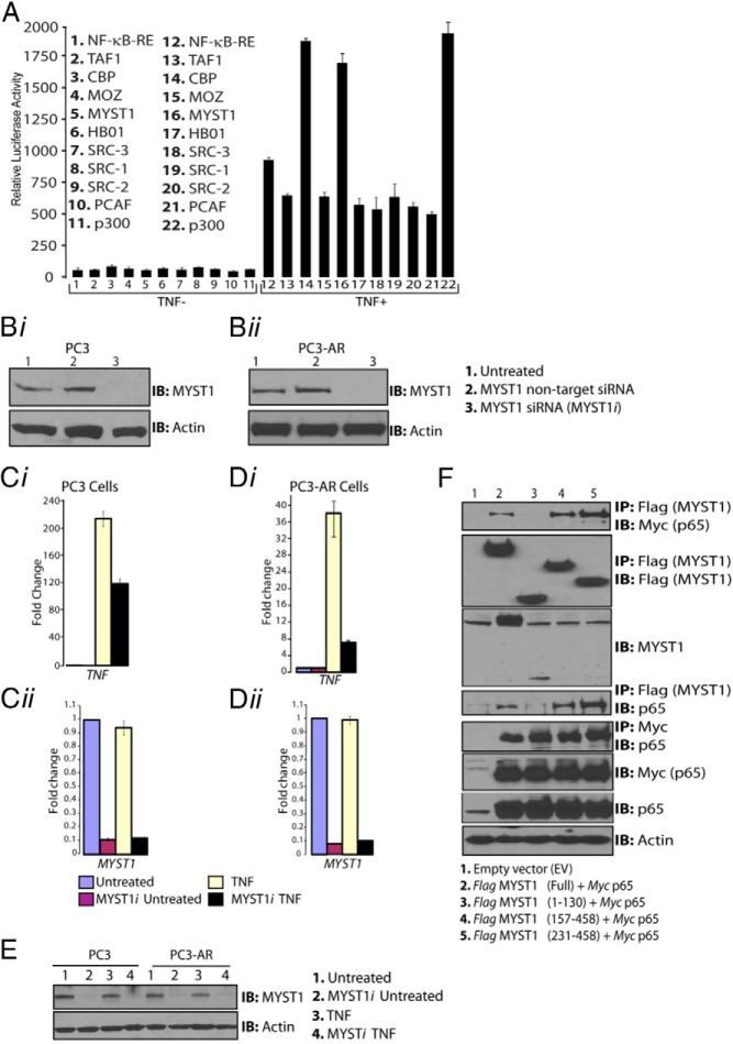
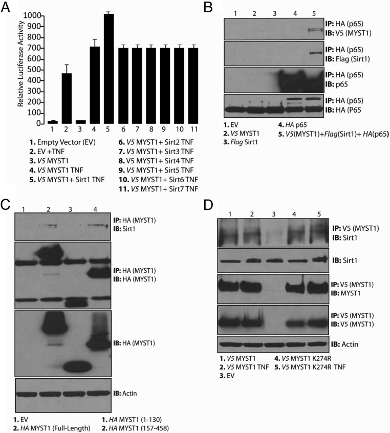
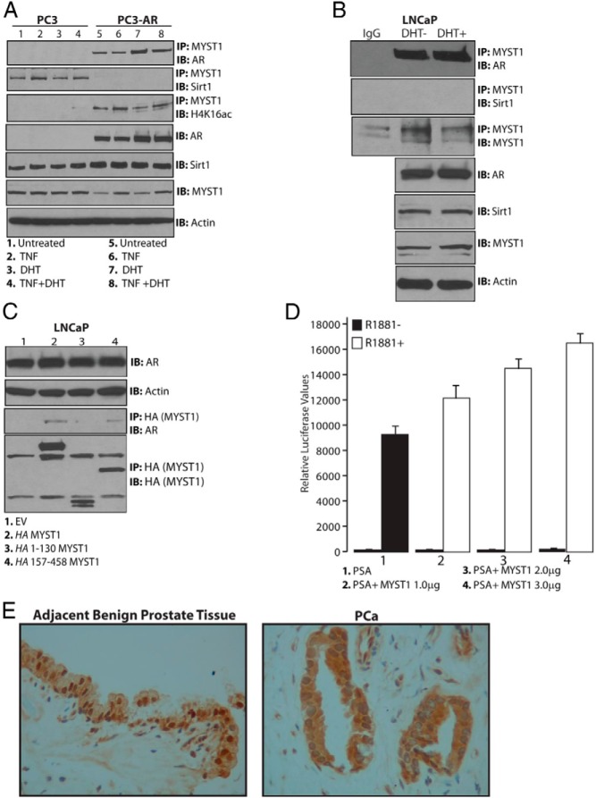
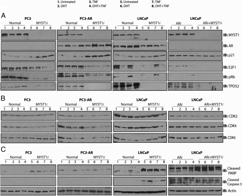
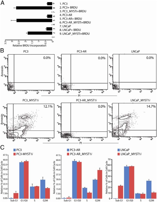
In prostate cancer (PCa), the functional synergy between androgen receptor (AR) and nuclear factor-κ B (NF-κB) escalates the resistance to therapeutic regimens and promotes aggressive tumor growth. Although the underlying mechanisms are less clear, gene regulatory abilities of coactivators can bridge the transcription functions of AR and NF-κB. The present study shows that MYST1 (MOZ, YBF2 and SAS2, and TIP60 protein 1) costimulates AR and NF-κB functions in PCa cells. We demonstrate that activation of NF-κB promotes deacetylation of MYST1 by sirtuin 1. Further, the mutually exclusive interactions of MYST1 with sirtuin 1 vs AR regulate the acetylation of lysine 16 on histone H4. Notably, in AR-lacking PC3 cells and in AR-depleted LNCaP cells, diminution of MYST1 activates the cleavage of poly(ADP-ribose) polymerase and caspase 3 that leads to apoptosis. In contrast, in AR-transformed PC3 cells (PC3-AR), depletion of MYST1 induces cyclin-dependent kinase (CDK) N1A/p21, which results in G2M arrest. Concomitantly, the levels of phospho-retinoblastoma, E2F1, CDK4, and CDK6 are reduced. Finally, the expression of tumor protein D52 (TPD52) was unequivocally affected in PC3, PC3-AR, and LNCaP cells. Taken together, the results of this study reveal that the functional interactions of MYST1 with AR and NF-κB are critical for PCa progression.


Figures

Figure 1.
Figure 1.
MYST1 is a potential coactivator of NF-κB transcriptional activity. A, PC3 cells were cotransfected with a battery of HAT-containing coactivators and tested by a luciferase-based reporter assay for activation of NF-κB-RE. After 24 hours of transfection, cells were treated with 25 ng of TNF/mL of medium for 30 minutes. After cell lysis and determination of luciferase activity, results showed that the activation of NF-κB-RE was augmented by MYST1, CBP, and p300. Bi and Bii, IB analysis confirmed the down-regulation of MYST1 expression by siRNA (MYST1i) in parental PC3 and PC3-AR cells without affecting the expression of the housekeeping protein actin. Ci and Di, qRT-PCR data showed an increased level of TNF transcript, which was down-regulated by 50% as a result of the knockdown of MYST1 in parental PC3 cells and was reduced by 80% in PC3-AR cells. Cii, Dii, and E, No changes were observed in the levels of MYST1 mRNA (Cii and Dii) and protein (E) in PC3 and PC3-AR cells. F, After 24 hours, cells were harvested and lysed, and IP was performed with equal quantities of cell lysates by incubation with anti-Flag antibody. The IB analysis revealed that p65 interacted with the 157–458 and 231–458 regions of MYST1, which represents the HAT domain.
Figure 2.
Figure 2.
Deacetylation of MYST1 activates NF-κB functions. A, After 24 hours of transfection, the cells were harvested and lysed, and, subsequently, respective cell lysates were subjected to IP with anti-V5 followed by IB with Kac antibodies. Autoacetylation of V5-tagged wild-type MYST1 was dramatically abrogated upon the mutagenesis of lysine 274 to arginine (MYST1K274R). B, qRT-PCR analysis revealed that MYST1K274R was more potent than wild-type MYST1 in activating the TNF expression in PC3 cells after treatment with TNF. C, Similarly, qRT-PCR analysis revealed that MYST1K274R was more potent in activating the TNF expression in PC3-AR cells after treatment with TNF. D, V5-MYST1 in untreated PC3 cells showed autoacetylation and interaction with p65, which was completely lost upon treatment with TNF.
Figure 3.
Figure 3.
Sirt1 regulates autoacetylation of MYST1. A, A luciferase assay was performed to confirm that Sirt1 potentiated the MYST1-mediated activation of NF-κB after TNF treatment of PC3 cells that were cotransfected with MYST1, Sirt1, and NF-κB RE. B, Molecular interaction of MYST1, p65, and Sirt1 in PC3 cells transfected with V5-MYST1, HA-p65, and Flag-Sirt1 after IP with HA followed by IB with V5 and Flag antibodies. C, In PC3 cells, the endogenous Sirt1 showed interaction with the full-length and C terminus of MYST1 (157–458). D, Endogenous Sirt1 interacted with both wild-type and mutant MYST1K274R independent of TNF treatment of PC3 cells.
Figure 4.
Figure 4.
Mutually exclusive interactions of MYST1 with Sirt1 or AR in PCa cells. A and B, Endogenous MYST1 interacted with Sirt1 in PC3 cells but not in PC3-AR and LNCaP cells wherein MYST1 interacts with AR. C, Endogenous AR interacted with the full-length and the 157–458 region of MYST1. D, Induction of PSA luciferase activity in LNCaP cells by increasing amounts of MYST1-expressing plasmid upon treatment with 10 nM R1881 treatment confirms the role of MYST1 as a coactivator of AR. E, Immunohistochemical analysis with anti-MYST1 antibodies of formalin-fixed human prostate cancer tissues (n = 5) revealed that MYST1 was nuclear in the benign prostate epithelium adjacent to PCa cells, in which MYST1 was cytoplasmic as well as nuclear. Original magnification, ×200.
Figure 5.
Figure 5.
MYST1 depletion affects the expression of cell cycle regulatory genes. PC3 and PC3-AR cells were treated either alone with 25 ng/mL TNF for 30 minutes or 100 nM DHT for 24 hours and in combination, in which TNF was added after 24 hours of DHT treatment. A, B, and C, MYST1 was depleted (MYST1i) by siRNA in PC3, PC3-AR, and LNCaP cells. Then, the parental and MYST1i cells were examined for the expression of E2F1, PSA, CDKN1A, TPD52, and ICAM1. ***, No PSA was detected in PC3 and PC3-AR cells. The fold changes between DHT+TNF and DHT+TNF+MYSTi are expressed as the means ± SEM (n = 3): #, P < .05; ##, P < .01.
Figure 6.
Figure 6.
MYST1 differentially regulates the level of proteins that regulate cell cycle and apoptosis. A, All the IB analyses were performed on the same batch of biological samples. Panel 1 demonstrates effective depletion of MYST1 (MYST1i), ARi, and ARi+MYST1i proteins in PC3, PC3-AR, and LNCaP cells compared with that in the parental lines. Panel 2 shows the levels of AR in all the parental and siRNA-treated cell lines. Panels 3, 4, and 5 show that down-regulation of MYST1 in PC3 and PC3-AR cells enhanced the level of p21 (panel 3), which was concomitant with the dramatic decline in the levels of E2F1 (panel 3) and p-Rb (panel 4). Note that the dilution of anti-p21 antibody used for PC3-AR cells was 1:2000, which did not detect any p21 protein in PC3 cells until a 1:500 dilution was used to probe the respective membrane. Slight increases in p21 levels could be observed in parental LNCaP cells as a result of the MYST1 depletion that was not seen in AR alone (ARi)– or ARi+MYSTi–depleted LNCaP cells. Panel 6 shows that the levels of TPD52 are negatively affected in all of the respective cell lines because of the down-regulation of MYST1. B, No significant alterations in the levels of CDK2 (panel 1), CDK4 (panel 2), and CDK6 (panel 3) were observed in parental and MYST1i PC3 cells. However, the PC3-AR cells showed an overall reduction in the levels of CDK2, CDK4, and CDK6 upon MYST1 down-regulation. Although the levels of CDK2, CDK4, and CDK6 are detectable in MYST1-depleted PC3-AR cells after treatments, their levels are still lower than those of the parental PC3-AR cells. The LNCaP cells showed a decline in the level of CDK6, depending on the expression of MYST1. However, the levels of CDK2, CDK4, and CDK6 remained unchanged in ARi and ARi+MYST1i LNCaP cells. C, Although PC3 and LNCaP cells showed substantial increases in the levels of cleaved PARP (panel 1) due to MYST1 depletion, cleaved caspase 3 (panel 2) was only detectable in MYST1i, ARi, and ARi+MYST1i LNCaP cells.
Figure 7.
Figure 7.
MYST1 mediates apoptosis or G2M arrest in PC3 and PC3-AR cells. A, BrdU assay reveals that MYST1-depleted cells exhibited reduced incorporation of BrdU compared with that in the parental PC3, PC3-AR, and LNCaP cells. B, Annexin/PI staining by flow cytometry revealed that the lower BrdU incorporation in PC3 and LNCaP cells could be due to the loss of cells by apoptosis. The values have been added to the quadrant that represents double-positive cells, which indicates that these cells demonstrated annexin as well as PI positivity. These panels show a representative of 3 experiments performed independently. C, Apoptosis and the cell cycle were analyzed by flow cytometry after staining of the cells with PI and RNase treatment. Depletion of MYST1 in PC3 and LNCaP cells led to accumulation of cells in the sub-G1 phase, which confirms apoptosis. In PC3-AR cells, MYST1 down-regulation that activates p21 leads to G2M arrest. Means and SD were calculated from the data acquired from 3 independent experiments, which were performed in triplicate with each cell type. The percentage of cells in each phase of the cell cycle was calculated from the total number of gated cells, which was set to 100%.
Figure 8.
Figure 8.
A model depicting the functional and molecular interplay of MYST1 with AR, NF-κB, and Sirt1 in PCa. MYST1 interacts with the key transcription factors and also plays a critical role in the expression of key cell cycle regulatory proteins. The model shows that the proinflammatory milieu, such as TNF treatment, causes deacetylation of MYST1. The green ball represents the acetyl moiety, which is removed by Sirt1 after TNF treatment. Further, we advocate that because of the mutually exclusive nature of MYST1 interactions, the MYST1–p65-Sirt1 complex act as a repressor complex and the MYST1–p65-AR complex functions as an activator complex, which together enhance proliferation and inhibit growth arrest or apoptosis of PCa cells.

Similar articles

Cited by

References

    1. Shiota M, Yokomizo A, Masubuchi D, et al. Tip60 promotes prostate cancer cell proliferation by translocation of androgen receptor into the nucleus. Prostate. 2010;70:540–554. - PubMed
    1. Cronauer MV, Culig Z. Molecular aspects of prostate cancer. World J Urol. 2012;30:277–278. - PubMed
    1. Culig Z, Santer FR. Androgen receptor co-activators in the regulation of cellular events in prostate cancer. World J Urol. 2012;30:297–302. - PubMed
    1. Gaughan L, Logan IR, Cook S, Neal DE, Robson CN. Tip60 and histone deacetylase 1 regulate androgen receptor activity through changes to the acetylation status of the receptor. J Biol Chem. 2002;277:25904–25913. - PubMed
    1. Halkidou K, Gnanapragasam VJ, Mehta PB, et al. Expression of Tip60, an androgen receptor coactivator, and its role in prostate cancer development. Oncogene. 2003;22:2466–2477. - PubMed

Publication types

MeSH terms