Review
. 2014 Jul 20;21(3):396-413.
doi: 10.1089/ars.2014.5851. Epub 2014 Jun 12.

Endoplasmic reticulum stress and oxidative stress in cell fate decision and human disease

Affiliations
Review

Endoplasmic reticulum stress and oxidative stress in cell fate decision and human disease

Stewart Siyan Cao et al. Antioxid Redox Signal. .

Abstract

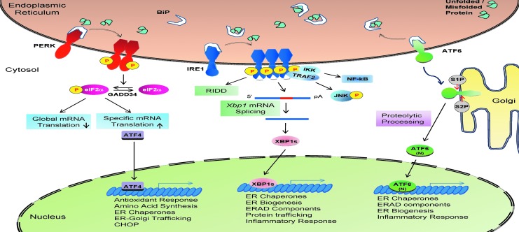
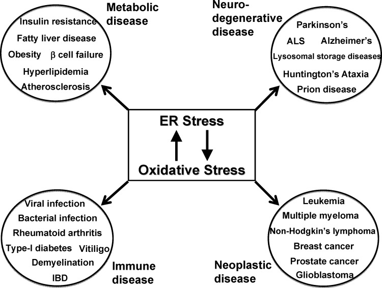
Significance: The endoplasmic reticulum (ER) is a specialized organelle for the folding and trafficking of proteins, which is highly sensitive to changes in intracellular homeostasis and extracellular stimuli. Alterations in the protein-folding environment cause accumulation of misfolded proteins in the ER that profoundly affect a variety of cellular signaling processes, including reduction-oxidation (redox) homeostasis, energy production, inflammation, differentiation, and apoptosis. The unfolded protein response (UPR) is a collection of adaptive signaling pathways that evolved to resolve protein misfolding and restore an efficient protein-folding environment.

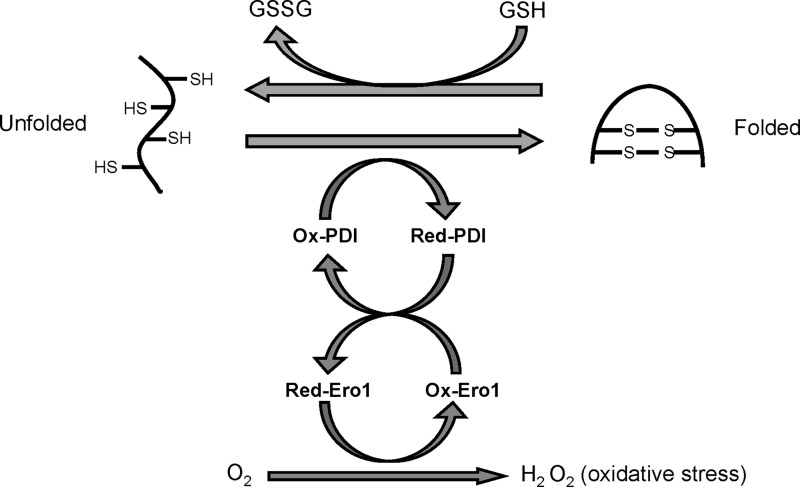
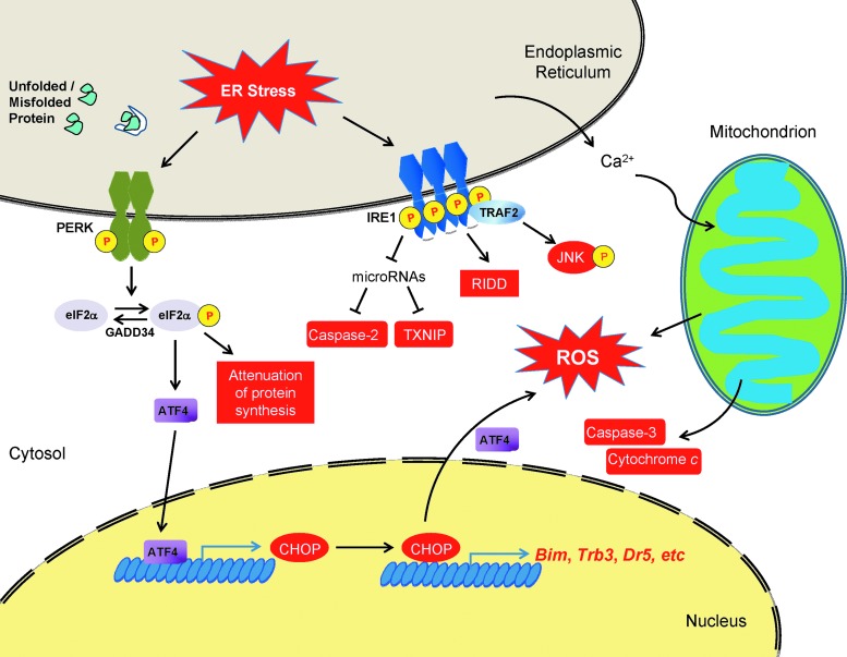
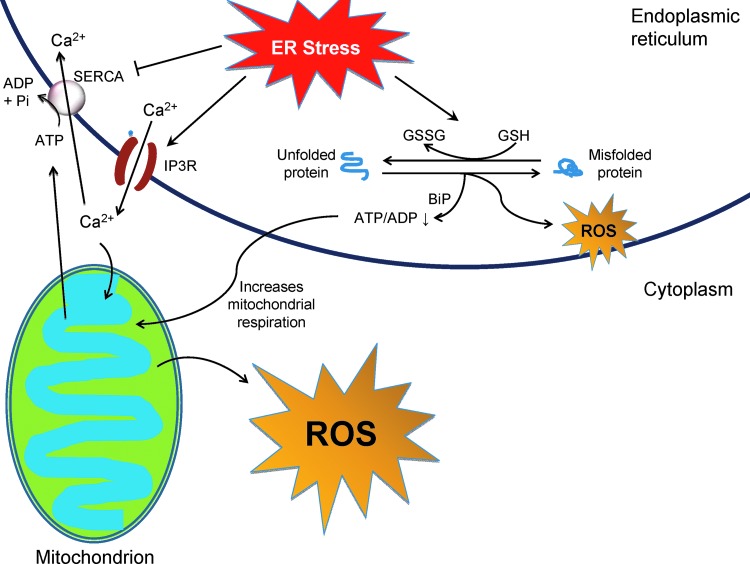
Recent advances: Production of reactive oxygen species (ROS) has been linked to ER stress and the UPR. ROS play a critical role in many cellular processes and can be produced in the cytosol and several organelles, including the ER and mitochondria. Studies suggest that altered redox homeostasis in the ER is sufficient to cause ER stress, which could, in turn, induce the production of ROS in the ER and mitochondria.

Critical issues: Although ER stress and oxidative stress coexist in many pathologic states, whether and how these stresses interact is unknown. It is also unclear how changes in the protein-folding environment in the ER cause oxidative stress. In addition, how ROS production and protein misfolding commit the cell to an apoptotic death and contribute to various degenerative diseases is unknown.

Future directions: A greater fundamental understanding of the mechanisms that preserve protein folding homeostasis and redox status will provide new information toward the development of novel therapeutics for many human diseases.


Figures

<b>FIG. 1.</b>
FIG. 1.
Oxidative protein folding in the ER. Oxidative protein folding of eukaryotic cells occurs in the ER, which is mediated by ER protein PDI and ERO1. ROS are generated as a byproduct of oxidative protein folding. Improperly paired disulfide bonds formed during protein folding can be reduced at the expense of glutathione, an essential antioxidant in the ER. See Introduction section for details. ER, endoplasmic reticulum; ROS, reactive oxygen species.
<b>FIG. 2.</b>
FIG. 2.
The mammalian UPR. In most mammalian cells, three UPR branches were identified: the PERK-eIF2α-ATF4-CHOP pathway, the IRE1α-XBP1 pathway, and the ATF6 pathway. The functions of the three pathways overlap and are redundant in many cell types. However, complete ablation of any of the three branches causes embryonic/perinatal death in mice, suggesting their unique and essential role at the physiological level. See “ER Stress and the UPR” section for details. ATF6, activating transcription factor 6; PERK, pancreatic ER eIF2α kinase; UPR, unfolded protein response.
<b>FIG. 3.</b>
FIG. 3.
ER stress-mediated cell death. ER stress leads to apoptotic cell death at transcriptional, post-transcriptional, translational, and post-translational levels. Pro-apoptotic components induced during ER stress are labeled red. See “ER Stress and Oxidative Stress in Cellular Homeostasis and Apoptosis” for details.
<b>FIG. 4.</b>
FIG. 4.
ER stress-induced ROS production in the cell. ROS are usually generated by cellular processes, including oxidative protein folding and mitochondrial respiration, which can be augmented to disrupt cell function and survival during ER stress. See “Cross-talk between ER stress and oxidative stress in apoptosis” for details. To see this illustration in color, the reader is referred to the web version of this article at www.liebertpub.com/ars
<b>FIG. 5.</b>
FIG. 5.
ER stress and oxidative stress in human diseases. ER stress and oxidative stress are linked to multiple human pathologies, including metabolic, neurodegenerative, immune/inflammatory, and neoplastic diseases. Studies on the two cellular stresses have not only contributed to our understanding of the pathogenesis, but also opened new avenues to next-generation therapies for these debilitating illnesses. See “ER Stress and Oxidative Stress in Human Diseases” for details.

References

    1. Almenier HA, Al Menshawy HH, Maher MM, and Al Gamal S. Oxidative stress and inflammatory bowel disease. Front Biosci (Elite Ed) 4: 1335–1344, 2012 - PubMed
    1. Ambade A. and Mandrekar P. Oxidative stress and inflammation: essential partners in alcoholic liver disease. Int J Hepatol 2012: 853175, 2012 - PMC - PubMed
    1. Atkins C, Liu Q, Minthorn E, Zhang SY, Figueroa DJ, Moss K, Stanley TB, Sanders B, Goetz A, Gaul N, Choudhry AE, Alsaid H, Jucker BM, Axten JM, and Kumar R. Characterization of a novel PERK kinase inhibitor with antitumor and antiangiogenic activity. Cancer Res 73: 1993–2002, 2013 - PubMed
    1. This reference has been deleted.
    1. Avivar-Valderas A, Salas E, Bobrovnikova-Marjon E, Diehl JA, Nagi C, Debnath J, and Aguirre-Ghiso JA. PERK integrates autophagy and oxidative stress responses to promote survival during extracellular matrix detachment. Mol Cell Biol 31: 3616–3629, 2011 - PMC - PubMed

Publication types

Substances