

Molecular profiles of pyramidal neurons in the superior temporal cortex in schizophrenia
- PMID: 24702465
- PMCID: PMC4196521
- DOI: 10.3109/01677063.2014.882918
Molecular profiles of pyramidal neurons in the superior temporal cortex in schizophrenia
Abstract
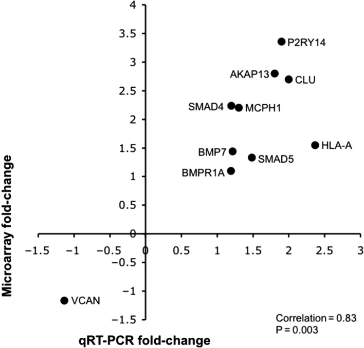
Disrupted synchronized oscillatory firing of pyramidal neuronal networks in the cerebral cortex in the gamma frequency band (i.e., 30-100 Hz) mediates many of the cognitive deficits and symptoms of schizophrenia. In fact, the density of dendritic spines and the average somal area of pyramidal neurons in layer 3 of the cerebral cortex, which mediate both long-range (associational) and local (intrinsic) corticocortical connections, are decreased in subjects with this illness. To explore the molecular pathophysiology of pyramidal neuronal dysfunction, we extracted ribonucleic acid (RNA) from laser-captured pyramidal neurons from layer 3 of Brodmann's area 42 of the superior temporal gyrus (STG) from postmortem brains from schizophrenia and normal control subjects. We then profiled the messenger RNA (mRNA) expression of these neurons, using microarray technology. We identified 1331 mRNAs that were differentially expressed in schizophrenia, including genes that belong to the transforming growth factor beta (TGF-β) and the bone morphogenetic proteins (BMPs) signaling pathways. Disturbances of these signaling mechanisms may in part contribute to the altered expression of other genes found to be differentially expressed in this study, such as those that regulate extracellular matrix (ECM), apoptosis, and cytoskeletal and synaptic plasticity. In addition, we identified 10 microRNAs (miRNAs) that were differentially expressed in schizophrenia; enrichment analysis of their predicted gene targets revealed signaling pathways and gene networks that were found by microarray to be dysregulated, raising an interesting possibility that dysfunction of pyramidal neurons in schizophrenia may in part be mediated by a concerted dysregulation of gene network functions as a result of the altered expression of a relatively small number of miRNAs. Taken together, findings of this study provide a neurobiological framework within which specific hypotheses about the molecular mechanisms of pyramidal cell dysfunction in schizophrenia can be formulated.
Keywords: cerebral cortex; gene expression profiling; laser-capture microdissection; microRNA; schizophrenia.
Figures


Similar articles
-
Molecular profiles of parvalbumin-immunoreactive neurons in the superior temporal cortex in schizophrenia.J Neurogenet. 2014 Mar-Jun;28(1-2):70-85. doi: 10.3109/01677063.2013.878339. Epub 2014 Mar 17. J Neurogenet. 2014. PMID: 24628518 Free PMC article.
-
Distinctive transcriptome alterations of prefrontal pyramidal neurons in schizophrenia and schizoaffective disorder.Mol Psychiatry. 2015 Nov;20(11):1397-405. doi: 10.1038/mp.2014.171. Epub 2015 Jan 6. Mol Psychiatry. 2015. PMID: 25560755 Free PMC article.
-
Transcriptional profile of pyramidal neurons in chronic schizophrenia reveals lamina-specific dysfunction of neuronal immunity.Mol Psychiatry. 2021 Dec;26(12):7699-7708. doi: 10.1038/s41380-021-01205-y. Epub 2021 Jul 16. Mol Psychiatry. 2021. PMID: 34272489 Free PMC article.
-
When cortical development goes wrong: schizophrenia as a neurodevelopmental disease of microcircuits.J Anat. 2010 Oct;217(4):324-33. doi: 10.1111/j.1469-7580.2010.01231.x. J Anat. 2010. PMID: 20408906 Free PMC article. Review.
-
Altered cortical glutamate neurotransmission in schizophrenia: evidence from morphological studies of pyramidal neurons.Ann N Y Acad Sci. 2003 Nov;1003:102-12. doi: 10.1196/annals.1300.007. Ann N Y Acad Sci. 2003. PMID: 14684438 Review.
Cited by
-
Expression quantitative trait loci (eQTLs) in microRNA genes are enriched for schizophrenia and bipolar disorder association signals.Psychol Med. 2015;45(12):2557-69. doi: 10.1017/S0033291715000483. Epub 2015 Mar 30. Psychol Med. 2015. PMID: 25817407 Free PMC article.
-
Screening of schizophrenia associated miRNAs and the regulation of miR-320a-3p on integrin β1.Medicine (Baltimore). 2019 Feb;98(8):e14332. doi: 10.1097/MD.0000000000014332. Medicine (Baltimore). 2019. PMID: 30813134 Free PMC article.
-
Widespread transcriptional disruption of the microRNA biogenesis machinery in brain and peripheral tissues of individuals with schizophrenia.Transl Psychiatry. 2020 Nov 4;10(1):376. doi: 10.1038/s41398-020-01052-5. Transl Psychiatry. 2020. PMID: 33149139 Free PMC article.
-
Plasma exosomes carrying mmu-miR-146a-5p and Notch signalling pathway-mediated synaptic activity in schizophrenia.J Psychiatry Neurosci. 2024 Aug 29;49(4):E265-E281. doi: 10.1503/jpn.230118. Print 2024 Jul-Aug. J Psychiatry Neurosci. 2024. PMID: 39209459 Free PMC article.
-
Increased extracellular clusterin in the prefrontal cortex in schizophrenia.Schizophr Res. 2015 Dec;169(1-3):381-385. doi: 10.1016/j.schres.2015.10.002. Epub 2015 Oct 21. Schizophr Res. 2015. PMID: 26482819 Free PMC article.
References
-
- Allen N. C., Bagade S., McQueen M. B., Ioannidis J. P., Kavvoura F. K., Khoury M. J., et al. Systematic meta-analyses and field synopsis of genetic association studies in schizophrenia: The SzGene database. Nat Genet. (2008);40:827–834. - PubMed
-
- Ananth H., Popescu I., Critchley H. D., Good C. D., Frackowiak R. S., Dolan R. J. Cortical and subcortical gray matter abnormalities in schizophrenia determined through structural magnetic resonance imaging with optimized volumetric voxel-based morphometry. Am J Psychiatry. (2002);159:1497–1505. - PubMed
Publication types
MeSH terms
Substances
Grants and funding
LinkOut - more resources
Full Text Sources
Other Literature Sources
Medical
Molecular Biology Databases