. 2014 Oct;21(7):593-605.
doi: 10.1111/micc.12138.

Involvement of H1 and H2 receptors and soluble guanylate cyclase in histamine-induced relaxation of rat mesenteric collecting lymphatics

Affiliations

Involvement of H1 and H2 receptors and soluble guanylate cyclase in histamine-induced relaxation of rat mesenteric collecting lymphatics

Kristine H Kurtz et al. Microcirculation. 2014 Oct.

Abstract

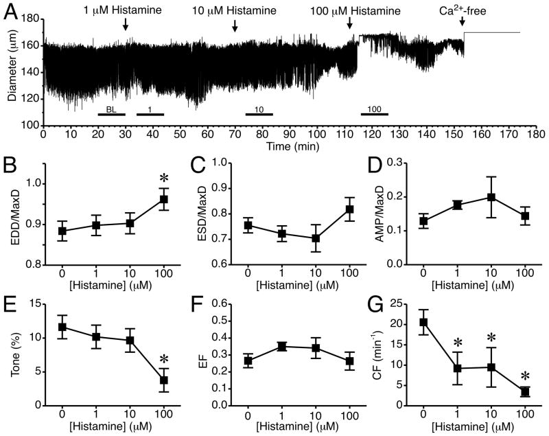
Objective: This study investigated the roles of the H1 and H2 histamine receptors, NO synthase, and sGC cyclase in histamine-induced modulation of rat mesenteric collecting lymphatic pumping.

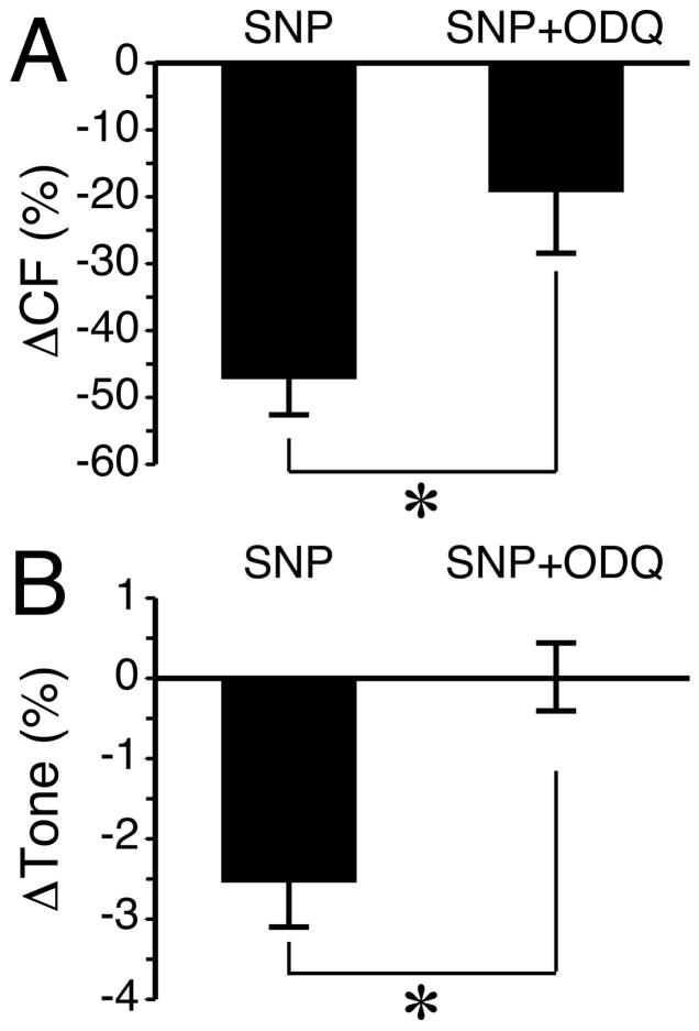
Methods: Isolated rat mesenteric collecting lymphatics were treated with 1- to 100-μM histamine. Histamine receptors were blocked with either the H1 antagonist mepyramine or the H2 antagonist cimetidine. The role of NO/sGC signaling was tested using the arginine analog L-NAME, the sGC inhibitor ODQ, and SNP as a positive control.

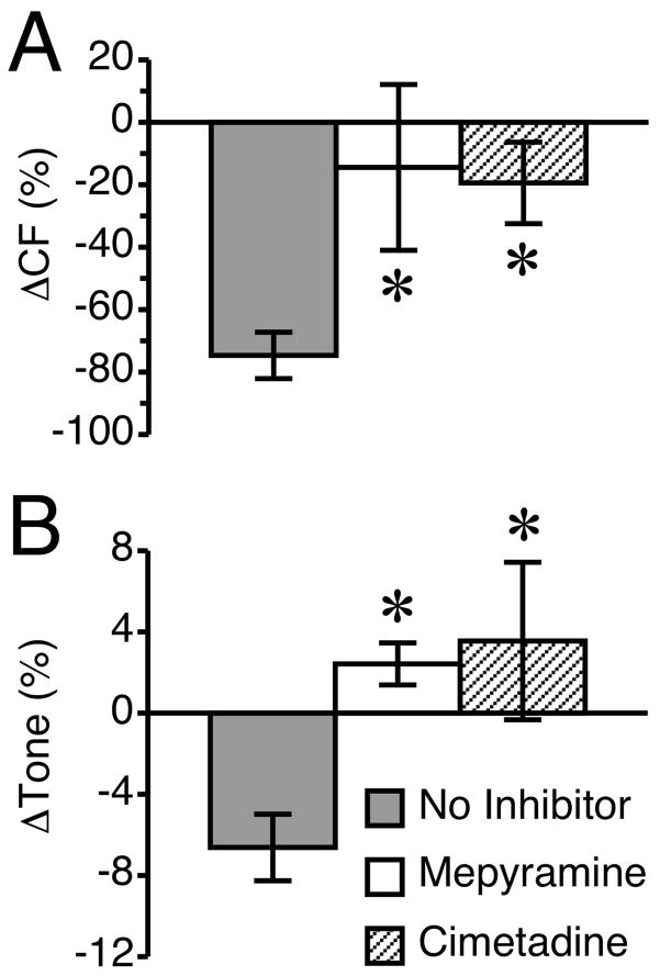
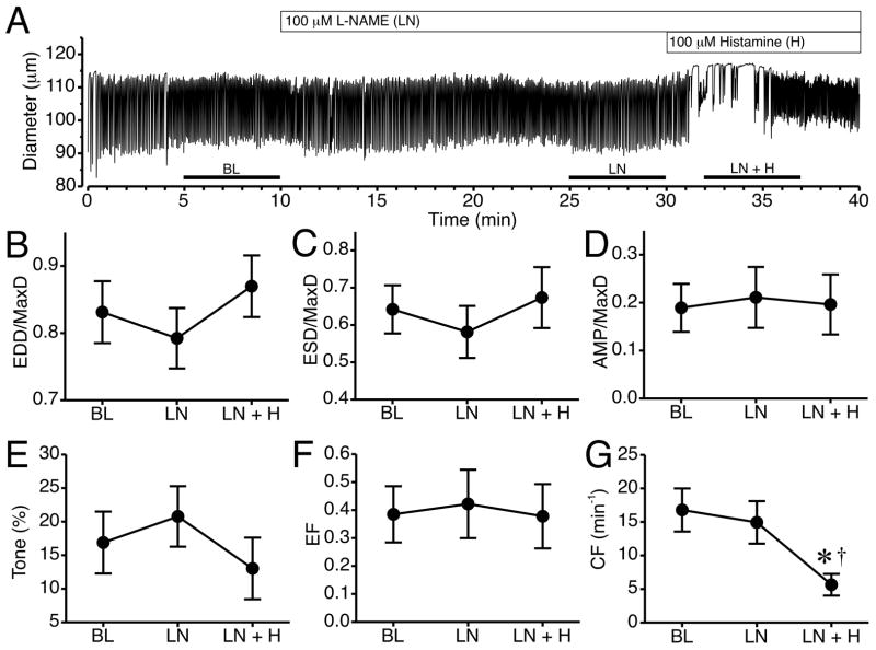
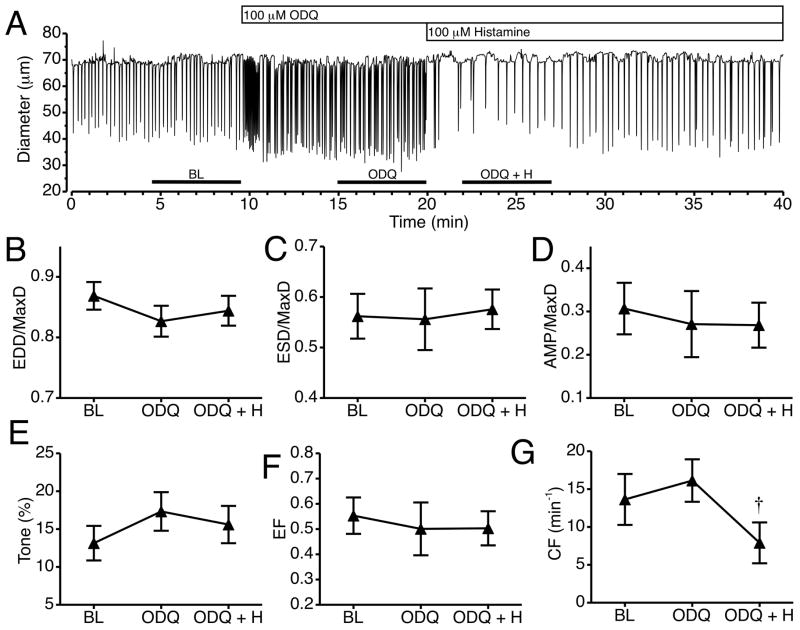
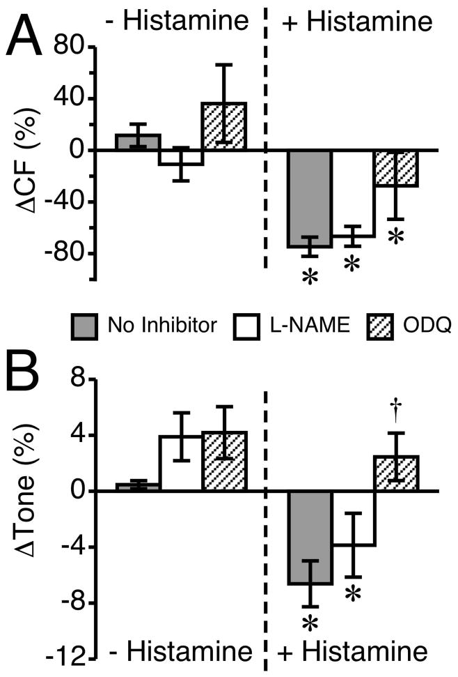
Results: Histamine applied at 100 μM decreased tone and CF of isolated rat mesenteric collecting lymphatics. Pharmacologic blockade of either H1 or H2 histamine receptors significantly inhibited the response to histamine. Pretreatment with ODQ, but not L-NAME, completely inhibited the histamine-induced decrease in tone. ODQ pretreatment also significantly inhibited SNP-induced lymphatic relaxation.

Conclusions: H1 and H2 histamine receptors are both involved in histamine-induced relaxation of rat mesenteric collecting lymphatics. NO synthesis does not appear to contribute to the histamine-induced response. However, sGC is critical for the histamine-induced decrease in tone and contributes to the drop in CF.

Keywords: endothelium; lymph flow; lymphatic pump; signal transduction.


Figures

Fig. 1
Fig. 1
Histamine and rat lymphatic mesenteric vessel pumping. (A) Time course of the changes in diameter after the addition of 1, 10, or 100 μM histamine to the isolated lymphatic vessel bath for a representative lymphatic vessel. The bath solution was changed to Ca2+-free APSS at the end of the experiment to determine MaxD. The subsequent panels (B–G) show the changes in mean values for (B) EDD/MaxD, (C) ESD/MaxD, (D) AMP/MaxD, (E) Tone, (F) EF, and (G) CF. The bars just above the x-axis show the time periods used for baseline (BL) and each concentration of histamine to determine the summarized data in panels B–G. (*P<0.05 versus baseline, prior to the addition of histamine; N=5 lymphatic vessels studied).
Fig. 2
Fig. 2
Western blotting of H1 and H2 receptors from isolated rat mesenteric lymphatic protein lysate. Each lane was loaded with 40 μg protein. Blots are representative of three separate experiments for each antibody. Control blots (no primary antibody) showed no bands (data not shown).
Fig. 3
Fig. 3
Confocal images of H1 histamine receptors in an isolated rat mesenteric lymphatic vessel. A. Images from a single confocal plane show labeling of the H1 receptor (H1R; green), VE-cadherin (red), and smooth muscle (SM) actin (blue) in the lymphatic walls. An overlay of these three channels, plus nuclei (white) is also shown. B. Close up images taken at 8, 10, and 12 μm from the start of the z-stack, featuring an en face view of the lymphatic wall. C. Close up images from a single z-section taken at 48 μm from the start of the z-stack, featuring a cross-sectional view of the lymphatic wall. Representative of three separate labeling experiments.
Fig. 4
Fig. 4
Confocal images of H2 histamine receptors in an isolated rat mesenteric lymphatic vessel. A. Images from a single confocal plane show labeling of the H2 receptor (H2R; green), VE-cadherin (red), and smooth muscle (SM) actin (blue) in the lymphatic walls. An overlay of these three channels, plus nuclei (white) is also shown. B. Close up view of z-sections obtained at 6, 8, and 10 μm from the start of the series, featuring an en face view of the lymphatic wall. C. Close up images from a single confocal plane taken at 18 μm from the start of the z-stack, featuring a cross-sectional view of the lymphatic wall. Representative of three separate labeling experiments.
Fig. 5
Fig. 5
Antagonists of either the H1 or H2 receptor block histamine-induced lymphatic relaxation. The H1 receptor blocker, mepyramine (100 μM) and the selective H2 blocker cimetidine (100 μM) were each applied 20 min prior to the application of 100 μM histamine. A time-matched “no inhibitor” group was used for comparison. Both blockers significantly inhibited the histamine-induced relaxation of lymphatics. (A) The histamine-induced reduction in CF was significantly inhibited by either H1 or H2 blockade. (B) The histamine-induced reduction in tone was significantly inhibited by both H1 and H2 receptor blockers. *P<0.05 versus no inhibitor, post addition of histamine. N=7 lymphatics studied for the “no inhibitor” group and N=5 lymphatic vessels each for the mepyramine and cimetidine groups.
Fig. 6
Fig. 6
Impact of NOS inhibition on histamine-induced collecting lymphatic relaxation. A. Representative time course of changes in lymphatic luminal diameter in response to the NOS inhibitor L-NAME (LN) and subsequent addition of histamine (LN+H). Changes in mean EDD/MaxD (B), ESD/MaxD (C), AMP/MaxD (D), Tone (E), EF (F), and CF (G) are also shown. The bars just above the x-axis show the time periods used to calculate the means in panels B–G. *P<0.05 versus baseline. P<0.05 versus L-NAME alone. N=6 lymphatic vessels studied.
Fig. 7
Fig. 7
Impact of sGC inhibition on histamine-induced collecting lymphatic relaxation. A. Representative time course of changes in lymphatic luminal diameter after the addition of the sGC inhibitor ODQ and subsequent addition of histamine. Changes in mean EDD/MaxD (B), ESD/MaxD (C), AMP/MaxD (D), Tone (E), EF (F), and CF (G) are also shown. The bars just above the x-axis show the time periods used to calculate the means in panels B–G. P<0.05 versus ODQ alone. N=6 lymphatic vessels studied.
Fig. 8
Fig. 8
Inhibition of the NO/sGC pathway attenuates histamine-induced reductions in lymphatic CF and tone. This figure compares the same data from the lymphatics that received pretreatment with L-NAME or ODQ with lymphatics that had no inhibitors applied prior to the addition of 100 μM histamine. (A) The change in CF is shown for 100 μM L-NAME or 100 μM ODQ versus no treatment (left set of bars), and histamine alone versus histamine treatment in the presence of L-NAME or ODQ (right set of bars). (B) The change in tone is shown in response to the same inhibitors before and after histamine treatment. The “no inhibitor” group baseline measurement was taken during a 5-min period ending 20 min before the addition of histamine, and the change from baseline prior to histamine represents a 5-min period just before the addition of histamine. *P<0.05 versus baseline, same group. P<0.05 versus no inhibitor + histamine. N=7 for the “no inhibitor” group and N=6 for the L-NAME and ODQ groups.
Fig. 9
Fig. 9
SNP-mediated percent changes in (A) CF and (B) Tone are inhibited by ODQ. For both variables, the changes are calculated from the five-minute period before SNP was added and the five-minute period after SNP was added. *P<0.01 between groups. For each treatment, N=4 lymphatic vessels studied.

Comment in

References

    1. Alitalo K. The lymphatic vasculature in disease. Nat Med. 2011;17:1371–1380. - PubMed
    1. Beermann S, Seifert R, Neumann D. Commercially available antibodies against human and murine histamine H(4)-receptor lack specificity. Naunyn Schmiedebergs Arch Pharmacol. 2012;385:125–135. - PubMed
    1. Cary SP, Winger JA, Marletta MA. Tonic and acute nitric oxide signaling through soluble guanylate cyclase is mediated by nonheme nitric oxide, ATP, and GTP. Proc Natl Acad Sci U S A. 2005;102:13064–13069. - PMC - PubMed
    1. Chambers R, Zweifach BW. Intercellular cement and capillary permeability. Physiol Rev. 1947;27:436–463. - PubMed
    1. Chatterjee V, Gashev AA. Aging-associated shifts in functional status of mast cells located by adult and aged mesenteric lymphatic vessels. Am J Physiol Heart Circ Physiol. 2012;303:H693–702. - PMC - PubMed

Publication types

MeSH terms

LinkOut - more resources