

Rare variants in NR2F2 cause congenital heart defects in humans
- PMID: 24702954
- PMCID: PMC3980509
- DOI: 10.1016/j.ajhg.2014.03.007
Rare variants in NR2F2 cause congenital heart defects in humans
Erratum in
- Am J Hum Genet. 2014 Jul 3;95(1):126
-
Rare Variants in NR2F2 Cause Congenital Heart Defects in Humans.Am J Hum Genet. 2016 Mar 3;98(3):592. doi: 10.1016/j.ajhg.2016.02.016. Epub 2016 Mar 3. Am J Hum Genet. 2016. PMID: 28863274 Free PMC article. No abstract available.
Abstract
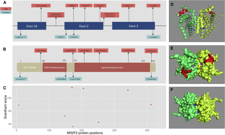
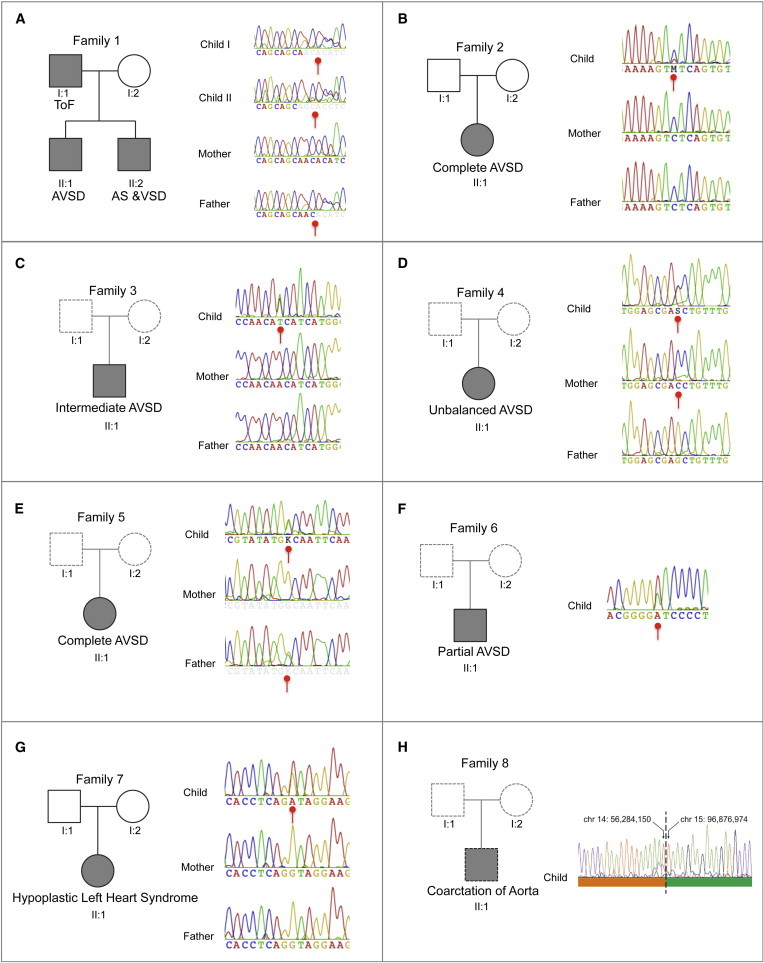
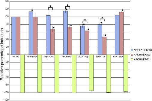
Congenital heart defects (CHDs) are the most common birth defect worldwide and are a leading cause of neonatal mortality. Nonsyndromic atrioventricular septal defects (AVSDs) are an important subtype of CHDs for which the genetic architecture is poorly understood. We performed exome sequencing in 13 parent-offspring trios and 112 unrelated individuals with nonsyndromic AVSDs and identified five rare missense variants (two of which arose de novo) in the highly conserved gene NR2F2, a very significant enrichment (p = 7.7 × 10(-7)) compared to 5,194 control subjects. We identified three additional CHD-affected families with other variants in NR2F2 including a de novo balanced chromosomal translocation, a de novo substitution disrupting a splice donor site, and a 3 bp duplication that cosegregated in a multiplex family. NR2F2 encodes a pleiotropic developmental transcription factor, and decreased dosage of NR2F2 in mice has been shown to result in abnormal development of atrioventricular septa. Via luciferase assays, we showed that all six coding sequence variants observed in individuals significantly alter the activity of NR2F2 on target promoters.
Copyright © 2014 The Authors. Published by Elsevier Inc. All rights reserved.
Figures
Similar articles
-
Novel de novo pathogenic variant in the NR2F2 gene in a boy with congenital heart defect and dysmorphic features.Am J Med Genet A. 2018 Jun;176(6):1423-1426. doi: 10.1002/ajmg.a.38700. Epub 2018 Apr 16. Am J Med Genet A. 2018. PMID: 29663647
-
A novel NR2F2 loss-of-function mutation predisposes to congenital heart defect.Eur J Med Genet. 2018 Apr;61(4):197-203. doi: 10.1016/j.ejmg.2017.12.003. Epub 2017 Dec 6. Eur J Med Genet. 2018. PMID: 29222010
-
NR2F2 loss‑of‑function mutation is responsible for congenital bicuspid aortic valve.Int J Mol Med. 2019 Apr;43(4):1839-1846. doi: 10.3892/ijmm.2019.4087. Epub 2019 Jan 31. Int J Mol Med. 2019. PMID: 30720060
-
De novo frameshift mutation in COUP-TFII (NR2F2) in human congenital diaphragmatic hernia.Am J Med Genet A. 2016 Sep;170(9):2457-61. doi: 10.1002/ajmg.a.37830. Epub 2016 Jul 1. Am J Med Genet A. 2016. PMID: 27363585 Free PMC article.
-
De novo variants in congenital diaphragmatic hernia identify MYRF as a new syndrome and reveal genetic overlaps with other developmental disorders.PLoS Genet. 2018 Dec 10;14(12):e1007822. doi: 10.1371/journal.pgen.1007822. eCollection 2018 Dec. PLoS Genet. 2018. PMID: 30532227 Free PMC article.
Cited by
-
De Novo and Rare Variants at Multiple Loci Support the Oligogenic Origins of Atrioventricular Septal Heart Defects.PLoS Genet. 2016 Apr 8;12(4):e1005963. doi: 10.1371/journal.pgen.1005963. eCollection 2016 Apr. PLoS Genet. 2016. PMID: 27058611 Free PMC article.
-
Unraveling cardiomyocyte responses and intercellular communication alterations in primary carnitine deficiency cardiomyopathy via single-nucleus RNA sequencing.Heliyon. 2024 Jun 24;10(13):e33581. doi: 10.1016/j.heliyon.2024.e33581. eCollection 2024 Jul 15. Heliyon. 2024. PMID: 39091928 Free PMC article.
-
COUP-TFII regulates early bipotential gonad signaling and commitment to ovarian progenitors.Cell Biosci. 2024 Jan 4;14(1):3. doi: 10.1186/s13578-023-01182-5. Cell Biosci. 2024. PMID: 38178246 Free PMC article.
-
COUP-TFII in Kidneys, from Embryos to Sick Adults.Diagnostics (Basel). 2022 May 9;12(5):1181. doi: 10.3390/diagnostics12051181. Diagnostics (Basel). 2022. PMID: 35626336 Free PMC article. Review.
-
A genome-wide by PM10 exposure interaction study for blood pressure in Korean adults.Sci Rep. 2023 Aug 11;13(1):13060. doi: 10.1038/s41598-023-40155-z. Sci Rep. 2023. PMID: 37567956 Free PMC article.
References
-
- Blue G.M., Kirk E.P., Sholler G.F., Harvey R.P., Winlaw D.S. Congenital heart disease: current knowledge about causes and inheritance. Med. J. Aust. 2012;197:155–159. - PubMed
-
- Pierpont M.E., Basson C.T., Benson D.W., Jr., Gelb B.D., Giglia T.M., Goldmuntz E., McGee G., Sable C.A., Srivastava D., Webb C.L., American Heart Association Congenital Cardiac Defects Committee, Council on Cardiovascular Disease in the Young Genetic basis for congenital heart defects: current knowledge: a scientific statement from the American Heart Association Congenital Cardiac Defects Committee, Council on Cardiovascular Disease in the Young: endorsed by the American Academy of Pediatrics. Circulation. 2007;115:3015–3038. - PubMed
-
- Bentham J., Bhattacharya S. Genetic mechanisms controlling cardiovascular development. Ann. N Y Acad. Sci. 2008;1123:10–19. - PubMed
-
- Hoffman J.I. Incidence of congenital heart disease: I. Postnatal incidence. Pediatr. Cardiol. 1995;16:103–113. - PubMed
Publication types
MeSH terms
Substances
Grants and funding
- RC2 HL102926/HL/NHLBI NIH HHS/United States
- MC_PC_U127561093/MRC_/Medical Research Council/United Kingdom
- 090532/WT_/Wellcome Trust/United Kingdom
- RC2 HL103010/HL/NHLBI NIH HHS/United States
- 100140/WT_/Wellcome Trust/United Kingdom
- RC2 HL102923/HL/NHLBI NIH HHS/United States
- UC2 HL102926/HL/NHLBI NIH HHS/United States
- PG/07/045/22690/BHF_/British Heart Foundation/United Kingdom
- UC2 HL103010/HL/NHLBI NIH HHS/United States
- RC2 HL102924/HL/NHLBI NIH HHS/United States
- MC_U127561093/MRC_/Medical Research Council/United Kingdom
- RG/13/10/30376/BHF_/British Heart Foundation/United Kingdom
- RG/10/17/28553/BHF_/British Heart Foundation/United Kingdom
- UC2 HL102923/HL/NHLBI NIH HHS/United States
- RG/07/010/23676/BHF_/British Heart Foundation/United Kingdom
- UC2 HL102924/HL/NHLBI NIH HHS/United States
- RC2 HL102925/HL/NHLBI NIH HHS/United States
- UC2 HL102925/HL/NHLBI NIH HHS/United States
- WT098051/WT_/Wellcome Trust/United Kingdom
LinkOut - more resources
Full Text Sources
Other Literature Sources
Medical
Molecular Biology Databases
