

The crucial role of mechanical heterogeneity in regulating follicle development and ovulation with engineered ovarian microtissue
- PMID: 24702961
- PMCID: PMC4016819
- DOI: 10.1016/j.biomaterials.2014.03.028
The crucial role of mechanical heterogeneity in regulating follicle development and ovulation with engineered ovarian microtissue
Abstract
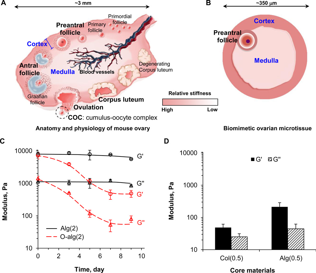
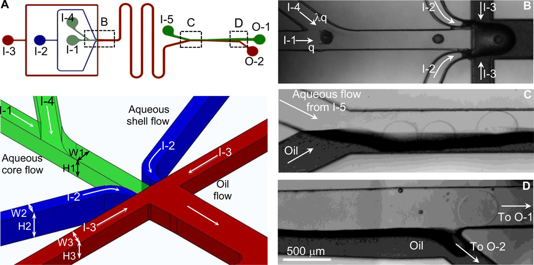
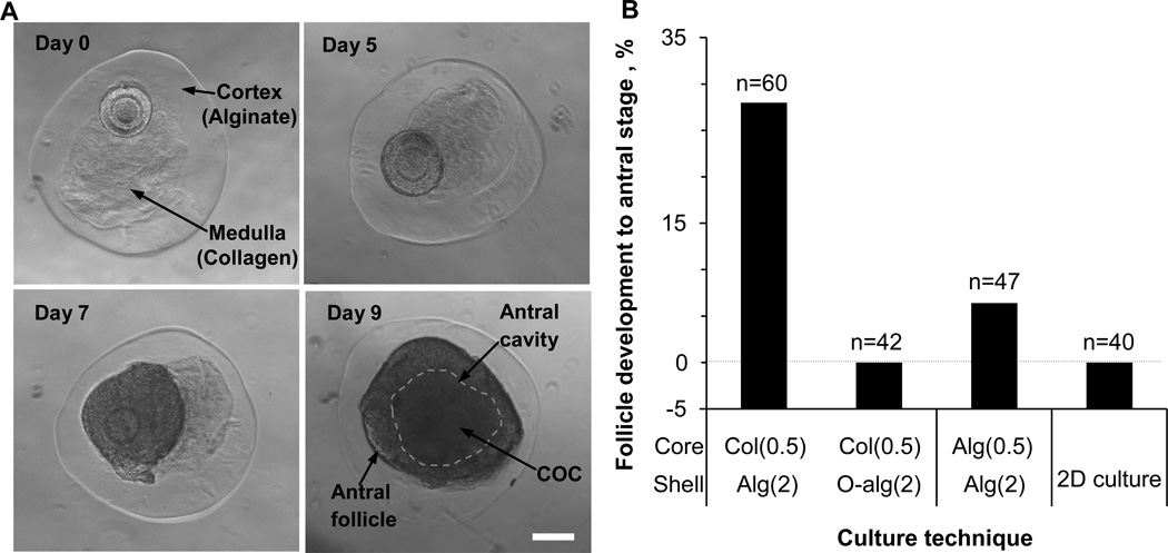
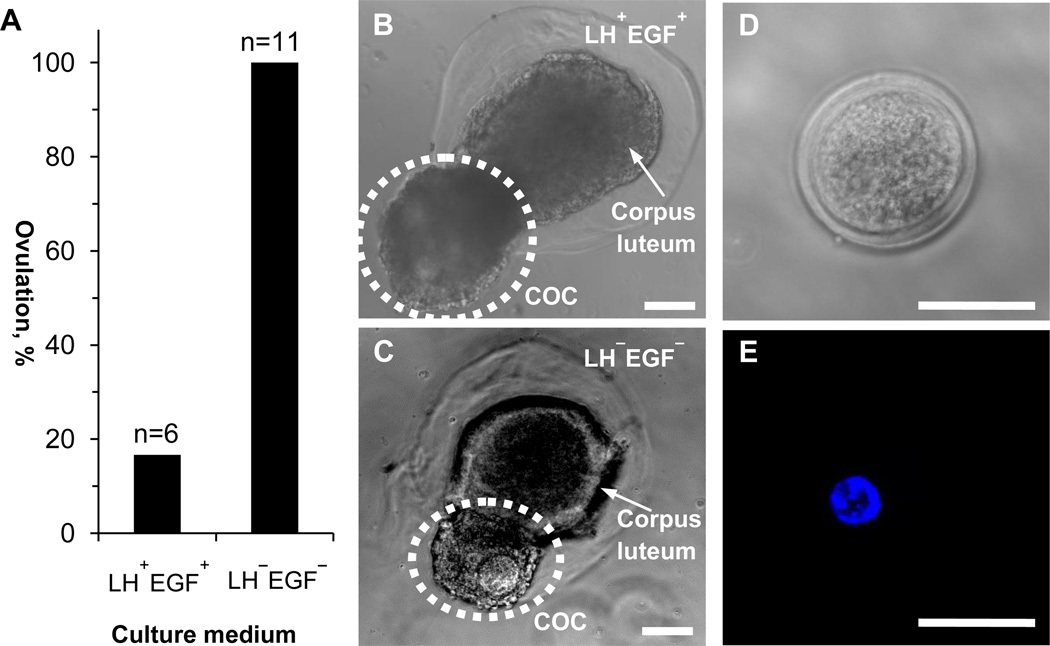
Contemporary systems for in vitro culture of ovarian follicles do not recapitulate the mechanical heterogeneity in mammalian ovary. Here we report microfluidic generation of biomimetic ovarian microtissue for miniaturized three-dimensional (3D) culture of early secondary preantral follicles by using alginate (harder) and collagen (softer) to fabricate the ovarian cortical and medullary tissues, respectively. This biomimetic configuration greatly facilitates follicle development to antral stage. Moreover, it enables in vitro ovulation of cumulus-oocyte complex (COC) from the antral follicles in the absence of luteinizing hormone (LH) and epidermal growth factor (EGF) that are well accepted to be responsible for ovulation in contemporary literature. These data reveal the crucial role of mechanical heterogeneity in the mammalian ovary in regulating follicle development and ovulation. The biomimetic ovarian microtissue and the microfluidic technology developed in this study are valuable for improving in vitro culture of follicles to preserve fertility and for understanding the mechanism of follicle development and ovulation to facilitate the search of cures to infertility due to ovarian disorders.
Keywords: Alginate; Follicle; Mechanobiology; Microfluidic; Microtissue; Ovulation.
Copyright © 2014 Elsevier Ltd. All rights reserved.
Figures
Similar articles
-
In vitro culture of ovarian follicles from Peromyscus.Semin Cell Dev Biol. 2017 Jan;61:140-149. doi: 10.1016/j.semcdb.2016.07.006. Epub 2016 Jul 7. Semin Cell Dev Biol. 2017. PMID: 27397871 Free PMC article. Review.
-
Microfluidic Encapsulation of Ovarian Follicles for 3D Culture.Ann Biomed Eng. 2017 Jul;45(7):1676-1684. doi: 10.1007/s10439-017-1823-7. Epub 2017 Mar 20. Ann Biomed Eng. 2017. PMID: 28321583 Free PMC article. Review.
-
Encapsulated three-dimensional culture supports development of nonhuman primate secondary follicles.Biol Reprod. 2009 Sep;81(3):587-94. doi: 10.1095/biolreprod.108.074732. Epub 2009 May 27. Biol Reprod. 2009. PMID: 19474063 Free PMC article.
-
Utilizing Fibrin-Alginate and Matrigel-Alginate for Mouse Follicle Development in Three-Dimensional Culture Systems.Biopreserv Biobank. 2018 Apr;16(2):120-127. doi: 10.1089/bio.2017.0087. Epub 2018 Jan 24. Biopreserv Biobank. 2018. PMID: 29363997
-
Mouse preantral follicle growth in 3D co-culture system using human menstrual blood mesenchymal stem cell.Reprod Biol. 2018 Mar;18(1):122-131. doi: 10.1016/j.repbio.2018.02.001. Epub 2018 Feb 15. Reprod Biol. 2018. PMID: 29454805
Cited by
-
Creating an Artificial 3-Dimensional Ovarian Follicle Culture System Using a Microfluidic System.Micromachines (Basel). 2021 Mar 4;12(3):261. doi: 10.3390/mi12030261. Micromachines (Basel). 2021. PMID: 33806282 Free PMC article.
-
3D culture applied to reproduction in females: possibilities and perspectives.Anim Reprod. 2024 Mar 8;21(1):e20230039. doi: 10.1590/1984-3143-AR2023-0039. eCollection 2024. Anim Reprod. 2024. PMID: 38510565 Free PMC article. Review.
-
Stiffness-Independent Highly Efficient On-Chip Extraction of Cell-Laden Hydrogel Microcapsules from Oil Emulsion into Aqueous Solution by Dielectrophoresis.Small. 2015 Oct 28;11(40):5369-74. doi: 10.1002/smll.201501388. Epub 2015 Aug 21. Small. 2015. PMID: 26297051 Free PMC article.
-
Deep Learning-Enabled Label-Free On-Chip Detection and Selective Extraction of Cell Aggregate-Laden Hydrogel Microcapsules.Small. 2021 Jun;17(23):e2100491. doi: 10.1002/smll.202100491. Epub 2021 Apr 25. Small. 2021. PMID: 33899299 Free PMC article.
-
Efficient One-Step Production of Microencapsulated Hepatocyte Spheroids with Enhanced Functions.Small. 2016 May;12(20):2720-30. doi: 10.1002/smll.201502932. Epub 2016 Apr 1. Small. 2016. PMID: 27038291 Free PMC article.
References
Publication types
MeSH terms
Substances
Grants and funding
LinkOut - more resources
Full Text Sources
Other Literature Sources
