. 2014 May 8;54(3):431-44.
doi: 10.1016/j.molcel.2014.03.006. Epub 2014 Apr 3.

Structure of a eukaryotic RNase III postcleavage complex reveals a double-ruler mechanism for substrate selection

Affiliations

Structure of a eukaryotic RNase III postcleavage complex reveals a double-ruler mechanism for substrate selection

Yu-He Liang et al. Mol Cell. .

Abstract

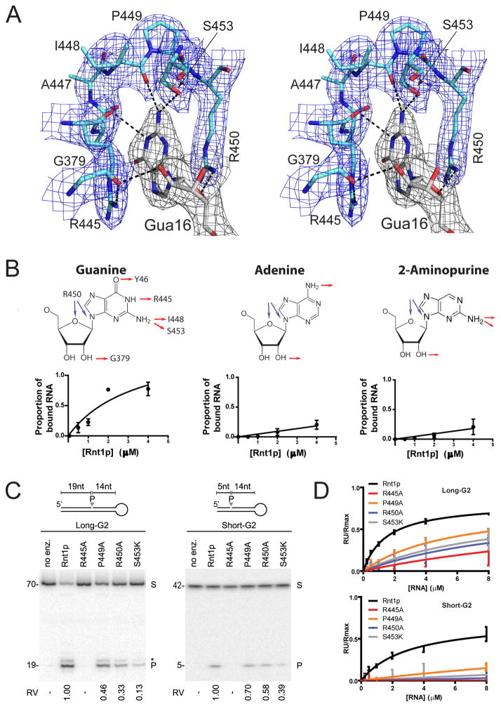
Ribonuclease III (RNase III) enzymes are a family of double-stranded RNA (dsRNA)-specific endoribonucleases required for RNA maturation and gene regulation. Prokaryotic RNase III enzymes have been well characterized, but how eukaryotic RNase IIIs work is less clear. Here, we describe the structure of the Saccharomyces cerevisiae RNase III (Rnt1p) postcleavage complex and explain why Rnt1p binds to RNA stems capped with an NGNN tetraloop. The structure shows specific interactions between a structural motif located at the end of the Rnt1p dsRNA-binding domain (dsRBD) and the guanine nucleotide in the second position of the loop. Strikingly, structural and biochemical analyses indicate that the dsRBD and N-terminal domains (NTDs) of Rnt1p function as two rulers that measure the distance between the tetraloop and the cleavage site. These findings provide a framework for understanding eukaryotic RNase IIIs.


Figures

Figure 1
Figure 1. New Mode of Substrate Recognition by RNase III
(A) Domain structures are illustrated for Homo sapiens Dicer (HsDicer, UniProtKB Q9UPY3), Giardia intestinalis Dicer (GiDicer, UniProtKB A8BQJ3), Homo sapiens Drosha (HsDrosha, UniProtKB Q9NRR4), Saccharomyces cerevisiae Rnt1p (ScRnt1p, UniProtKB Q02555), Kluyveromyces polysporus Dcr1 (KpDcr1, UniProtKB A7TR32), Aquifex aeolicus RNase III (AaRNase III, UniProtKB O67082), and Bacillus subtilis Mini-III (BsMini-III, UniProtKB O31418). The scale on top indicates the polypeptide lengths. The red box in RIIID represents the RNase III signature sequence. (B) The Rnt1p:RNA complex contains two NTD dimers (blue/red), one RIIID dimer (cyan/orange), two dsRBDs (pale cyan/light orange), and two 34-nt RNAs (grey/dark grey) in addition to Mg2+ ions and solvent molecules (not shown). (C) Distinct modes of dsRNA recognition by ScRnt1p (left) and AaRNase III (right) are illustrated. The RIIIDs are shown as ribbon diagrams outlined with transparent molecular surfaces, dsRBDs as solid molecular surfaces, and RNAs as tube-and-stick models. The span of the two dsRBDs along dsRNA is indicated with a double-headed arrow. See also Figure S1.
Figure 2
Figure 2. Cleavage Site Assembly of Eukaryotic RNase III
(A) Structure-based sequence alignment of ScRnt1p (this work), KpDcr1 (PDB entry 3RV0), GiDicer (PDB entry 2FFL), HsDicer (PDB entry 2EB1), and AaRNase III, (PDB entry 2NUG). The RIIIID signature sequence is indicated and the conserved amino acid residues in the cleavage site highlighted (in red). The boxed residues, N5 and K6, are unique for eukaryotic enzymes. (B) The dsRNA-processing center of Rnt1p has two cleavage sites (left). Stereoview on the right shows the cleavage site assembly. Electron density (annealed omit Fo-Fc, contoured at 4.5 σ) is shown as grey nets for the Mg2+ ions (black spheres) and coordinating water molecules (red spheres). Residues are illustrated as sticks in atomic colors [N in blue, C in green (protein) or orange (RNA), O in red, P in orange, and Mg in black]. Mg2+ coordination is indicated by solid lines, hydrogen bonds by dashed lines, and the distance between the 3′ hydroxyl oxygen of Cyt34 and the phosphorous of Cyt1 (3.1 Å) by a double-headed arrow. (C–F) Stereoviews show the superposition of the Rnt1p cleavage site assembly (in green) on that of AaRNase III (C), KpDcr1 (D), GiDicer (E), or HsDicer (F). The AaRNase III, KpDcr1, GiDicer, and HsDicer structures were adopted from the PDB as specified in (A) and shown in magenta. See also Figure S2.
Figure 3
Figure 3. New RNA-Binding Motif, RBM0, Identifies the NGNN Tetraloop
(A) The arrangement of the RBMs of AaRNase III (PDB entry 2EZ6) is shown on the left and that of ScRnt1p (this work) on the right. Only one subunit of the protein dimer is shown. The RIIID is shown in cyan and dsRBD in pale cyan. The RBMs are in blue and the linker between the two domains in red. Stem-loop RNA is shown as a molecular surface in grey or light grey. (B) RBM0 identifies the AGUC tetraloop. The dsRBD, outlined with a transparent molecular surface, is shown as a ribbon diagram with the RBM0 highlighted in blue. The RNA backbone is shown as a ribbon diagram with the four nucleotides in the tetraloop as sticks colored by atom (N in blue, O in red, C in grey, and P in orange). (C) On the left, dsRBD proteins used in structural analysis (in cyan/blue, this work; in yellow, PDB entry 1T4L; in pink, PDB entry 2LBS) are illustrated with RBMs indicated with dashed boxes. On the right, the three dsRBD structures are superimposed. (D) Interaction map for the binding of RBMs 0 and 1 to the AGUC tetraloop via interaction(s) with the base, ribose, and/or backbone of the RNA. Residues of RBMs 0 and 1 are colored in blue and cyan, respectively. (E) RBM0 is required for substrate cleavage. The Long- or Short-G2 substrate was incubated alone (no enz), with recombinant Rnt1p (Rnt1p), or with enzymes lacking the RBM0 (ΔRBM0). The reactions were carried out in multiple turnover (substrate excess) and physiological salt (150 mM KCl) conditions. S and P indicate the position of intact substrate and cleavage product, respectively. The asterisk indicates a secondary cleavage product. The sizes of the different RNA fragments are indicated on the left. The substrates are illustrated on top. (F) RBM0 is required for substrate binding. Increasing amounts of Long- or Short-G2 were injected into surface-bound, full-length or ΔRBM0 enzyme. Shown is the binding curve from surface plasmon resonance. The ratio of the resonance unit change (RU) over the theoretical maximal RU (Rmax) obtained for each binding assay is presented in the form of a graph. See also Figure S3 and Table S2.
Figure 4
Figure 4. G-clamp Is Tailored for the Recognition of Gua16
(A) Stereoview showing that the conserved Gua16 base is buried in the G-clamp formed by eight residues from RMBs 0 and 1. Residues are illustrated as sticks [N in blue, O in red, P in orange, and C in pale cyan (amino acids) or grey (nucleotide)] outlined with electron density map (blue/grey nets, 2Fo-Fc, contoured at 1.2 σ). Dashed lines indicate hydrogen bonds. (B) Guanine-specificity of the G-clamp. Rnt1p substrates carrying adenine or 2-aminopurine in the second position of the NGNN loop were tested for binding to Rnt1p using electrophoretic mobility gel shift assay (EMSA). The top panels show the structure of the three bases; hydrogen bonds formed between each base and the G-clamp are indicated by red and blue arrows. The bottom panels show the binding curves of the three substrates derived from gel shift assays. (C) Contribution of G-clamp residues to RNA cleavage. Four G-clamp residues directly interacting with Gua16 of the tetraloop were individually mutated and the impact on enzyme activities was tested using Long- or Short-G2 as described in Figure 3E. Relative velocities (RV), the cleavage rates obtained with the mutants relative to that obtained by the wild type, are indicated at bottom. (D) Impact of G-clamp mutations on substrate binding. The binding kinetics of different mutations to Long- or Short-G2 were assessed using surface plasmon resonance and the ratio of the resonance unit (RU) over the theoretical maximal RU (Rmax) obtained for each binding assay is presented in the form of a graph. See also Figure S4 and Tables S1 and S2.
Figure 5
Figure 5. dsRBD Interacts with the Stem down to the 9th Base Pair below the Tetraloop
(A) Stereoview showing the interactions between RBM1 and the first four base pairs below the tetraloop and that between the α2 K421 and the 5th base pair. Residues are shown as sticks in atomic colors [N in blue, C in green (protein) or grey (RNA), O in red, and P in orange]. Dashed lines indicate hydrogen bonds. A “zoomed out” view indicating the position of the RBMs within the Rnt1p:RNA structure is shown on the right. (B) Stereoview showing the interactions between the 9th base pair below the tetraloop and residues K392 from RBM2 and K311 from RBM4. (C) Interaction map between the Rnt1p dsRBD and the first five base pairs below the tetraloop. (D and E) Disruption of RBM2 interaction with the 9th base pair below the tetraloop alters RNA binding and cleavage. Residue K392 was mutated and the impact of the mutation was tested on substrate cleavage and binding as described in Figures 4C and 4D. See also Tables S1 and S2.
Figure 6
Figure 6. NTD Dimer Interacts with RNA to Increase Binding Affinity and the Precision of Cleavage Site Selection
(A) The NTD dimer of Rnt1p interacts with three nucleotides within or near the AGUC tetraloop. NTD1 (blue) and NTD2 (red) are shown as ribbons. RNA backbone is shown in orange. Contacting residues are shown as sticks. Dashed lines indicate hydrogen bonds. (B, C) Deletion of the NTD impairs cleavage site selection of G2 substrates. The substrates were labeled at their 5′ (B) or 3′ (C) ends. Wild-type enzyme (Rnt1p), enzyme lacking the RBM0 (ΔRBM0), or that lacking the NTD (ΔNTD) were incubated with either Long- or Short-G2 and the cleavage products visualized under single turnover (enzyme excess) and low salt (10 mM KCl) conditions. Alternative cleavage sites observed in the absence of the NTD are indicated by “#”. The cleavage rates are indicated as relative velocities (RV) at bottom. (D) The double-ruler architecture ensures the cleavage accuracy. The protein is illustrated as a molecular surface and color-coded as in (A). The RNA is shown as a cartoon in grey for the tetraloop and in blue and red for the stem. Gua16 is highlighted as a ball-and-stick illustration. See also Figure S5 and Tables S1 and S2.
Figure 7
Figure 7. Double-Ruler Mechanism for Substrate Selection by Rnt1p
(A) Schematic representation of Rnt1p interactions with G2 substrates. Nucleotides are shown as rectangles, while RBMs and NTDs as ellipsoids. C1 and C2 indicate the two canonical cleavage sites. The four boxes of the RNA substrate are outlined with dashed lines. Sites of hydrogen bonds formed between proteins and RNA backbone are shown as shaded rectangles and labeled with residue numbers only, whereas nucleotides forming base-specific hydrogen bonds are labeled by both one-letter code and residue number. Lowercase letters c, f, and s indicate, respectively, positions of chemical interference (Ghazal and Elela, 2006), hydrogen bonds requirement for cleavage (Lavoie and Abou Elela, 2008), and sites where hydrogen bonds were observed with the ribose 2′-OH in the dsRBD:RNA structure (PDB entry 1T4L). (B) Schematic representation of the ΔNTD interactions with the G2 substrates. C1 and C2 indicate the two canonical cleavage sites by Rnt1p. C* signifies additional cleavage sites produced by the ΔNTD.

References

    1. Abou Elela S, Ares M., Jr Depletion of yeast RNase III blocks correct U2 3′ end formation and results in polyadenylated but functional U2 snRNA. EMBO J. 1998;17:3738–3746. - PMC - PubMed
    1. Abou Elela S, Igel H, Ares M., Jr RNase III cleaves eukaryotic preribosomal RNA at a U3 snoRNP-dependent site. Cell. 1996;85:115–124. - PubMed
    1. Catala M, Tremblay M, Samson E, Conconi A, Abou Elela S. Deletion of Rnt1p alters the proportion of open versus closed rRNA gene repeats in yeast. Mol Cell Biol. 2008;28:619–629. - PMC - PubMed
    1. Chanfreau G, Buckle M, Jacquier A. Recognition of a conserved class of RNA tetraloops by Saccharomyces cerevisiae RNase III. Proc. Natl. Acad. Sci. USA. 2000;97:3142–3147. - PMC - PubMed
    1. Chanfreau G, Legrain P, Jacquier A. Yeast RNase III as a key processing enzyme in small nucleolar RNAs metabolism. J Mol Biol. 1998;284:975–988. - PubMed

Publication types

Associated data

LinkOut - more resources