. 2014 Jun 5;14(6):781-95.
doi: 10.1016/j.stem.2014.03.004. Epub 2014 Apr 3.

Pathways disrupted in human ALS motor neurons identified through genetic correction of mutant SOD1

Affiliations

Pathways disrupted in human ALS motor neurons identified through genetic correction of mutant SOD1

Evangelos Kiskinis et al. Cell Stem Cell. .

Abstract

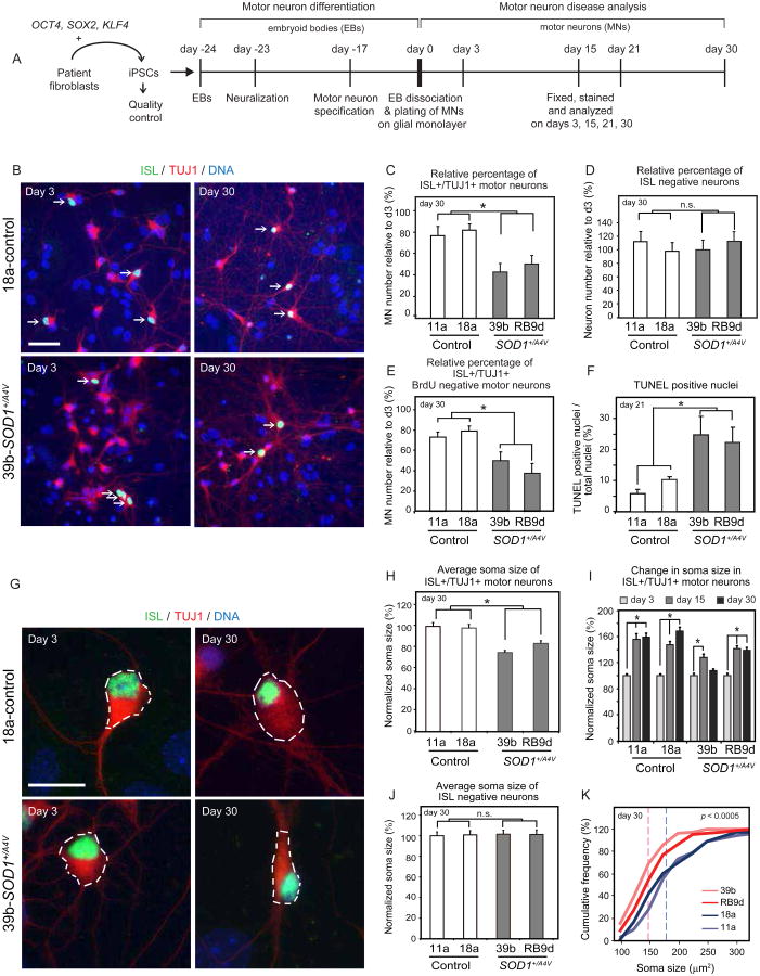
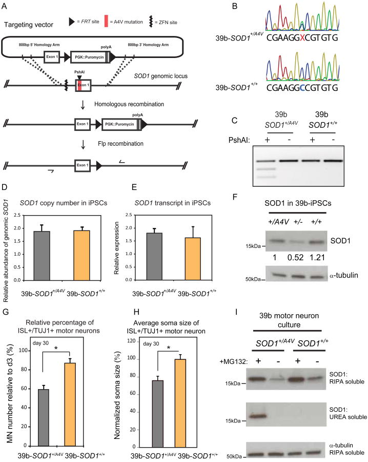
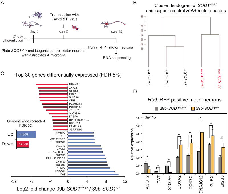
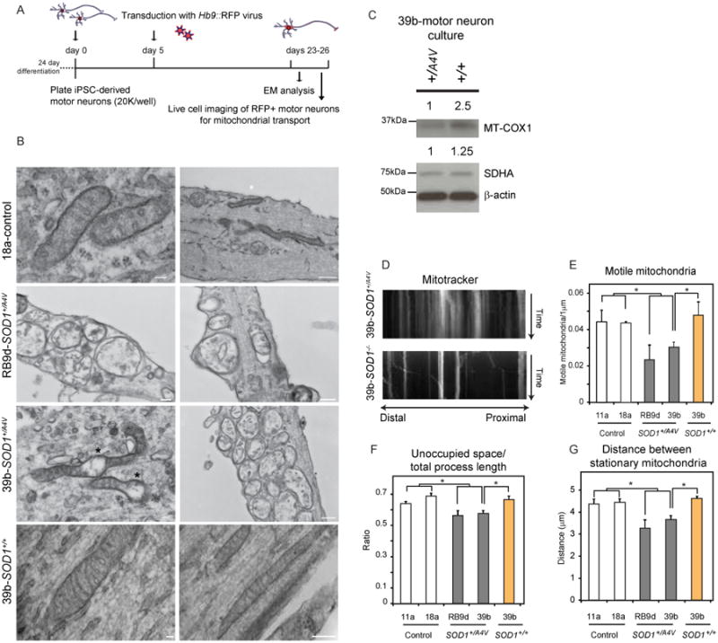
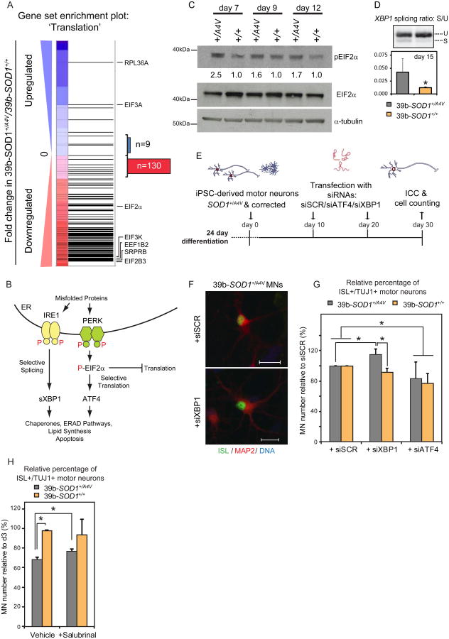
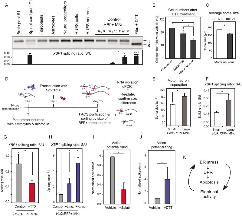
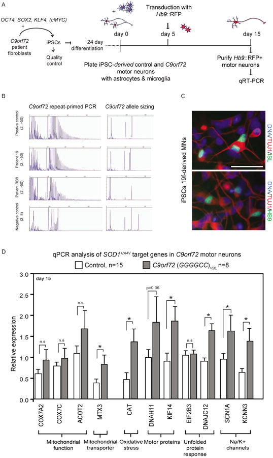
Although many distinct mutations in a variety of genes are known to cause Amyotrophic Lateral Sclerosis (ALS), it remains poorly understood how they selectively impact motor neuron biology and whether they converge on common pathways to cause neuronal degeneration. Here, we have combined reprogramming and stem cell differentiation approaches with genome engineering and RNA sequencing to define the transcriptional and functional changes that are induced in human motor neurons by mutant SOD1. Mutant SOD1 protein induced a transcriptional signature indicative of increased oxidative stress, reduced mitochondrial function, altered subcellular transport, and activation of the ER stress and unfolded protein response pathways. Functional studies demonstrated that these pathways were perturbed in a manner dependent on the SOD1 mutation. Finally, interrogation of stem-cell-derived motor neurons produced from ALS patients harboring a repeat expansion in C9orf72 indicates that at least a subset of these changes are more broadly conserved in ALS.


Figures

Figure 1
Figure 1. iPSC-Derived Motor Neurons From SOD1+/A4V ALS Patients Exhibit Survival and Morphometric Differences Relative to Healthy Controls
(A) Experimental outline: patient fibroblasts were reprogrammed to generate iPSCs, which were differentiated into MNs and assessed for ALS-related phenotypes. (B) Neuronal cultures on glial monolayers 3 and 30 days post-differentiation from control and SOD1+/A4V iPSCs. MNs co-stained for ISL and TUJ1 are indicated by white arrows (scale bar=50μm). (C) Quantifications of ISL positive MNs (n=3, m>8000, +/-SEM, P<0.05), and (D) ISL negative neurons (n=3, m>25000, +/-SEM, P<0.05), after 30 days in culture. (E) Quantifications of MN numbers that are BrdU negative after 30 days in culture (n=1, m>3600, +/-SD, P<0.05). Differential motor neurogenesis does not explain the lower numbers in SOD1+/A4V cases. (F) Quantifications of TUNEL positive nuclei of neuronal cultures without glia after 21 days in culture, (n=2, m>13000, +/-SD, P<0.05). (G) Representative images of measured soma size (white-dotted circumference) of control and SOD1+/A4V MNs, (scale bar=20μm). (H) Quantifications of ISL positive MN soma size, values normalized to controls (n=3, m=340, +/-SEM, P<0.01). (I) MN soma size after 3, 15 and 30 days in culture normalized to day 3 for each cell line. Although MNs increase in size over time in all 4 cell lines, they do less so in SOD1+/A4V cases. (J) Quantifications of ISL negative neuron soma size, values normalized to controls (n=3, m=446, +/-SEM, P: n.s.). (K) Cumulative frequency graphs of MN soma size after 30 days in culture. Dotted lines indicate averages for control and disease. n.s: not significant; n=experiment, m=cell number.
Figure 2
Figure 2. Genetic Correction of the SOD1A4V Mutant Allele Rescues Motor Neuron Survival and Soma Size Deficits
(A) Gene targeting strategy used to generate isogenic 39b-SOD1+/+ iPSC line. Nucleases targeting the SOD1 locus created a double strand break upstream of Exon1; homologous recombination of the genomic locus with a targeting plasmid with control sequence of Exon1 coupled to PGK∷Puromycin replaced the SOD1A4V mutant allele; after antibiotic selection, the resistance cassette was removed by Flp-mediated recombination, leaving only an FRT footprint.; ZFN: Zinc Finger Nuclease, FRT: Flippase Recognition Target. (B) Sequencing chromatograms of Exon1 of SOD1 in iPSC line 39b before and after targeting, demonstrate the correction of the A4V mutation. (C) PshAI restriction digestion of amplified SOD1 cDNA before and after gene targeting. The mutation creates a PshAI restriction site that is absent in the corrected line. (D) qPCR of genomic SOD1 shows that there are no extra copies of the gene in the corrected line and (E) qRT-PCR of cDNA shows that the expression levels of SOD1 are the same in the corrected line (n=3, +/-SEM). (F) SOD1 protein levels assessed by western blot (WB) analysis in parental (39b-SOD1+/A4V), targeted hemizygous knockout (39b-SOD1+/-) and corrected (39b-SOD1+/+) iPSC clones. (G) Isogenic 39b-SOD1+/+ MNs exhibit increased cell survival (n=6, m>13000, +/-SEM, P<0.05) and (H) soma size (n=3, m=280, +/-SEM, P<0.01). (I) WB analysis of SOD1 protein in detergent- soluble (RIPA) and detergent-insoluble (UREA) fractions in day 12 neuronal cultures. Insoluble SOD1 is not detected under normal conditions. After proteasome inhibition by MG132 treatment, insoluble SOD1 selectively accumulates only in SOD1+/A4V MNs and not in the corrected line.
Figure 3
Figure 3. Transcriptional Analysis of 39b-SOD1+/A4V and Isogenic Control Motor Neurons by RNA-Seq Reveals Genotypic Regulatory Changes
(A) Experimental outline: SOD1+/A4V and isogenic control MN cultures were co-cultured with primary glial cells, transduced with an Hb9∷RFP lentivirus and FACS-purified on day 15 for RNA isolation and sequencing. (B) Dendogram demonstrating clear genotypic clustering based on transcriptional changes measured by RNA sequencing. (C) Total number of genes and top 30 genes (based on fold change) misregulated in SOD1+/A4V MNs with an FDR 5%. (D) qRT-PCR validation of misregulated genes in independent samples (n=3, +/-SEM).
Figure 4
Figure 4. SOD1+/A4V Motor Neurons Exhibit Mitochondrial Defects That are Rescued by Genetic Correction of the SOD1A4V Mutation
(A) Experimental outline: SOD1+/A4V and isogenic control MN cultures were plated at low densities, transduced with an Hb9∷RFP lentivirus on day 5 and analyzed on days 23-26. (B) Electron microscopy analysis demonstrating that in contrast to control neurons (top panel), SOD1+/A4V neurons (middle panels) often exhibited swelling, morphological disorganization and clustering in neurites. Mitochondria in genetically corrected neurons (bottom panel) exhibited normal morphological characteristics. Representative images out of 3 independent experiments are shown. (C) WB analysis of mitochondrial proteins. (D) Representative kymographs of MitoTracker-stained mitochondria in MNs after live cell imaging. Distal and proximal orientation relative to cell body is indicated. SOD1+/A4V MNs exhibit mitochondria transportation deficiencies relative to controls with (E) fewer motile mitochondria, (F) less space unoccupied by mitochondria relative to axon length and (G) shorter distances between stationary mitochondria (n=4, +/-SEM, P<0.05).
Figure 5
Figure 5. SOD1+/A4V Motor Neurons Exhibit Signatures of an Unfolded Protein Response and are Selectively Vulnerable to ER Stress Induction
(A) Gene set enrichment analysis of transcriptional changes in SOD1+/A4V MNs shows strong downregulation (normalized enrichment score: -3.31) for genes involved in translational capacity. Horizontal black bars represent individual genes with representative examples indicated. Blue or red represents genes which were up or downregulated respectively in SOD1+/A4V relative to isogenic control MNs. Out of 139 genes detected that are annotated as involved in regulation of translation, 130 were downregulated, consistent with activation of the UPR pathway. (B) Diagram illustrating the canonical unfolded protein response. (C) WB analysis demonstrates increased levels of phosphorylated EIF2α, a marker for activation of the UPR pathway, in SOD1+/A4V MN cultures. Percentage relative to control samples for each time point is shown. (D) SOD1+/A4V MNs exhibit increased levels of XBP1 splicing, a marker of ER stress. RNA was isolated from purified Hb9∷RFP MNs after 15 days in culture (n=3, +/-SEM, P<0.05). U: unspliced, S: spliced. (E) Experimental strategy used to assess the contribution of XBP1 and ATF4 in the survival of SOD1+/A4V MNs. (F) Representative images of untreated and treated MN cultures are shown. (G) SOD1+/A4V MN numbers selectively increase after XBP1 knockdown, while ATF4 knockdown significantly decreases numbers in both control and SOD1+/A4V cases (n=3, m>9800 and m>8500, P<0.05), +/-SEM, P<0.05). (H) Salubrinal has a modest but positive effect on survival of SOD 1+/A4V MNs (n=2, m>10000, +/-SEM, P<0.05).
Figure 6
Figure 6. Human Motor Neurons Exhibit Increased Levels of Basal ER Stress That is Dependent on Their Physiological Activity
(A) Purified Hb9∷GFP control MNs show higher levels of spliced XBP1 relative to other human cell types. Human spinal cord RNA also shows higher levels relative to brain RNA. U: unspliced, S: spliced. (B) Control MNs are more vulnerable to acute ER stress induction (DTT, 2mM) relative to fibroblasts or astrocytes, while (C) DTT treatment leads to a reduction in MN soma size (n=2, +/-SD, P<0.05). (D) Experimental strategy used to isolate MNs based on cell size. Control MN cultures were infected with the Hb9∷RFP virus on day 5. On day 15, RFP+ MNs were FACS-purified and sorted by size to separate small and large cells. (E) A subset of cells were re-plated to confirm soma size by measuring MAP2+ cell bodies (F). Basal levels of XBP1 splicing were assessed in RNA isolated from the remaining purified MNs showing that larger MNs had higher spliced XBP1 than smaller ones (n=3, +/-SEM, P<0.05). Treatment of MN cultures with (G) TTX reduces while (H) linopiridine and kainate increase XBP1 splicing respectively. Treatment with (I) salubrinal reduces, while (J) DTT increases action potential firing (n=3, +/-SEM, P<0.05). (K) ER stress, the UPR and electrical activity of MNs are connected.
Figure 7
Figure 7. Transcriptional Changes Detected in SOD1+/A4V Motor Neurons are Partially Conserved in Motor Neurons With C9orf72 Expansions
(A) Experimental outline: ALS patient fibroblasts harboring C9orf72 repeat expansions were reprogrammed to generate iPSCs, which were differentiated into MNs and plated in co-culture with glial cells. On day 5 cultures were infected with an Hb9∷RFP lentivirus and on day 15, RFP-labeled MNs were FACS-purified for RNA isolation. (B) Genotyping of samples by repeat-primed PCR and allele sizing for C9orf72.Patient samples RB8 and 19 exhibited more than 50 GGGGCC repeats. (C) Representative images of MN cultures differentiated from C9orf72 ALS patients (scale bar=50μm). (D) A subset of transcripts found to be differentially expressed in SOD1+/A4V MNs also exhibited significant transcriptional changes in MNs derived from C9orf72 (n=2 lines, n=8 replicates, +/-SEM, P<0.05) compared to a large number of control samples (n=6 lines, n=15 replicates, +/-SEM, P<0.05).

Comment in

  • The ER under rapid fire.
    Schwenk BM, Edbauer D. Schwenk BM, et al. EMBO J. 2014 Jun 2;33(11):1195-7. doi: 10.1002/embj.201488692. Epub 2014 Apr 30. EMBO J. 2014. PMID: 24788411 Free PMC article.

References

    1. Abecasis GR, Auton A, Brooks LD, DePristo MA, Durbin RM, Handsaker RE, Kang HM, Marth GT, McVean GA. An integrated map of genetic variation from 1,092 human genomes. Nature. 2012;491:56–65. - PMC - PubMed
    1. Ash PE, Bieniek KF, Gendron TF, Caulfield T, Lin WL, Dejesus-Hernandez M, van Blitterswijk MM, Jansen-West K, Paul JW, 3rd, Rademakers R, et al. Unconventional translation of C9ORF72 GGGGCC expansion generates insoluble polypeptides specific to c9FTD/ALS. Neuron. 2013;77:639–646. - PMC - PubMed
    1. Bilican B, Serio A, Barmada SJ, Nishimura AL, Sullivan GJ, Carrasco M, Phatnani HP, Puddifoot CA, Story D, Fletcher J, et al. Mutant induced pluripotent stem cell lines recapitulate aspects of TDP-43 proteinopathies and reveal cell-specific vulnerability. Proc Natl Acad Sci U S A. 2012;109:5803–5808. - PMC - PubMed
    1. Boulting GL, Kiskinis E, Croft GF, Amoroso MW, Oakley DH, Wainger BJ, Williams DJ, Kahler DJ, Yamaki M, Davidow L, et al. A functionally characterized test set of human induced pluripotent stem cells. Nat Biotechnol. 2011;29:279–286. - PMC - PubMed
    1. Boyce M, Bryant KF, Jousse C, Long K, Harding HP, Scheuner D, Kaufman RJ, Ma D, Coen DM, Ron D, et al. A selective inhibitor of eIF2alpha dephosphorylation protects cells from ER stress. Science. 2005;307:935–939. - PubMed

Publication types

MeSH terms

Associated data