Review
. 2014 Jul:41:64-89.
doi: 10.1016/j.preteyeres.2014.03.002. Epub 2014 Apr 4.

Cholesterol in the retina: the best is yet to come

Affiliations
Review

Cholesterol in the retina: the best is yet to come

Irina A Pikuleva et al. Prog Retin Eye Res. 2014 Jul.

Abstract

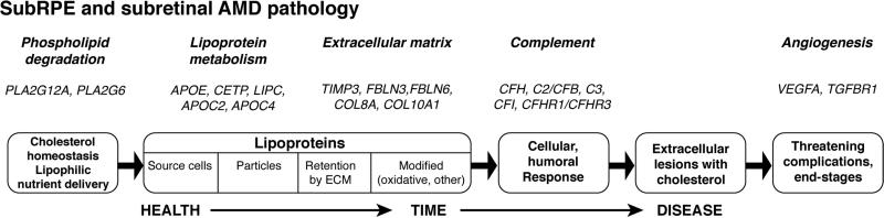
Historically understudied, cholesterol in the retina is receiving more attention now because of genetic studies showing that several cholesterol-related genes are risk factors for age-related macular degeneration (AMD) and because of eye pathology studies showing high cholesterol content of drusen, aging Bruch's membrane, and newly found subretinal lesions. The challenge before us is determining how the cholesterol-AMD link is realized. Meeting this challenge will require an excellent understanding these genes' roles in retinal physiology and how chorioretinal cholesterol is maintained. In the first half of this review, we will succinctly summarize physico-chemical properties of cholesterol, its distribution in the human body, general principles of maintenance and metabolism, and differences in cholesterol handling in human and mouse that impact on experimental approaches. This information will provide a backdrop to the second part of the review focusing on unique aspects of chorioretinal cholesterol homeostasis, aging in Bruch's membrane, cholesterol in AMD lesions, a model for lesion biogenesis, a model for macular vulnerability based on vascular biology, and alignment of AMD-related genes and pathobiology using cholesterol and an atherosclerosis-like progression as unifying features. We conclude with recommendations for the most important research steps we can take towards delineating the cholesterol-AMD link.

Keywords: Age-related macular degeneration; Cholesterol; Choroid; Drusen; Lipoproteins; Retina.


Figures

Fig. 1
Fig. 1. Two different views of cholesterol
A. Chemical structrure and numbering of atoms. B. planarity of the molecule. The oxygen atom in the 3β-hydroxyl is shown in red.
Fig. 2
Fig. 2. Lipoprotein essentials
A lipoprotein particle (upper left) is a multimolecular assembly that solubilizes oil droplets rich in esterified cholesterol (EC) and triglyceride (TG) for transport through an aqueous environment within a thin surface of phospholipid, unesterified cholesterol (UC), and apolipoproteins that are recognized by receptors and serve as cofactors for enzymes. Lipoproteins are secreted by the liver, intestine, brain, heart, placenta, kidney, and RPE. BrM-LP produced by the RPE represents a distinct class of lipoproteins as compared to the particles present in systemic circulation (CM, VLDL, LDL, and HDL). BrM-LP is composed of apolipoproteins B, E, and AI, and is large like VLDL, yet rich in EC like LDL. BrM-LP provides abundant cholesterol and apolipoproteins (including apolipoproteins B, E, A-I, C-I, C-II) to aging BrM and drusen. Adapted with permission from (Curcio et al., 2011a).
Fig. 3
Fig. 3. Lipoprotein bio-transformations and trafficking in human circulation
Lipoprotein particles are shown as circles of different color with the cirlcle diameter reflecting a particle size (not at a scale). Receptor (LDL-R), transporter (ABCA1), and enzymes pertinent to lipoproteins (LIPC, ACAT, CETP, and lipoprotein lipase) are also indicated. CHOL, either esterified or unesterified cholesterol or both; EC, esterified cholesterol; FFA, free fatty acids; PL, phospholipids; TG, triglycerides.
Fig. 4
Fig. 4. Chorioretinal layers and major cell types
Modified from (Zheng et al., 2012). The the neurosensory retina has nine distinct layers (from top to bottom): inner limiting membrane (ILM), nerve fiber layer (NFL), ganglion cell layer (GCL), inner plexiform layer (IPL), inner nuclear layer (INL), outer plexiform layer (OPL), outer nuclear layer (ONL), external limiting membrane (ELM), photoreceptor inner segments (IS), and photoceptor outer segments (OS). The tenth layer, the retinal pigment epithelium (RPE), lies outside the neurosensory retina but is considered a part of the retina. The major retinal cell types are ganglion cells (G), diffuse amacrine cells (DA), amacrine cells (Am), Müller cells (M), bipolar cells (B), horizontal cells (H), rods (R), and cones (C).
Fig. 5
Fig. 5. Cholesterol distribution in human macula, localized by filipin
This staining came from a set of experiments described in (Curcio et al., 2005a). Labeling of retinal layers is the same as in Fig. 4. Syn/ped, synapses of photoreceptors with post-receptoral neurons (inner), and layer of cone pedicles (outer); He, Henle fiber layer. Arrowheads point to cone photoreceptor outer segments.
Fig. 6
Fig. 6. Immunohistochemistry localizations of cholesterol-related proteins in human retina and RPE
SREBPs, SCAP, Insigs control the expression of HMGCR and LDLR playing key roles in cellular cholesterol input: HMGCR is the rate-limiting enzyme in cholesterol biosynthesis, whereas LDLR uptakes cholesterol-rich LDL. LXRs regulate the expression of ABCA1, a cholesterol efflux transporter, as well as many other genes involved in cellular cholesterol output and other cellular processes. CYPs 27A1, 46A1, and 11A1 are the only three enzymes that initiate the quantitatively significant pathways of cholesterol metabolism in non-hepatic organs including the retina and RPE. Phase contrast images (on the left of each panel) are given for comparison. Nuclei were stained by DAPI (blue), and immunoreactivity was detected by DyLight 649 conjugated secondary Abs (red). Staining with serum from non-immunized animal (rabbit or goat) served as a negative control. Labeling of retinal layers is the same as in Fig. 4. Scale bars are 30 μm. Taken from (Zheng et al., 2012).
Fig. 7
Fig. 7. Localization of cholesterol in human BrM and isolated drusen
All drusen shown are considered hard. Drusen in E-F are mechanically isolated. Bars in C and F are 20 μm. A, Oil red O (ORO) binds to lipids in BrM and presumed retinoids in RPE lipofuscin. B. Filipin staining reveals intense fluorescence for EC, the predominant component of BrM lipid per direct assay of isolated lipoproteins (Curcio et al., 2009b). RPE lipofuscin is slightly autofluorescent at ultraviolet excitation wavelengths used for filipin visualization. C. Cholesterol localizes to lipoproteins in BrM and membranes of RPE and choroidal cells. RPE fluorescence is due to lipofuscin plus additional signal due to intracellular cholesterol. D. In drusen ORO binding EC shows a scalloped pattern with EC-poor cores at the base of many lesions, i.e., near BrM. E. A similar pattern is visible by filipin staining, plus dots signifying EC-rich lakes. F. In contrast, UC is particularly prominent in cores at druse base, perhaps signifying extracellular neutral pH cholesterol esterase activity that hydrolyzes EC, leaving cholesterol behind.
Fig. 8
Fig. 8. Proposed RPE lipid inputs-outputs and a model for AMD lesion formation
BLinD/soft drusen and SDD are localized in two different compartments (below and above RPE, respectively). Normal aging RPE is at the left and center. AMD is at right. The input-output pathways are suggested based on available data. These pathways serve as a basis for the model (2L2C model) that identifies RPE-based lipid recycling pathways for rods and cones as biologic processes that drive the formation of AMD extracellular lesions. Different cholesterol content of rod and cone OS membranes is of key importance The sub-RPE lesions are plausibly formed when passage of constitutively secreted products from RPE are either overproduced or retained instead of being efficiently cleared. The evidence for this mechanism is good and involves the following steps: 1) Plasma lipoproteins delivering lipophilic essentials, including vitamins E, A, lutein, and cholesterol (UC), enter basolateral RPE (Tserentsoodol et al., 2006b). 2) ApoB,E lipoproteins secreted basolaterally by RPE (Johnson et al., 2011) (gold circles) are assembled from multiple lipid sources. Fatty acids in lipoproteins isolated from BrM are dominated by linoleate, implicating internalized plasma lipoproteins (from step 1) as a major source. UC from all sources is esterified to EC. 3) Lipoproteins are retained by interacting with BrM extracellular matrix and accumulate throughout adulthood, creating pre-BLinD on BrM's inner surface. 4) Reactive oxygen species from nearby mitochondria promote appearance of pro-inflammatory and toxic moieties. Lipoproteins fuse and form lipid pools and UC-rich liposomes within BLinD/soft drusen, rendering them biomechanically unstable, pro-inflammatory, and cytotoxic. 5) Disks in rod OS lose UC and gain docosahexaenoate in transit from OS base to tip (Albert and Boesze-Battaglia, 2005) (shown as loss of white). OS-derived docosahexaenoate stored as triacylglycerol in RPE after phagocytosis return to OS (Rodriguez de Turco et al., 1999). HDL particles cycling between RPE and photoreceptors (Tserentsoodol et al., 2006a) could handle both transfers as part of a vectorial lipid flow retainable within interphotoreceptor matrix as UC-containing SDD, especially under rod-rich perifovea. 6) Cone OS maintain high UC content along their length, because their disks are comb-like projections of plasma membrane (Albert and Boesze-Battaglia, 2005). Cone OS UC enters RPE via disk shedding, lysosomal uptake, and acid lipase activity (Elner, 2002). UC is released for intracellular transfer, esterification, and assembly into basolaterally-secreted lipoproteins, especially under cone-rich fovea. The mechanism for the sub-retinal lesion (SDD) formation is not yet known. Adapted with permission from (Curcio et al., 2013).
Fig. 9
Fig. 9. Layers of AMD pathology
Histology of the outer retina shows distinctive AMD pathology on either aspect of the retinal pigment epithelium (RPE). SDD is subretinal drusenoid deposit (bright yellow), an extracellular lesion surrounded by delicate RPE apical processes containing melanosomes. The RPE is at stage of degeneration featuring melanosomes and lipofuscin granules shed into underlying BLamD (pale yellow). BLamD is basal laminar deposit, a stereotypically thickened RPE basal lamina, often containing basal mounds. The latter are an aggregation of soft druse/BLinD material in transit from the RPE to BrM. Pre-BLinD is layer of lipoprotein particles internal to the inner collagenous layer of BrM and an immediate precursor to BLinD (basal linear deposit). BLinD (not shown here) is pooled lipoprotein-derived debris, in a thin layer. A lump large enough to elevate the RPE is a soft druse. BrM is the inner surface of the choroidal vasculature, and thus a vessel wall in addition to a stratum for RPE attachment. It has inner and outer collagenous layers that are calcified and glassy-appearing in this example. ChC, choriocapillaries with fenestrated endothelium, are partly atrophied in this example. Other layers of the neurosensory retina: OPL, outer plexiform layer; HFL, Henle fibers; ONL, outer nuclear layer; IS, inner segments; OS, outer segments. From a 85 yr old Caucasian male with geographic atrophy, 3 mm from the foveal center, 0.8 μm section, osmium tannic acid paraphenylenediamine post-fixation, toluidine blue stain. With permission from the Annual Review of Genomics and Human Genetics, Volume 15 © 2014 by Annual Reviews, http://www.annualreviews.org (Fritsche et al., 2014).
Fig. 10
Fig. 10. Genes and pathobiology align
Shown at the top are biological processes encompassing genetic associations of AMD recently published by the AMD Gene Consortium Meta-analysis (Fritsche et al., 2013) comprising 17,000 late AMD cases. In the boxes is an atherosclerosis-like “response to retention of lipoproteins” theory of pathogenesis (Tabas et al., 2007; Williams and Tabas, 1995, 1998) that has been re-tooled for sub-RPE AMD pathology involving BrM, soft drusen, and BLinD, and neovascularization (Curcio et al., 2009a; Spaide et al., 2003). Evidence supporting the pathobiology model was recently called compelling (Miller, 2013). Newly characterized AMD pathology in the subretinal space (SDD) (Curcio et al., 2013) can be accommodated within this multi-pathway scheme if cholesterol and lipoproteins are considered the unifying features. Although a very large genetic association ARMS2/HTRA1 is not yet included, because the function of its encoded gene product is uncertain, 19/24 genes identified by this comprehensive meta-analysis (Fritsche et al., 2013) can be assigned to biological processes along this scheme. The processes are weighted towards formation and sequelae of the extracellular lesions, and including both sub-RPE and sub-retinal compartments accounts for the most number of genes.

References

    1. A randomized, placebo-controlled, clinical trial of high-dose supplementation with vitamins C and E, beta carotene, and zinc for age-related macular degeneration and vision loss. AREDS Report No. 8. Arch. Ophthalmol. 2001;119:1417–1436. - PMC - PubMed
    1. Third Report of the National Cholesterol Education Program (NCEP) Expert Panel on Detection, Evaluation, and Treatment of High Blood Cholesterol in Adults (Adult Treatment Panel III) final report. Circulation. 2002;106:3143–3421. - PubMed
    1. Lutein + zeaxanthin and omega-3 fatty acids for age-related macular degeneration: the Age-Related Eye Disease Study 2 (AREDS2) randomized clinical trial. J. Amer. Med. Assoc. 2013;309:2005–2015. - PubMed
    1. Ablonczy Z, et al. Human retinal pigment epithelium cells as functional models for the RPE in vivo. Investigative ophthalmology & visual science. 2011;52:8614–8620. - PMC - PubMed
    1. Acton JH, et al. Drusen detection in retro-mode imaging by a scanning laser ophthalmoscope. Acta Ophthalmol. 2011;89:e404–411. - PubMed

Publication types