Review
. 2014 Jul:181:40-55.
doi: 10.1016/j.chemphyslip.2014.03.003. Epub 2014 Apr 2.

The three lives of viral fusion peptides

Affiliations
Review

The three lives of viral fusion peptides

Beatriz Apellániz et al. Chem Phys Lipids. 2014 Jul.

Abstract

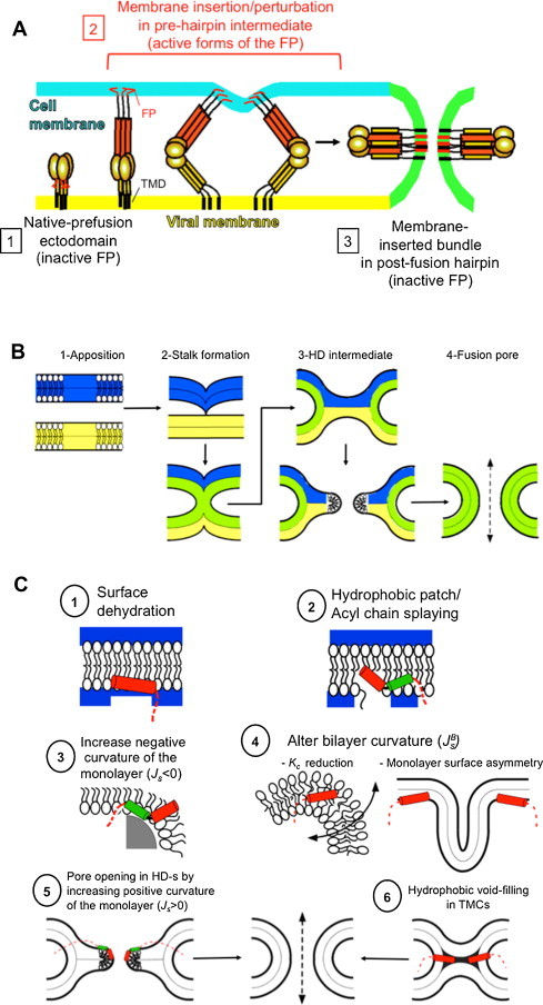
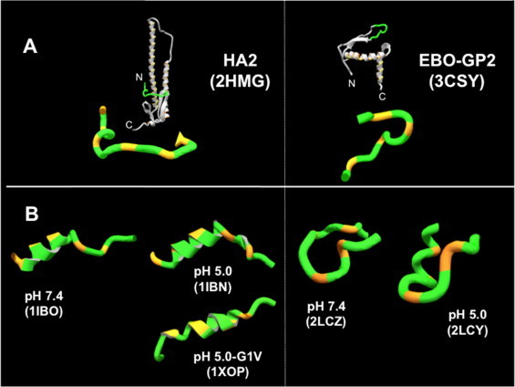
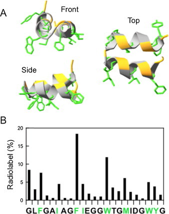
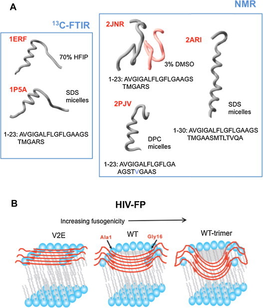
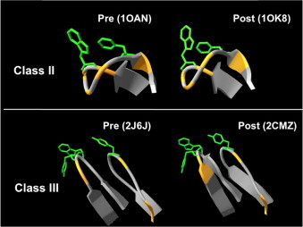
Fusion peptides comprise conserved hydrophobic domains absolutely required for the fusogenic activity of glycoproteins from divergent virus families. After 30 years of intensive research efforts, the structures and functions underlying their high degree of sequence conservation are not fully elucidated. The long-hydrophobic viral fusion peptide (VFP) sequences are structurally constrained to access three successive states after biogenesis. Firstly, the VFP sequence must fulfill the set of native interactions required for (meta) stable folding within the globular ectodomains of glycoprotein complexes. Secondly, at the onset of the fusion process, they get transferred into the target cell membrane and adopt specific conformations therein. According to commonly accepted mechanistic models, membrane-bound states of the VFP might promote the lipid bilayer remodeling required for virus-cell membrane merger. Finally, at least in some instances, several VFPs co-assemble with transmembrane anchors into membrane integral helical bundles, following a locking movement hypothetically coupled to fusion-pore expansion. Here we review different aspects of the three major states of the VFPs, including the functional assistance by other membrane-transferring glycoprotein regions, and discuss briefly their potential as targets for clinical intervention.

Keywords: Fusion peptide; Membrane fusion; Peptide-lipid interaction; Viral entry.


Figures

None
Graphical abstract
Fig. 1
Fig. 1
Viral glycoprotein-induced membrane fusion and proposed functions for the FP in the process. (A) General model of membrane fusion promoted by Class I viral fusion glycoproteins. (B) Stages of the process from a lipid-centric perspective (see text). (C) Putative effects of FPs on target membrane rupture and deformation during fusion (see text).
Fig. 2
Fig. 2
Native and membrane-inserted structures of IFV and EBOV fusion peptides. (A) X-ray structures disclosing several turns in the native fusogenic subunits. Structures on top disclose their position within the ectodomain. (B) NMR structures of peptides in DPC micelles reflecting features of α-helix. In both panels the orange stretches mark the positions of Gly/Pro residues. Structures with the PDB codes in brackets have been used to render the Figure.
Fig. 3
Fig. 3
Helical hairpin model for IFV-FP. (A) NMR structure of the synthetic HA-FP1–23 peptide in DPC micelles (PDB entry code: 2KXA). Side-chains of hydrophobic residues are displayed in green, while orange stretches in the ribbon mark positions of Gly residues. (B) Residue labeling in the sequence covered by HA-FP1–23 upon hydrophobic photolabeling of BHA incubated at low-pH in the presence of membranes. Percentages of label in the residues were calculated based on the data reported by (Harter et al., 1989). Side-chains of residues in green characters are depicted in the previous panel.
Fig. 4
Fig. 4
Conformational plasticity of HIV-FP. (A) Gallery of structures of synthetic peptides solved by IR (Gordon et al., 2002, Gordon et al., 2004) and NMR (Jaroniec et al., 2005, Li and Tamm, 2007, Munch et al., 2007). Corresponding PDB entry codes are displayed in red. 2JNR corresponds to the FP in complex with VIRIP (reddish chain). (B) Models for the FP inserted into Chol-containing membranes adopting 6-strand, anti-parallel β-sheet structures, as inferred from SS-NMR determinations. The most fusion-active version inserts deeper (see text). (For interpretation of the references to color in this figure legend, the reader is referred to the web version of this article.)
Fig. 5
Fig. 5
Fusion loops derived from Class II and Class III glycoproteins. X-ray structures of FLs in pre- and post-fusion states of Class II DENV-E and Class III VSV-G glycoproteins are displayed (top and bottom panels, respectively). In both panels the exposed side-chains of aromatic residues are shown in green, while orange stretches mark the positions of Gly/Pro residues. Structures with the PDB codes in brackets have been used to render the figure.

References

    1. Adam B., Lins L., Stroobant V., Thomas A., Brasseur R. Distribution of hydrophobic residues is crucial for the fusogenic properties of the Ebola virus GP2 fusion peptide. J. Virol. 2004;78:2131–2136. - PMC - PubMed
    1. Agirre A., Flach C., Goni F.M., Mendelsohn R., Valpuesta J.M., Wu F., Nieva J.L. Interactions of the HIV-1 fusion peptide with large unilamellar vesicles and monolayers. A cryo-TEM and spectroscopic study. Biochim. Biophys. Acta. 2000;1467:153–164. - PubMed
    1. Apellaniz B., Garcia-Saez A., Nir S., Nieva J.L. Destabilization exerted by peptides derived from the membrane-proximal external region of HIV-1 gp41 in lipid vesicles supporting fluid phase coexistence. Biochim. Biophys. Acta. 2011;1808:1797–1805. - PubMed
    1. Apellaniz B., Ivankin A., Nir S., Gidalevitz D., Nieva J.L. Membrane-proximal external HIV-1 gp41 motif adapted for destabilizing the highly rigid viral envelope. Biophys. J. 2011;101:2426–2435. - PMC - PubMed
    1. Apellaniz B., Nieva J.L., Schwille P., Garcia-Saez A.J. All-or-none versus graded: single-vesicle analysis reveals lipid composition effects on membrane permeabilization. Biophys. J. 2010;99:3619–3628. - PMC - PubMed

Publication types