Review
. 2014 Apr;25(100):104-10.
doi: 10.1016/j.sbi.2014.02.003. Epub 2014 Apr 2.

Telomerase structure

Affiliations
Review

Telomerase structure

Sara Sandin et al. Curr Opin Struct Biol. 2014 Apr.

Abstract

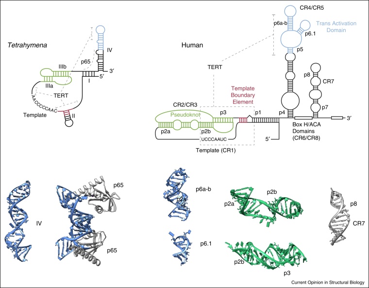
The telomerase reverse transcriptase has an essential role in telomere maintenance and in cancer biology. Progress during the last year has revealed the three-dimensional architecture of both human and ciliate telomerase at about 25Å resolution, obtained using single particle electron microscopy (EM). The structural analysis of the two holoenzyme complexes isolated from cells shows that whilst the ciliate telomerase is monomeric, the human telomerase is dimeric and only functional as a dimer. We critically discuss the approaches taken to assign the location of protein and RNA subunits, as well as fitting the crystal structure of the catalytic protein subunit in the medium resolution EM density maps. Comparison of the two structural interpretations reveals not only a common RNA/reverse transcriptase core, but also significant differences due to different RNA subunit size and protein composition. These differences suggest that the oligomeric state and subunit composition of telomerase in evolutionary distant organism have evolved.


Figures

Figure 1
Figure 1
Human telomere structure and telomerase recruitment. Telomeric DNA consists of arrays of the TTAGGG telomeric repeat, forming a long region of double stranded DNA terminating in the single stranded G-rich overhang. In addition to being packaged by histones into chromatin, telomeric DNA is bound by the sheltering complex consisting of six telomere binding proteins: TRF1, TRF2, Rap1, TIN2, TPP1 and POT1. TRF1 and TRF2 bind sequence specifically as homodimers to double-stranded telomeric DNA, TIN2 forms a bridging interaction with TRF1 and TRF2 and TPP1. POT1 and TPP1 are localized to the G-overhang via the binding of POT1 to the single-stranded G-overhang. Telomerase is recruited to the tip of telomeres through the interaction of TPP1 with the N-terminal domain of the telomerase catalytic subunit TERT and through base pairing between the template region in the telomerase RNA subunit TER and the G-overhang.
Figure 2
Figure 2
Domain structure of the telomerase RNA subunit TER. Secondary structures of the Tetrahymena and human telomerase TER subunits in which regions corresponding to the pseudo-knot (green), template boundary element (red), transactivation domain (blue) are highlighted. The interaction of TERT with TER domains is indicated (grey dotted line). The NMR structures of TER fragments are from the protein data bank and coloured according to the secondary structures: IV (pdb number: 2FEY), IV-p65 C-terminal domain (pdb number: 4ERD), p6a-b (pdb number: 2Z31), p6.1 (pdb number: 1OQ0), p2a/p2b (pdb number: 2LE3), p2b/p3 (pdb number: 2K96) and p8/CR7 (pdb number: 2QH2).
Figure 3
Figure 3
Domain structure of the TERT catalytic subunit. TERT contains a telomerase essential N-terminal (TEN) domain, a flexible linker region, a telomerase RNA-binding (TRBD) domain, a reverse transcriptase (RT) domain and a C-terminal domain (CTE). The TEN domain (pink) binds single-stranded telomeric DNA upstream of the primer-template. The TRBD domain (dark blue) binds the pseudo-knot and the transactivation domain of TER. Crystal structures of isolated domains from Tetrahymena: TEN domain (pdb number: 2B2A) is depicted in pink and TRDB (pdb number: 4R4G) domain in dark blue. The crystal structure of the beetle TERT (dark and light blue) shows a ring-like structure in complex with a RNA-DNA hairpin (pdb number: 3KYL).
Figure 4
Figure 4
3D structures of human and Tetrahymena telomerases. Single particle analyses of full-length human (a–d) and Tetrahymena (e–g) holoenzyme complexes in negative stain. (a) The 3D reconstruction of the human telomerase dimer at 30 Å resolution showing two monomers connected by a flexible hinge region. (b) Independent refinement of one half of the dimer, at a resolution of 23 Å, superimposed with the dimer density (wireframe). (c and d) Two views, related by a 90° rotation around the horizontal axis, of the refined monomer together with the docked crystal structure of beetle TERT. TERT is coloured blue, with the TRBD domain in dark blue, and the DNA strand in orange. (e and f) Two views of the Tetrahymena telomerase. 3D reconstruction at 25 Å resolution. The two views are related by a 90° rotation around the vertical axis. The location of TERT, TER and accessory proteins p75, p19, p45, p50, Teb and p65 are indicated. (g) Docking of the atomic structures p65, Teb1C, homology model Tetrahymena TERT (blue), and RNA model of TER (black) into the EM density map, reproduced with permission from [12].

References

    1. Blackburn E.H., Collins K. Telomerase: an RNP enzyme synthesizes DNA. Cold Spring Harb Perspect Biol. 2011;3 - PMC - PubMed
    1. Pfeiffer V., Lingner J. Replication of telomeres and the regulation of telomerase. Cold Spring Harb Perspect Biol. 2013;5:a010405. - PMC - PubMed
    1. de Lange T. How telomeres solve the end-protection problem. Science. 2009;326:948–952. - PMC - PubMed
    1. Gunes C., Rudolph K.L. The role of telomeres in stem cells and cancer. Cell. 2013;152:390–393. - PubMed
    1. Greider C.W., Blackburn E.H. Identification of a specific telomere terminal transferase activity in Tetrahymena extracts. Cell. 1985;43:405–413. - PubMed

Publication types

LinkOut - more resources