. 2014 May;21(5):443-8.
doi: 10.1038/nsmb.2807. Epub 2014 Apr 6.

Structural basis of recognition of interferon-α receptor by tyrosine kinase 2

Affiliations

Structural basis of recognition of interferon-α receptor by tyrosine kinase 2

Heidi J A Wallweber et al. Nat Struct Mol Biol. 2014 May.

Abstract

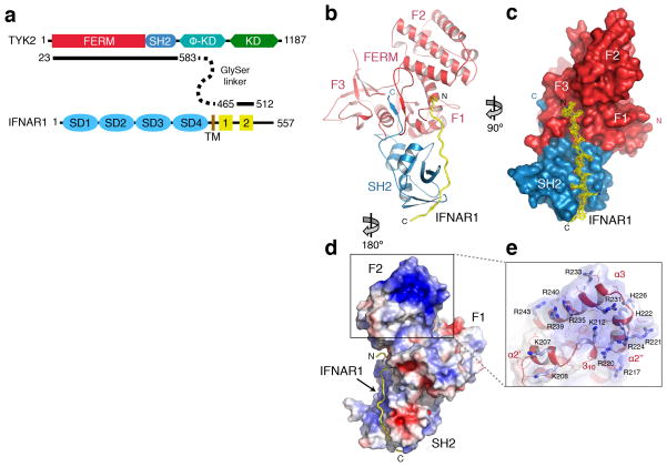
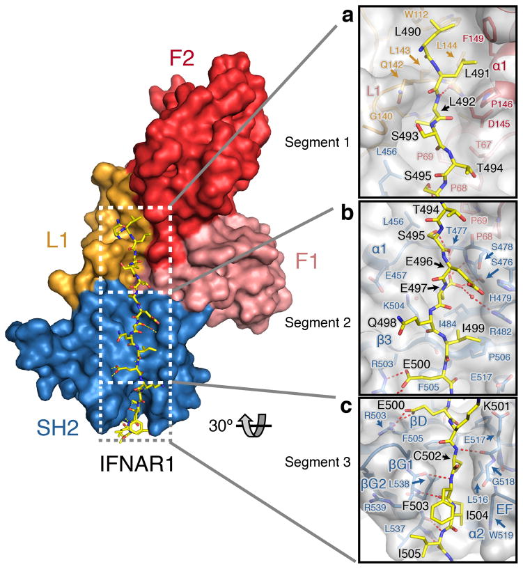
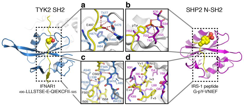
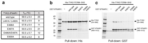
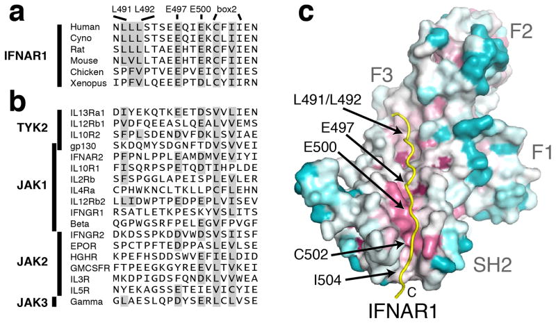
Tyrosine kinase 2 (TYK2) is a member of the Janus kinase (JAK) family of nonreceptor tyrosine kinases, which are essential for proper signaling in immune responses and development. Here we present a 2.0-Å-resolution crystal structure of a receptor-binding fragment of human TYK2, encompassing the FERM and SH2 domains, in complex with a so-called 'box2'-containing intracellular peptide motif from the interferon-α receptor chain 1 (IFNAR1). The TYK2-IFNAR1 interface reveals an unexpected receptor-binding mode that mimics a SH2 domain-phosphopeptide interaction, with a glutamate replacing the canonical phosphotyrosine residue. This structure provides the first view, to our knowledge, of a JAK in complex with its cognate receptor and defines the molecular logic through which JAKs have evolved to interact with divergent receptor sequences.


Conflict of interest statement

Competing interest statement: The authors declare no competing financial interests.

Figures

Figure 1
Figure 1. The structure of the TYK2 FERM–SH2 receptor binding module in complex with IFNAR1
(a) Schematic diagram of TYK2 and IFNAR1 showing the domains linked to generate expression constructs. TYK2 pseudokinase and kinase domains are noted ϕ–KD and KD. IFNAR1 FNIII-like subdomains are noted as SD1 to SD4, transmembrane region as TM, and box1 and box2 as 1 and 2, respectively. (b, c) The structure of the TYK2–IFNAR1 complex from two perspectives. In (b), a cartoon representation shows the interaction between TYK2 and IFNAR1. The FERM domain is colored in red, with the F1, F2, and F3 subdomains labeled. The SH2 domain is colored in blue, and IFNAR1 is colored in yellow. N- and C-termini are labeled N and C, respectively. Surface representation (c) of TYK2 FERM–SH2 is rotated 90° with the IFNAR1 peptide shown as a stick model with overlaid electron density (contoured at 1.0σ) displayed in yellow. (d) Electrostatic surface potential for the TYK2 FERM–SH2 structure, contoured at −5 to 5 kT/e. (e) The inset box shows a detailed view of the α2′, α2″, and α3 helices of the F2 subdomain, with basic sidechains shown as sticks and colored in light blue. Electrostatic surface potential is shown as a transparent surface.
Figure 2
Figure 2. IFNAR1 interacts with TYK2 in three segments
Detailed views of the (a) segment 1, (b) segment 2, and (c) segment 3 regions of the IFNAR1 peptide interfaces are shown. Key residues are labeled and shown as sticks, with IFNAR1 colored yellow. The TYK2 FERM F1 and F2 subdomains and L1 loop are colored pink, red, and orange respectively, and the TYK2 SH2 domain is colored blue. For the inset panels, TYK2 is also represented as a transparent grey surface. Polar contacts are represented as dashed lines.
Figure 3
Figure 3. The TYK2–IFNAR1 complex resembles a SH2–phosphopeptide interaction
Comparison of the TYK2 SH2 interaction with IFNAR1 side-by-side with the SHP2 SH2 domain bound to a phosphotyrosine peptide from IRS-1 (Protein Data Bank ID 1AYB). Detailed views of the (a) IFNAR1 glutamate and (c) box2 interactions with TYK2 are shown inset on the left, with (b) the IRS-1 phosphotyrosine and (d) C-terminal interactions with SHP2 are shown inset on the right. The TYK2 SH2 domain is colored blue and the SHP2 SH2 domain is colored purple, with bound peptides colored yellow. Key residues are shown as stick models and polar contacts shown as dashed lines.
Figure 4
Figure 4. IFNAR1 interface contacts stabilize TYK2 interaction
(a) Apparent melting temperatures (mean ± s.d. of >6 measurements, with n shown in third column) were determined for TYK2 bound to IFNAR1 wild type and mutant peptides using differential scanning fluorimetry. P-values from an unpaired, two-tailed students t-test are reported in Supplementary Fig. 9. (b, c) Reciprocal His- and GST-pulldown experiments were performed by expressing His-tagged TYK2 FERM–SH2 (residues 23–566) alone or with GST-tagged IFNAR1 variants (residues 465–512) in insect cells. Expressed proteins were purified by either (a) Ni-NTA or (b) glutathione affinity chromatography, and eluted proteins analyzed by SDS–PAGE. The two ~25kD background bands eluted from the glutathione column represent endogenous GST protein.
Figure 5
Figure 5. TYK2–IFNAR1 interface residues are conserved in other cytokine receptors and JAKs
(a, b) Sequence comparison of the TYK2-bound human IFNAR1 peptide to (a) IFNAR1 orthologs and (b) other human cytokine receptors that signal through JAK kinases. Sequences were aligned to IFNAR1 based on putative box2 sequences. (c) Conservation analysis of the TYK2 receptor-binding interface across the JAK family. Amino acid conservation among 150 JAK orthologs was mapped onto the surface of the TYK2 FERM–SH2, with residues colored from most (purple) to least (cyan) conserved.

Comment in

References

    1. Leonard WJ, O’Shea JJ. Jaks and STATs: biological implications. Annu Rev Immunol. 1998;16:293–322. - PubMed
    1. Haan C, Kreis S, Margue C, Behrmann I. Jaks and cytokine receptors--an intimate relationship. Biochem Pharmacol. 2006;72:1538–1546. - PubMed
    1. Casanova JL, Holland SM, Notarangelo LD. Inborn errors of human JAKs and STATs. Immunity. 2012;36:515–528. - PMC - PubMed
    1. Kontzias A, Kotlyar A, Laurence A, Changelian P, O’Shea JJ. Jakinibs: a new class of kinase inhibitors in cancer and autoimmune disease. Curr Opin Pharmacol. 2012;12:464–470. - PMC - PubMed
    1. Radtke S, et al. The Jak1 SH2 domain does not fulfill a classical SH2 function in Jak/STAT signaling but plays a structural role for receptor interaction and up-regulation of receptor surface expression. J Biol Chem. 2005;280:25760–25768. - PubMed

Publication types

Associated data