. 2015 Mar 19;34(12):1553-62.
doi: 10.1038/onc.2014.87. Epub 2014 Apr 7.

ICAM-2 confers a non-metastatic phenotype in neuroblastoma cells by interaction with α-actinin

Affiliations

ICAM-2 confers a non-metastatic phenotype in neuroblastoma cells by interaction with α-actinin

J M Feduska et al. Oncogene. .

Abstract

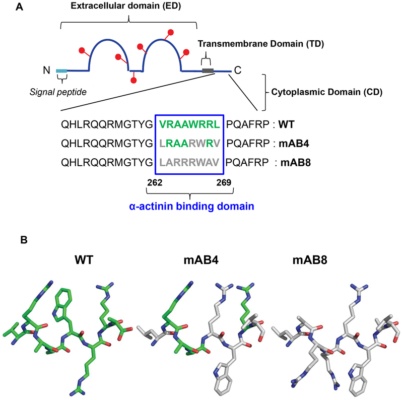
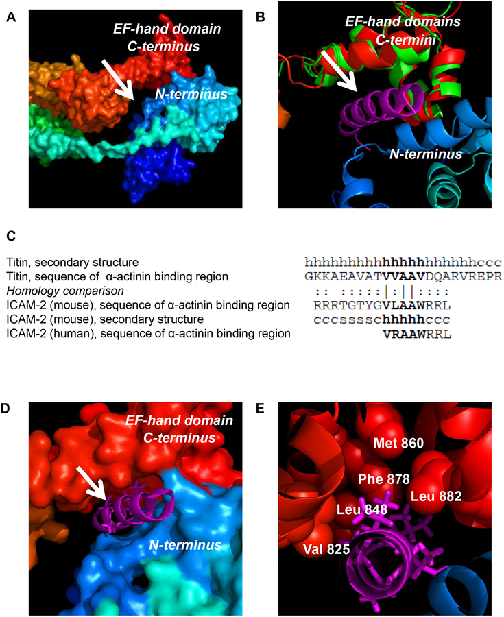
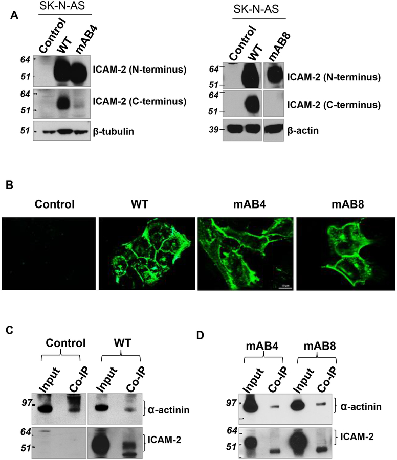
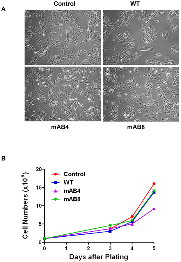
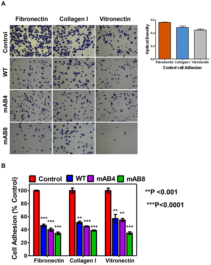
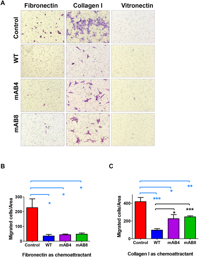
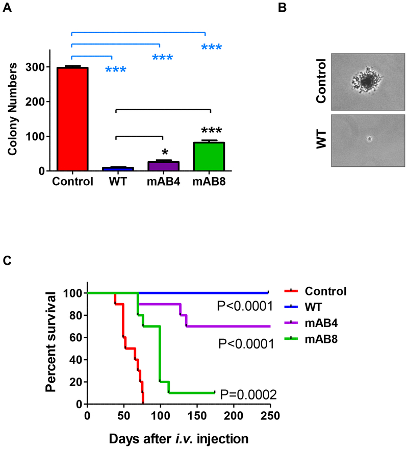
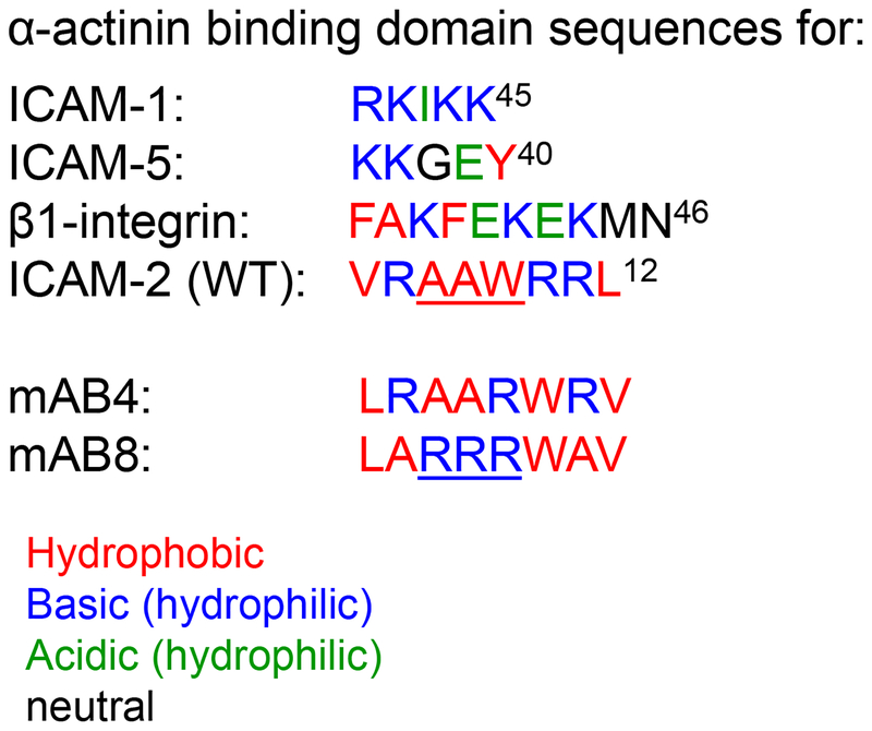
Progressive metastatic disease is a major cause of mortality for patients diagnosed with multiple types of solid tumors. One of the long-term goals of our laboratory is to identify molecular interactions that regulate metastasis, as a basis for developing agents that inhibit this process. Toward this goal, we recently demonstrated that intercellular adhesion molecule-2 (ICAM-2) converted neuroblastoma (NB) cells from a metastatic to a non-metastatic phenotype, a previously unknown function for ICAM-2. Interestingly, ICAM-2 suppressed metastatic but not tumorigenic potential in preclinical models, supporting a novel mechanism of regulating metastasis. We hypothesized that the effects of ICAM-2 on NB cell phenotype depend on the interaction of ICAM-2 with the cytoskeletal linker protein α-actinin. The goal of the study presented here was to evaluate the impact of α-actinin binding to ICAM-2 on the phenotype of NB tumor cells. We used in silico approaches to examine the likelihood that the cytoplasmic domain of ICAM-2 binds directly to α-actinin. We then expressed variants of ICAM-2 with mutated α-actinin-binding domains, and compared the impact of ICAM-2 and each variant on NB cell adhesion, migration, anchorage-independent growth, co-precipitation with α-actinin and production of localized and disseminated tumors in vivo. The in vitro and in vivo characteristics of cells expressing ICAM-2 variants with modified α-actinin-binding domains differed from cells expressing ICAM-2 wild type (WT) and also from cells that expressed no detectable ICAM-2. Like the WT protein, ICAM-2 variants inhibited cell adhesion, migration and colony growth in vitro. However, unlike the WT protein, ICAM-2 variants did not completely suppress development of disseminated NB tumors in vivo. The data suggest the presence of α-actinin-dependent and α-actinin-independent mechanisms, and indicate that the interaction of ICAM-2 with α-actinin is critical to conferring an ICAM-2-mediated non-metastatic phenotype in NB cells.


Conflict of interest statement

Conflict of interest.

The authors declare no conflict of interest.

Figures

Figure 1.
Figure 1.. Schematic structure of ICAM-2 and ICAM-2 variants.
(A) Sequence of the cytoplasmic domains of endogenous human ICAM-2 wild type (WT) protein and variants mAB4 and mAB8. The extracellular domain (ED) of the protein contains two immunoglobulin-like domains with six N-glycosylation sites (red lollipops), a 26-amino acid transmembrane domain (TD) and a 26-amino acid cytoplasmic (CD) domain. The N-terminus of the nascent protein contains a 21-amino acid signal peptide (sp) that is not present in the 254 amino acid mature protein. (B) Structures of the 8-mer proposed α-actinin binding domains of ICAM-2 WT, mAB4 and mAB8 were generated using PDB IJ19 and PyMol. The predicted structure of this domain of ICAM-2 WT is in green. The amino acid side chains of this domain of ICAM-2 mAB4 and mAB8 that have spatial orientations similar to those in the WT protein are also in green.
Figure 2.
Figure 2.. Modeling of ICAM-2 interaction with α-actinin.
(A) Surface representation of the full-length chicken α-actinin structure (PDB code 1SJJ) reveals a potential binding cavity (arrow) in the cleft between the actin binding domain (ABD, blue) and EF-hand (red) domains. The N-terminus to C-terminus is colored from blue (ABD) to red (EF-hand domain). (B) Structural alignment between full-length chicken α-actinin (red) and the EF-hand domain of human α-actinin (green)(PDB code 1H8B) demonstrated a high confidence alignment (RMSD of 1.47 Å). Structural alignment of the EF-hand domain of chicken α-actinin (red) with EF-hand domain of human α-actinin (residues 823–894, green) complexed to the α-actinin binding domain helix of rabbit titin (known structure, magenta) placed this helix in the putative binding pocket suggested by the structure in “A”. (C) The sequences of the α-actinin binding domains of rabbit titin, murine ICAM-2, and human ICAM-2 have high homology. The secondary structure of the rabbit and murine peptides are indicated as: “h” = helix, “s” = β-sheet and “c” = coil. Amino acid homology comparison is indicated as: “|” = identical; “:” = similar. (D) Modeling reveals a likely high-affinity interaction between the structurally conserved alpha-helix of titin (magenta) and the cytoplasmic domains of murine and human ICAM-2 (by structural and sequence homology) with the EF-hand domain of α-actinin (red). (E) Specific hydrophobic residues of the EF-hand domain of α-actinin and adjacent hydrophobic residues of a helical α-actinin binding structure suggest that ICAM-2 associates directly with α-actinin through hydrophobic interactions. The magenta stick structures represent the “VVAAV” component of the titin helix that is most conserved in ICAM-2.
Figure 3.
Figure 3.. ICAM-2 WT and variants localized to cell membranes.
(A) Immunoblots confirmed that tranfected SK-N-AS cells expressed readily detectable levels of ICAM-2 WT, mAB4 or mAB8. Data in both membranes were generated in the same experiment, but immunoblotted individually. (B) Immunofluorescence staining demonstrated that ICAM-2 WT, mAB4 and mAB8 localized to cell membranes. Scale bar represents 10 μm for all panels. Details of procedures used are in the Methods. (C, D) ICAM-2 WT, but not mAB4 and mAB8, co-precipitated with α-actinin. Immunoprecipitations (IP) were performed using whole cell lysates and a mouse monoclonal antibody to α-actinin (MAB1682, Millipore-Fisher Scientific). The presence of all three types of ICAM-2 proteins in the “input” whole cell lysates (wcl) was confirmed prior to IP.
Figure 4.
Figure 4.. Morphologies and growth rates of SK-N-AS cells transfected to express ICAM-2 WT, mAB4 or mAB8 are equivalent.
(A) All transfectants grew as attached monolayers and had nascent neuronal morphologies; all four transfectants grew as tight cell groups but groupings of cells transfected to express ICAM-2 WT appeared more cohesive and more angular than Control transfectants. (B) Cell proliferation assays showed that transfectancts had similar cell doubling times of 30.4 ± 4.7 hours.
Figure 5.
Figure 5.. ICAM-2 WT and mAB variants inhibit NB cell adhesion to ECM proteins.
(A) NB cells adherent to each ECM protein were imaged after staining. The optical density for Control cells (used in calculations and presented as 100%) were for fibronectin=0.5630 ± 0.0032, for collagen I=0.4855 ± 0.019, and for vitronectin=0.4443 ± 0.0160 (mean ± SEM), as shown in right panel. (B) NB cell adhesion was quantitated by determination of the optical density of crystal violet-stained, adherent cells by standard methods. Values for ICAM-2 transfectants were significantly different from Control transfectants (**P<0.001 or ***P<0.0001, as indicated).
Figure 6.
Figure 6.. ICAM-2 WT and mAB variants inhibit NB cell motility in Boyden chamber migration assays.
(A) Transfected NB cells that migrated to the distal side of Boyden chamber membranes were imaged after an 18-hour incubation. Representative images were acquired with a Zeiss Axio Observer Z.1 microscope and Zen 2011 Blue software. (B) Migrated cell numbers were determined manually, and analyzed by t-test. ICAM-2 WT, mAB4 and mAB8 inhibited cell motility toward fibronectin compared to cells that expressed no detectable ICAM-2 (Control) (P<0.05*), in an α-actinin-independent manner. (C) ICAM-2 WT, mAB4 and mAB8 inhibited cell migration using collagen I as chemoattractant in an α-actinin-dependent manner (*P=0.0365; **P=0.0018; ***P<0.0001; *P=0.0155; ***P<0.0001).
Figure 7.
Figure 7.. ICAM-2 inhibited colony growth in soft agar and development of disseminated tumors in vivo. ICAM-2 mAB8 delayed but did not inhibit development of disseminated tumors in an in vivo model of metastatic neuroblastoma.
(A) ICAM-2 WT, mAB4 and mAB8 suppressed anchorage-independent growth in vitro. Colonies of >20 cells were visualized 14–21 days after plating. Results were quantitated manually and analyzed using a two-tailed t-test and GraphPad Prism software (*P=0.0135 and ***P<0.0001). (B) Photomicrographs of representative colonies for Control and ICAM-2 WT transfectants were acquired at 400× magnification using a Dr-5 digital camera (Southern Microscope) mounted to a Nikon Eclipse TS100 inverted microscope and archived with TSView software (version 7.1.04, Tucsen Imaging Technology Co./Southern Microscope). (C) Kaplan-Meier survival plots of mice that received i.v. injections of SK-N-AS cell transfectants were analyzed by log-rank (Mantel-Cox) test using GraphPad Prism 5 software (Version 5.02) showed that mice receiving cells expressing mAB variants survived longer than mice receiving cells expressing no detectable ICAM-2, but not as long as mice receiving cells expressing ICAM-2 WT. *Data for control transfectants were previously published in BMC Cancer by BioMed Central. Reprinted with permission.
Figure 8.
Figure 8.. Disparate α-actinin binding sequences in proteins belonging to two families of cell adhesion molecules: ICAM-1, ICAM-2, ICAM-5 and β1-integrin.
Residues are indicated as hydrophobic (red); basic hydrophilic (blue); acidic hydrophilic (green), and neutral (black).

References

    1. Casasnovas JM, Springer TA, Liu JH, Harrison SC, Wang JH. Crystal structure of ICAM-2 reveals a distinctive integrin recognition surface. Nature 1997; 387: 312–315. - PubMed
    1. Nortamo P, Li R, Renkonen R, Timonen T, Prieto J, Patarroyo M et al. The expression of human intercellular adhesion molecule-2 is refractory to inflammatory cytokines. Eur J Immunol 1991; 21: 2629–2632. - PubMed
    1. de Fougerolles AR, Stacker SA, Schwarting R, Springer TA. Characterization of ICAM-2 and evidence for a third counter-receptor for LFA-1. J Exp Med 1991; 174: 253–267. - PMC - PubMed
    1. Kotovuori A, Pessa-Morikawa T, Kotovuori P, Nortamo P, Gahmberg CG. ICAM-2 and a peptide from its binding domain are efficient activators of leukocyte adhesion and integrin affinity. J Immunol 1999; 162: 6613–6620. - PubMed
    1. Yoon KJ, Phelps DA, Bush RA, Remack JS, Billups CA, Khoury JD. ICAM-2 expression mediates a membrane-actin link, confers a nonmetastatic phenotype and reflects favorable tumor stage or histology in neuroblastoma. PLoS One 2008; 3: e3629. - PMC - PubMed

Publication types

MeSH terms