. 2014 May:57-58:15-23.
doi: 10.1016/j.jchemneu.2014.03.001. Epub 2014 Apr 3.

Cellular profile of the dorsal raphe lateral wing sub-region: relationship to the lateral dorsal tegmental nucleus

Affiliations

Cellular profile of the dorsal raphe lateral wing sub-region: relationship to the lateral dorsal tegmental nucleus

Rani K Vasudeva et al. J Chem Neuroanat. 2014 May.

Abstract

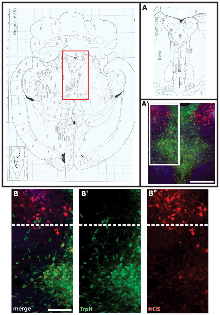
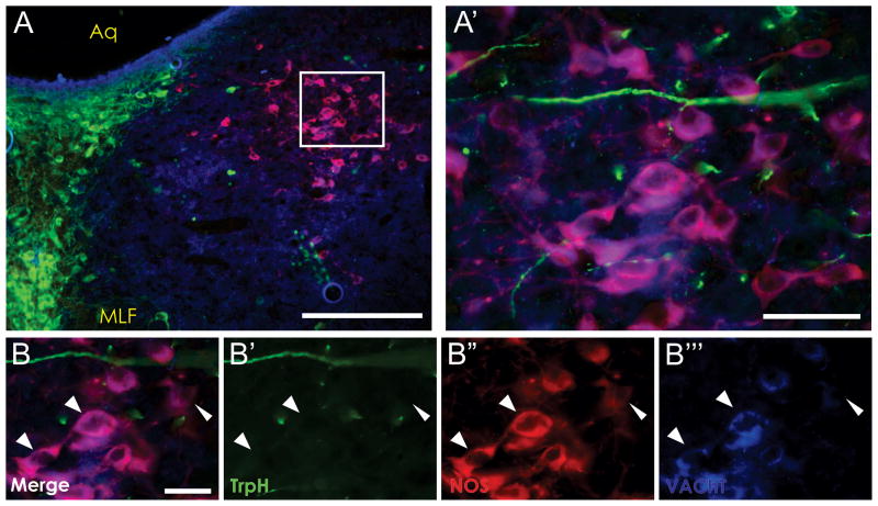
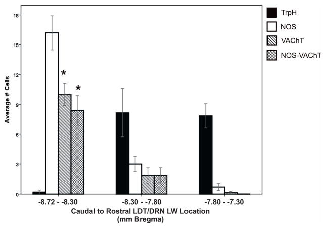
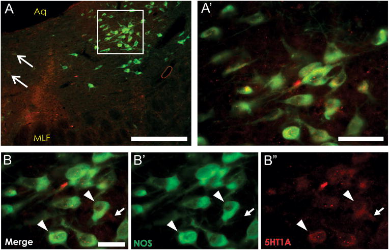
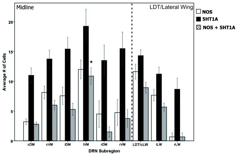
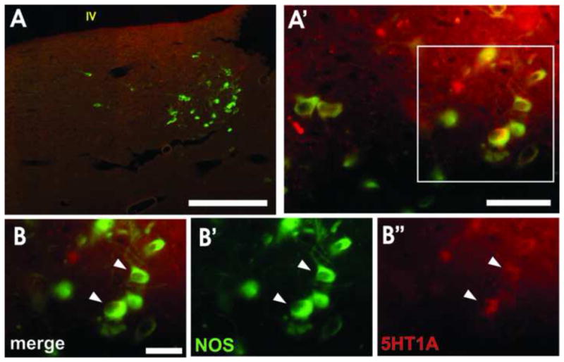
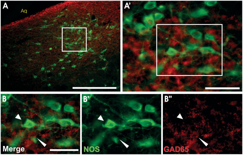
As one of the main serotonergic (5HT) projections to the forebrain, the dorsal raphe nucleus (DRN) has been implicated in disorders of anxiety and depression. Although the nucleus contains the densest population of 5HT neurons in the brain, at least 50% of cells within this structure are non-serotonergic, including a large population of nitric oxide synthase (NOS) containing neurons. The DRN has a unique topographical efferent organization and can also be divided into sub-regions based on rostro-caudal and medio-lateral dimensions. NOS is co-localized with 5HT in the midline DRN but NOS-positive cells in the lateral wing (LW) of the nucleus do not express 5HT. Interestingly, the NOS LW neuronal population is immediately rostral to and in line with the cholinergic lateral dorsal tegmental nucleus (LDT). We used immunohistochemical methods to investigate the potential serotonergic regulation of NOS LW neurons and also the association of this cell grouping to the LDT. Our results indicate that >75% of NOS LW neurons express the inhibitory 5HT1A receptor and are cholinergic (>90%). The findings suggest this assembly of cells is a rostral extension of the LDT, one that it is subject to regulation by 5HT release. As such the present study suggests a link between 5HT signaling, activation of cholinergic/NOS neurons, and the stress response including the pathophysiology underlying anxiety and depression.

Keywords: Acetylcholine; Dorsal raphe; Lateral dorsal tegmental nucleus; Nitric oxide synthase; Serotonin.


Figures

Figure 1
Figure 1. Horizontal Sections of NOS and VAChT Staining across the DRN
Horizontal sections of the rat brainstem at the level of the DRN and lateral dorsal tegmental nucleus (LDT) reveal the lateral wing sub-region of the DRN and the LDT (A′, white box) form a continuous column of cells along the rostral-caudal dimension. The caudal region of the LDT is bordered by the fourth ventricle (top of figure A and A′). Tryptophan hydroxylase (TrpH) positive neurons (B′, green) clearly intermingle with NOS positive cells (B″, red) along the caudal lateral wing and rostral LDT border (B, B′, B″, white dashed line). Scale bars: A′ = 500 μm, B = 250 μm. Abbreviations in A: 3-oculomotor nucleus, 3V- third ventricle, 4- trochlear nucleus, 4n- trochlear nerve root, 4V- fourth ventricle, CnF- cuneiform nucleus, DpMe- deep mesencephalic nucleus, DR- dorsal raphe nucleus, dtg-dorsal tegmental bundle, EW- nucleus of Edinger Westphal, fr- fasciculus retroflexus, LDTg- lateral dorsal tegmental nucleus, mlf- medial longitudinal fasciculus, PF- parafascicular thalamic nucleus, RI- rostral interstitial nucleus of the mlf.
Figure 2
Figure 2. Juxtaposition of NOS, TrpH, and VAChT in the lateral wing of the DRN
NOS-positive, tryptophan hydroxylase (TrpH)-negative cells located within the caudal portion of the DRN lateral wing (A, A′) express the vesicular acetylcholine transporter (VAChT). High magnification images (B – B‴, arrowheads) show NOS-positive neurons (B″, red) co-localize with VAChT (B‴, blue) but not TrpH (B′, green). This finding indicates that NOS-positive, 5HT-negative cells within the DRN lateral wing are actually the rostral-most extension of the cholinergic lateral dorsal tegmental nucleus. Scale bars: A = 250 μm, A′ = 50 μm, B = 30 μm. Aq- cerebral aqueduct, MLF- medial longitudinal fasciculus.
Figure 3
Figure 3. Distribution and cell counts of NOS, TrpH, and VAChT cells across the DRN
The average number of cells expressing tryptophan hydroxylase (TrpH, black bars), nitric oxide synthase (NOS, white bars), vesicular acetylcholine transferase (VAChT, diagonal bars), or NOS and VAChT (hashed bars) are compared caudal to rostral across the rostral LDT/caudal LW border (−8.72 to −8.30 Bremga) through to the rostral LW subregion of the DRN (−7.80 to −7.30 Bregma). The LDT/LW border region (left side data bars; −8.72 - −8.30 Bregma) contains significantly higher numbers of VAChT and NOS-VAChT expressing cells than more rostral regions of the LW or any DRN midline subregions. (* p < .001)
Figure 4
Figure 4. Co-localization of the 5HT1A receptor and NOS on lateral wing neurons in the caudal DRN
NOS-positive neurons located in the lateral wing DRN- rostral LDT border (A, A′) express the 5HT1A receptor. White arrows in (A) indicates NOS-positive cells in the DRN midline to confirm the caudal DRN location (Vasudeva et al., 2011). High magnification (B) demonstrates clear co-expression of NOS (green, B′) and the 5HT1A receptor (red, B″) on the same cells (arrowheads). A non-NOS cell expressing the 5HT1A receptor can also be seen (B-B″, white arrow). Scale bars: A = 500 μm, A′ = 50 μm, B = 30 μm. Aq- cerebral aqueduct, MLF= medial longitudinal fasciculus.
Figure 5
Figure 5. Distribution and cell counts of NOS cells and 5HT1A expression across the DRN
The average number of NOS (white bars), 5HT1A receptor (black bars) and double labeled cells (hashed bars) are shown across the midline and lateral wing DRN. Both 5HT1A-only cells and double labeled cells were seen in every DRN sub-region. With the exception of the caudal lateral wing (cLW)/LDT border, the intermediate ventromedial sub-region (iVM) contained the highest number of double labeled cells compared to all other subregions (* = p < .05). cDM- caudal dorsomedial, cLW-caudal lateral wing, cVM- caudal ventromedial, iDM- intermediate dorsomedial, iLW- intermediate lateral wing, iVM- intermediate ventromedial, LDT- lateral dorsal tegmental area, rDM- rostral dorsomedial, rLW- rostral lateral wing, rVM- rostral ventromedial.
Figure 6
Figure 6. Expression of NOS and the 5HT1A receptor in the LDT
NOS-positive, 5HT-lacking neurons of the lateral dorsal tegmental nucleus (LDT; A, A′) express the 5HT1A receptor. High magnification (B, arrowheads) shows NOS-positive cells (B′, green) co-expressing the 5HT1A receptor (B″, red). Scale bars: A = 500 μm, A′ = 50 μm, B = 30 μm. IV- fourth ventricle.
Figure 7
Figure 7. NOS LW cells are not GABAergic
NOS-positive cells in the DRN lateral wing-LDT transition zone (A, A′) are not GABAergic cells. High magnification (B) clearly shows NOS-positive cells (B′, green, arrowheads) are separate from GAD65-positive neurons (B″, red, pointed arrowheads). Scale bars: A = 250 μm, A′ = 50 μm, B = 50 μm. Aq- cerebral aqueduct.

Similar articles

Cited by

References

    1. Abrams JK, et al. Serotonergic systems associated with arousal and vigilance behaviors following administration of anxiogenic drugs. Neuroscience. 2005;133:983–997. - PubMed
    1. Bains JS, Ferguson AV. Nitric oxide regulates NMDA-driven GABAergic inputs to type I neurones of the rat paraventricular nucleus. J Physiol. 1997;499 (Pt 3):733–746. - PMC - PubMed
    1. Blier P, de Montigny C. Serotonin and drug-induced therapeutic responses in major depression, obsessive-compulsive and panic disorders. Neuropsychopharmacology. 1999;21:91S–98S. - PubMed
    1. Bonnavion P, et al. Heterogeneous distribution of the serotonin 5-HT(1A) receptor mRNA in chemically identified neurons of the mouse rostral brainstem: Implications for the role of serotonin in the regulation of wakefulness and REM sleep. J Comp Neurol. 2010;518:2744–2770. - PubMed
    1. Boucetta S, Jones BE. Activity profiles of cholinergic and intermingled GABAergic and putative glutamatergic neurons in the pontomesencephalic tegmentum of urethane-anesthetized rats. J Neurosci. 2009;29:4664–4674. - PMC - PubMed

MeSH terms

Substances

LinkOut - more resources