. 2014 Jan 24;58(1):2256.
doi: 10.4081/ejh.2014.2256.

Developmental immunolocalization of the Klotho protein in mouse kidney epithelial cells

Affiliations

Developmental immunolocalization of the Klotho protein in mouse kidney epithelial cells

J H Song et al. Eur J Histochem. .

Abstract

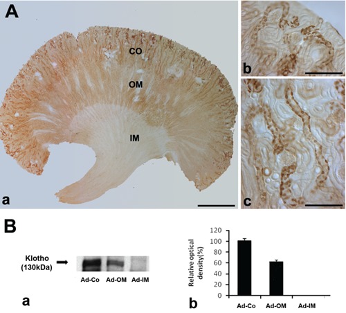
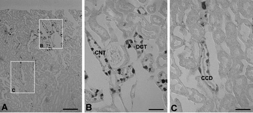
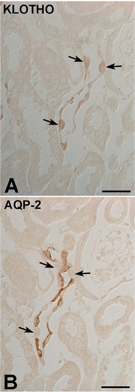
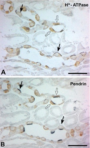
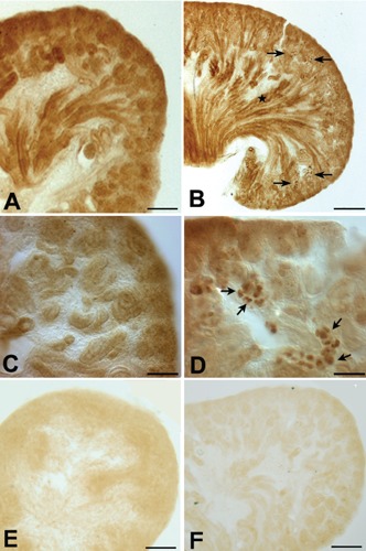
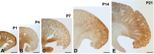
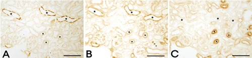
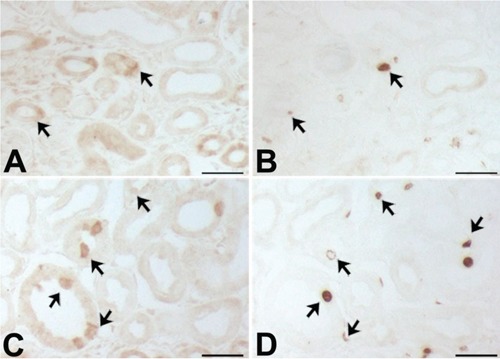
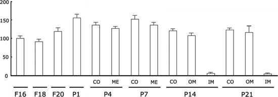
A defect in Klotho gene expression in the mouse results in a syndrome that resembles rapid human aging. In this study, we investigated the detailed distribution and the time of the first appearance of Klotho in developing and adult mouse kidney. Kidneys from 16-(F16), 18-(F18) and 20-day-old (F20) fetuses, 1- (P1), 4- (P4), 7- (P7), 14- (P14), and 21-day-old (P21) pups and adults were processed for immunohistochemistry and immunoblot analyses. In the developing mouse kidney, Klotho immunoreactivity was initially observed in a few cells of the connecting tubules (CNT) of 18-day-old fetus (F) and in the medullary collecting duct (MCD) and distal nephron of the F16 developing kidney. In F20, Klotho immunoreactivity was increased in CNT and additionally observed in the outer portion of MCD and tip of the renal papilla. During the first 3 weeks after birth, Klotho-positive cells gradually disappeared from the MCD due to apoptosis, but remained in the CNT and cortical collecting ducts (CCD). In the adult mouse, the Klotho protein was expressed only in a few cells of the CNT and CCD in cortical area. Also, Klotho immunoreactivity was observed in the aquaporin 2-positive CNT, CCD, and NaCl co-transporter-positive distal convoluted tubule (DCT) cells and type B and nonA-nonB intercalated cells of CNT, DCT, and CCD. Collectively, our data indicate that immunolocalization of Klotho is closely correlated with proliferation in the intercalated cells of CNT and CCD from aging, and may be involved in the regulation of tubular proliferation.


Conflict of interest statement

Conflict of interest: the authors declare no conflict of interests.

Figures

Figure 1.
Figure 1.
A) Light micrographs of 50 μm thick vibratome sections from the adult control mouse kidney illustrating single immunostaining for Klotho. a) Klotho immunoreactivity is located in the Cortex(Co) and outer medulla (OM); no Klotho immunostaining is observed in the inner medulla (IM). b,c) Higher magnification of Co; Klotho protein is expressed in cells of the CNT and CCD; scale bars: a) 100 μm; b,c) 10 μm. B and a) Western blot demonstrating Klotho protein expression in the Co and OM of adult mouse kidney; protein (30 μg) was applied to each lane. b) The relative optical density of the immunoblot band is presented (P<0.05).
Figure 2.
Figure 2.
A) Light micrographs of 5 μm wax sections from kidneys of adult mouse illustrating immunostaining for Klotho. B) Klotho immunoreactivity is located in the connecting tubule (CNT) and distal convoluted tubule (DCT) of cortex; C) Klotho immunoreactivity is located in the some cells of cortical collecting ducts (CCD). Scale bars: 5 μm.
Figure 3.
Figure 3.
DIC micrographs of the cortex from adult mouse kidney. Illustrating serial immunostaining for (A) Klotho and (B) AQP2 in the cortical collecting duct. AQP2-negative cells (arrows) are observed in the intercalated cells of the CCD. Scale bars: 2 μm.
Figure 4.
Figure 4.
Light micrographs of consecutive 1-μm thick sections from adult kidneys, illustrating double immunostaining for Klotho (brown) and H+-ATPase (blue), (A) klotho (brown) and (B) pendrin (blue); numerous Klotho-labeled cells are located in the intercalated cells of the CNTs and CCDs. Klotho-labeled cells are observed in apical pen-drin/basolateral H+-ATPase positive type B intercalated cells (arrows); also expressed in the apical pendrin and H+-ATPase/basolateral pendrin and H+-ATPase negative nonA-nonB intercalated cells (arrows). Scale bars: 5 μm.
Figure 5.
Figure 5.
Light micrographs of 50-μm thick vibratome sections from 16-day-old (A, C) and 19-day-old (B, D) fetal mouse kidneys, illustrating Klotho immunostaining. C, D) Higher magnification of A and B, respectively. A, C) Klotho immunostaining is observed in a broad area of the 16-day-old fetus. B) In the 19-day-old fetal kidney, Klotho immunoreactivity is increased in the distal nephron (arrows) and is also observed in the outer portion of the MCD (star) and in the tip of the renal papilla. D) Klotho immunoreactivity is observed in a few cells of the CNT. E) Negative control with no Klotho antibody in F16 kidney. F) Negative control with no Klotho antibody in F19 kidney. Scale bars: A,B) 100 μm; C,D) 10 μm.
Figure 6.
Figure 6.
Light micrographs of 50-μm thick vibratome sections from the kidneys of mouse pups at post-natal days 1 (A), 4 (B), 7 (C), 14 (D), and 21 (E); medullary Klotho immunoreactivity was gradually decreased; in contrast, cortical Klotho immunoreactivity gradually increases after birth; note a remarkable increase in the immunoreactivity of Klotho in the Co. Scale bars: 100 μm.
Figure 7.
Figure 7.
Light micrographs of consecutive 2-μm thick sections from kidneys of mouse pups at postnatal day 7, illustrating immunostaining of AQP2, Klotho, and NCC. A) Apical AQP2-positive immunoreactivity identifies the principal cells of the CD (stars). B) Klotho is expressed in the CNT, DCT (asterisks), and CCD (stars). C) Immunoperoxidase labeling of NCC in the DCT (asterisks) of the renal cortex. Scale bars: 20 μm.
Figure 8.
Figure 8.
Light micrographs of consecutive 2-μm thick sections from mouse cortex at post-natal days 4 (A,B) and 7 (C,D), illustrating labeling (A,C) Klotho and (B,D) BrdU positivity; some BrdU-labeled cell are observed among the Klotho-positive tubules (arrows). Scale bars: 5 μm.
Figure 9.
Figure 9.
Densitometric evaluation of Western blot analyses (n=5) demonstrating the expression of the 130 kDa Klotho protein in developing mouse kidneys; a gradual increase in the relative abundance of Klotho protein was observed from F16 to P1 in whole kidney; after birth, a strong decrease in Klotho expression was found, at P14 and P21, in inner medulla (IM).

References

    1. Kuro-o M, Matsumura Y, Aizawa H, Kawaguchi H, Suga T, Utsugi T, et al. Mutation of the mouse klotho gene leads to a syndrome resembling ageing. Nature 1997;390:45-51 - PubMed
    1. Kurosu H, Yamamoto M, Clark JD, Pastor JV, Nandi A, Gurnani P, et al. Suppression of aging in mice by the hormone Klotho. Science 2005;309:1829-33 - PMC - PubMed
    1. Yoshida T, Fujimori T, Nabeshima Y. Mediation of unusually high concentrations of 1,25-dihydroxyvitamin D in homozygous klotho mutant mice by increased expression of renal 1alpha-hydroxylase gene. Endocrinology 2002; 143:683-9 - PubMed
    1. Kurosu H, Ogawa Y, Miyoshi M, Yamamoto M, Nandi A, Rosenblatt KP, et al. Regulation of fibroblast growth factor-23 signaling by klotho. J Biol Chem 2006;281:6120-3 - PMC - PubMed
    1. Mitani H, Ishizaka N, Aizawa T, Ohno M, Usui S, Suzuki T, et al. In vivo klotho gene transfer ameliorates angiotensin II-induced renal damage. Hypertension 2002; 39:838-43 - PubMed

Publication types