. 2014 Apr 7:5:3571.
doi: 10.1038/ncomms4571.

An alternate binding site for PPARγ ligands

Affiliations

An alternate binding site for PPARγ ligands

Travis S Hughes et al. Nat Commun. .

Abstract

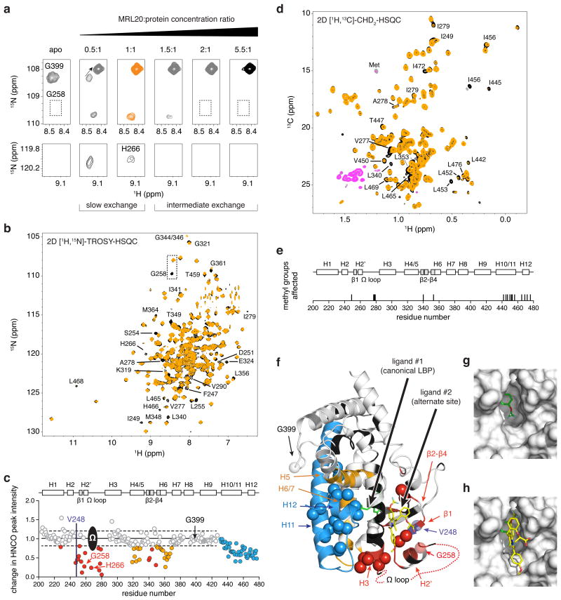
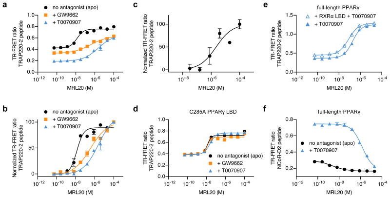
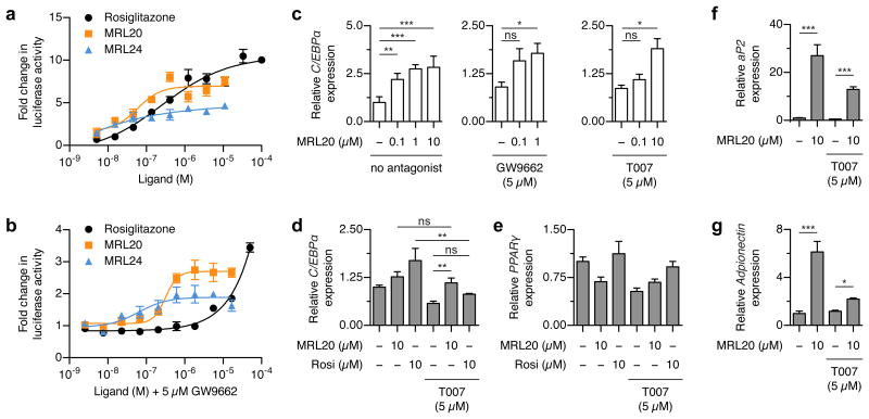
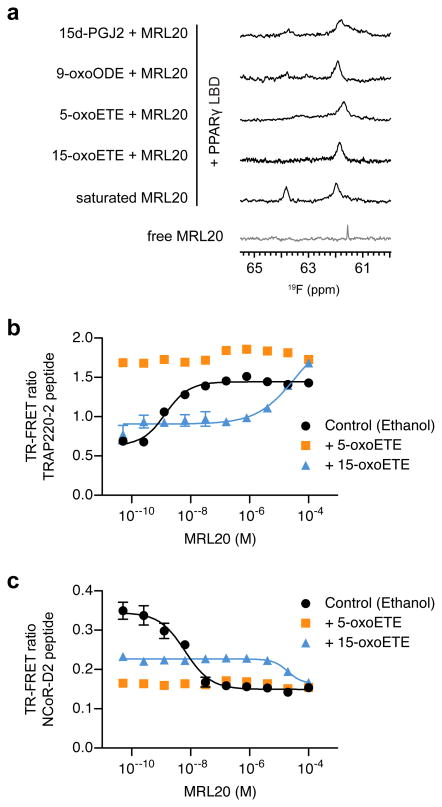
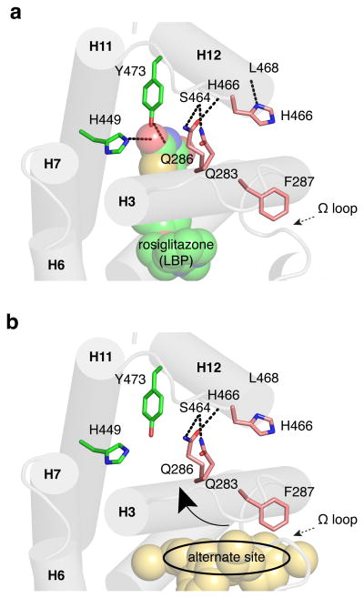
PPARγ is a target for insulin-sensitizing drugs such as glitazones, which improve plasma glucose maintenance in patients with diabetes. Synthetic ligands have been designed to mimic endogenous ligand binding to a canonical ligand-binding pocket to hyperactivate PPARγ. Here we reveal that synthetic PPARγ ligands also bind to an alternate site, leading to unique receptor conformational changes that impact coregulator binding, transactivation and target gene expression. Using structure-function studies we show that alternate site binding occurs at pharmacologically relevant ligand concentrations, and is neither blocked by covalently bound synthetic antagonists nor by endogenous ligands indicating non-overlapping binding with the canonical pocket. Alternate site binding likely contributes to PPARγ hyperactivation in vivo, perhaps explaining why PPARγ full and partial or weak agonists display similar adverse effects. These findings expand our understanding of PPARγ activation by ligands and suggest that allosteric modulators could be designed to fine tune PPARγ activity without competing with endogenous ligands.


Figures

Figure 1
Figure 1. MRL20 and MRL24 bind to PPARγ with a 2:1 stoichiometry
(a) Chemical structure of MRL20 and MRL24. Titration of (b,c) MRL20 or (d,e) MRL24 results in the population of two PPARγ LBD-bound 19F NMR resonances, where (c,e) are a plot of the total 19F peak area in (b) and (c), normalized to the area of the saturated canonical peak; concentrations beyond two molar equivalents of ligand are corrected (free ligand signal subtracted and adjusted for differential longitudinal relaxation; see methods for details). (f,g) MRL20 populates two 19F NMR resonances within the context of the (f) PPARγ/RXRα LBD heterodimer and (g) full-length PPARγ. (h) A single, sharp 19F resonance is observed for free MRL20 in buffer or added to lysozyme, indicating that alternate site binding of MRL20 to PPARγ occurs in a specific manner.
Figure 2
Figure 2. Mapping the alternate MRL20 binding site in PPARγ
(a) Titration of MRL20 into 15N-PPARγ LBD monitored by 2D [1H,15N]-TROSY-HSQC NMR reveals two binding transitions. The first slow exchange transition corresponds to the canonical LBP binding event (apo to 1:1), and the second intermediate exchange transition to the alternate site binding event (>1:1 stoichiometry). (b) Comparison of 2D [1H,15N]-TROSY-HSQC spectra for 15N-PPARγ LBD bound to 1 or 2 molecules of MRL20 (black and orange, respectively). (c) NMR chemical shift footprinting reveals a decrease in peak intensity between 3D TROSY-HNCO experiments collected for 2H,13C,15N-PPARγ LBD bound to 1 or 2 molecules of MRL20 (black/pink and orange/grey, respectively, for positive/negative peak amplitudes) and reveals residues affected by the alternate site binding event. (d) Comparison of 2D [1H,13C]-methyl CHD2-detected HSQC data for 2H,13C,15N-PPARγ LBD bound to 1 or 2 molecules of MRL20. (e) Residues with methyl NMR resonances affected upon binding a second MRL20 ligand. (f) NMR chemical shift footprinting changes mapped onto the PPARγ LBD structure reveals the site of interaction (red) and regions allosterically affected by alternate site binding (blue,orange); spheres represent methyl groups affected, and regions colored black have unassigned NMR chemical shifts likely due to dynamics on the NMR intermediate exchange regime. (g) The alternate site is formed by a solvent-accessible pocket on the PPARγ LBD surface. (h) Molecular docking of a second MRL20 ligand (yellow) into the alternate site, which is also shown in (f).
Figure 3
Figure 3. Mapping the alternate MRL24 binding site in PPARγ
(a) Comparison of 2D [1H,15N]-TROSY-HSQC spectra for 15N-PPARγ LBD bound to 1 or 2 molecules of MRL24 (black and orange, respectively). (b) Comparison of 2D [1H,13C]-methyl CHD2-detected HSQC data for 2H,13C,15N-PPARγ LBD bound to 1 or 2 molecules of MRL24. (c) NMR chemical shift footprinting reveals a decrease in peak intensity between 3D TROSY-HNCO experiments collected for 2H,13C,15N-PPARγ LBD bound to 1 or 2 molecules of MRL24 (black/pink and orange/grey, respectively, for positive/negative peak amplitudes) and reveals residues affected by the alternate site binding event.
Figure 4
Figure 4. Covalent antagonists do not block alternate site ligand binding to PPARγ
(a) Chemical structures of GW9662 and T0070907. (b) GW9662 (cyan) covalently attaches to PPARγ residue C285 (yellow). (c) When GW9662 (cyan) is covalently bound to PPARγ, it sterically blocks MRL20 (magenta) from binding to the LBP. (d,e) 19F NMR reveals (d) MRL20 and (e) MRL24 bind to PPARγ LBD bound to a covalent antagonist and populate a single resonance. (f) Molecular docking of MRL20 ligand into the alternate site of PPARγ LBD covalently bound to GW9662. (g) 2D [1H,15N]-TROSY-HSQC NMR confirms that MRL20 binds to 15N-PPARγ LBD covalently bound to GW9662. (h) Titration of MRL20 into GW9662 bound 15N-PPARγ LBD monitored by 2D [1H,15N]-TROSY-HSQC NMR reveals MRL20 binding effects occuring in slow exchange at residues remote from the alternate binding site. (i) HDX-MS analysis reveals that alternate site binding of MRL20 to PPARγ LBD covalently bound to GW9962 causes protection from HDX in the alternate site region and the PPARγ AF-2 surface.
Figure 5
Figure 5. Alternate site binding affects PPARγ-coregulator interaction
(a) TR-FRET assay showing the effect of MRL20 on TRAP220-2 peptide binding to PPARγ protein in the absence or presence of a covalent antagonist, performed in duplicate and plotted as the average (± s.d.). (b) Same data as (a) but normalized to illustrate the biphasic response for MRL20 to apo PPARγ and the EC50 differences between all conditions. (c) Same as in (b) but focused on the alternate site response for apo PPARγ. (d) PPARγ C285A mutation serves as a control to show that C285 is critical for covalent antagonist attachment and blocking MRL20 binding to the LBP in the TR-FRET assay. (e,f) TR-FRET assay shows an alternate-site response for MRL20 using (e) full-length PPARγ protein and full-length PPARγ protein heterodimerized to RXRα LBD covalently bound to T0070907 performed in duplicate and plotted as the average (± s.d.). (f) Covalent attachment of T0070907 increases the basal interaction of NCoR-3 peptide right shifts the MRL20 IC50 compared to apo PPARγ, performed in duplicate and plotted as the average (± s.d.). All data fit to a sigmoidal dose response curve.
Figure 6
Figure 6. Alternate site binding affects PPARγ transactivation and target gene expression
(a,b) Luciferase reporter assay showing the concentration-dependent effects of MRL20, MRL24 and rosiglitazone on PPARγ transactivation (a) without GW9662 pretreatment and (b) with GW9662 pretreatment, performed by cotransfection of Gal4-PPARγ LBD and UAS::luc reporter plasmid, performed in triplicate and plotted with the average (± s.e.m.) and fit to a sigmoidal dose response curve. (c) qRT-PCR analysis of C/EBPα expression in Jurkat cells, performed in quadruplicate, plotted with the average (± s.e.m.), and analyzed using Bonferroni post hoc comparison; ns = not significant.. (d-g) qRT-PCR analysis of PPARγ target genes in NIH-3T3-L1 cells harvested 3 days after inducing differentiation, performed in quadruplicate, plotted with the average (± s.e.m.), and analyzed using one-way ANOVA with Tukey post hoc comparison; ns = not significant. T007 = T0070907.
Figure 7
Figure 7. Alternate site binding of MRL20 to PPARγ bound to an endogenous ligand
(a) 19F NMR analysis reveals that MRL20 binds to PPARγ LBD covalently bound to endogenous ligands. (b,c) TR-FRET assay demonstrates that alternate site MRL20 binding affects the interaction between (b) TRAP220-2 and (c) NCoR-D2 for PPARγ LBD covalently bound to 15-oxoETE, but not 5-oxoETE. PPARγ LBD protein not covalently bound to an endogenous ligand but exposed to vehicle (ethanol) was used as a control, performed in duplicate and plotted as the average (± s.d.) and fit to a sigmoidal dose response curve.
Figure 8
Figure 8. Possible structural mechanism for indirect stabilization of the PPARγ AF-2 surface via the alternate site
(a) Ligands such as rosiglitazone that bind to the PPARγ canonical LBP form hydrogen bonds with residues in the helix 12 pocket (Y473 and H449) to directly stabilize helix 12 and the AF-2 surface. (b) Ligands that bind to the alternate site may indirectly stabilize the AF-2 surface by stabilizing helix 3, facilitating hydrogen bond formation between side chains of residues on helix 3 to residues in the helix 11-12 loop, particularly in the presence of a bound coregulator, which could affect helix 12/AF-2 stabilization.
Figure 9
Figure 9. Alternate site-bound ligands uniquely protrude around helix 3 to affect the conformation of the Ω loop
Ligands bound deep in the canonical LBP are colored orange, and the second bound ligand, whether it occupies the alternate site region or not, are colored blue; Ω loop region, if observed in the crystal structure, from M257-V277 is colored green. Alternate site bound ligands that affect the Ω loop conformation are also circled. (a) Co-crystal structure of the PPARγ LBD bound to one molecule of MRL20 (PDB: 2Q59). (b) Co-crystal structure of the PPARγ LBD bound to one molecule of T2384 (PDB: 3K8S; chain A). (c) Co-crystal structure of the PPARγ LBD with MRL20 (orange) (PDB: 2Q59) docked with a second MRL20 ligand (blue). (d) Co-crystal structure of the PPARγ LBD bound to two molecules of T2384 in the canonical LBP (orange) and alternate site (blue) (PDB: 3K8S; chain B). (e) Co-crystal structure of the PPARγ LBD bound to two molecules of 9-(S)-HODE (PDB: 2VSR). (f) Co-crystal structure of the PPARγ LBD bound to 5-methoxy-indole acetate (orange) and 15-oxoETE (blue) (PDB: 3ADW).

References

    1. Nwachukwu JC, Nettles KW. The nuclear receptor signalling scaffold: insights from full-length structures. EMBO J. 2012;31:251–3. - PMC - PubMed
    1. Chandra V, et al. Multidomain integration in the structure of the HNF-4alpha nuclear receptor complex. Nature. 2013;495:394–8. - PMC - PubMed
    1. Chandra V, et al. Structure of the intact PPAR-gamma-RXR-alpha nuclear receptor complex on DNA. Nature. 2008;456:350–356. - PMC - PubMed
    1. Orlov I, Rochel N, Moras D, Klaholz BP. Structure of the full human RXR/VDR nuclear receptor heterodimer complex with its DR3 target DNA. EMBO J. 2012;31:291–300. - PMC - PubMed
    1. Rochel N, et al. Common architecture of nuclear receptor heterodimers on DNA direct repeat elements with different spacings. Nat Struct Mol Biol. 2011;18:564–70. - PubMed

Publication types