. 2014 Apr 4;9(4):e93343.
doi: 10.1371/journal.pone.0093343. eCollection 2014.

Synthesis and propagation of complement C3 by microglia/monocytes in the aging retina

Affiliations

Synthesis and propagation of complement C3 by microglia/monocytes in the aging retina

Matt Rutar et al. PLoS One. .

Abstract

Introduction: Complement activation is thought to contribute to the pathogenesis of age-related macular degeneration (AMD), which may be mediated in part by para-inflammatory processes. We aimed to investigate the expression and localization of C3, a crucial component of the complement system, in the retina during the course of aging.

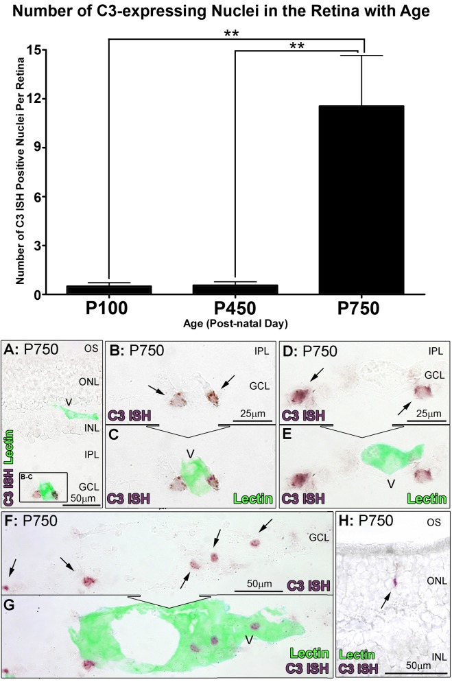
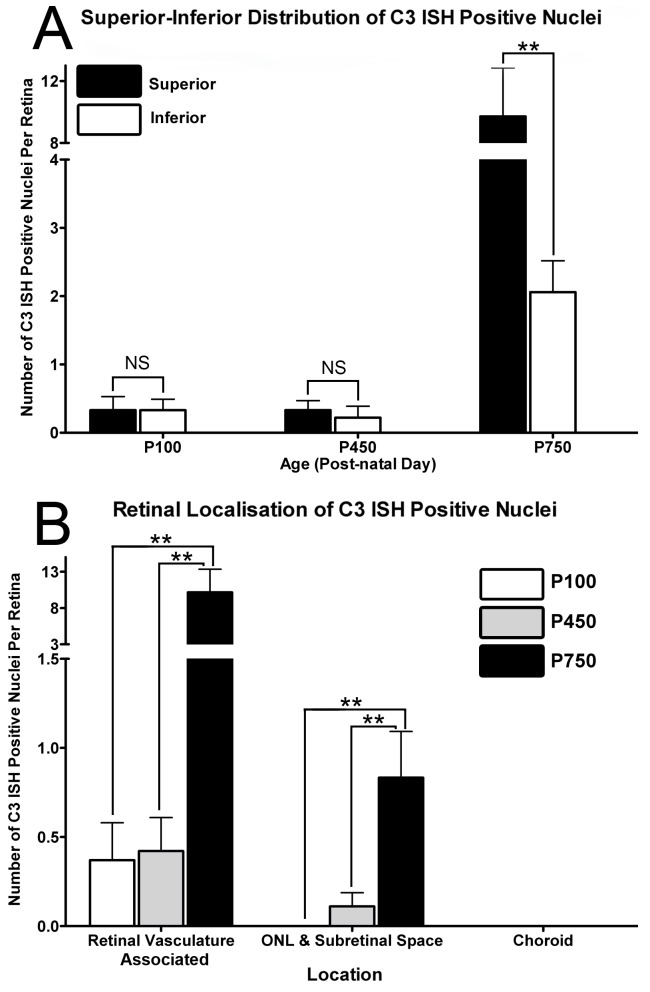
Methods: SD rats were born and reared in low-light conditions, and euthanized at post-natal (P) days 100, 450, or 750. Expression of C3, IBA1, and Ccl- and Cxcl- chemokines was assessed by qPCR, and in situ hybridization. Thickness of the ONL was assessed in retinal sections as a measure of photoreceptor loss, and counts were made of C3-expressing monocytes.

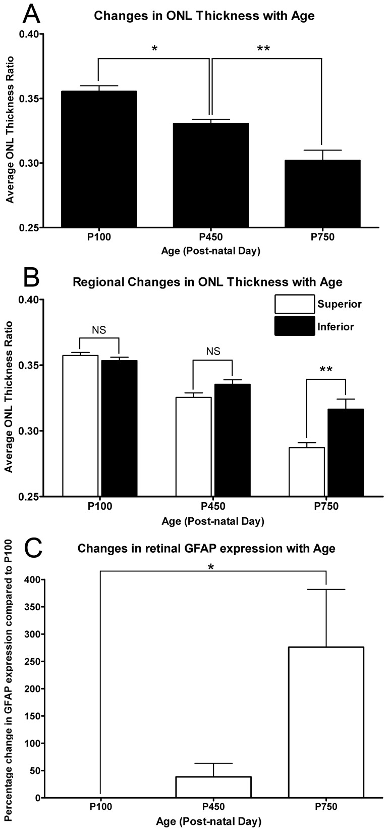
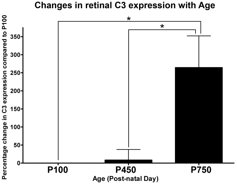
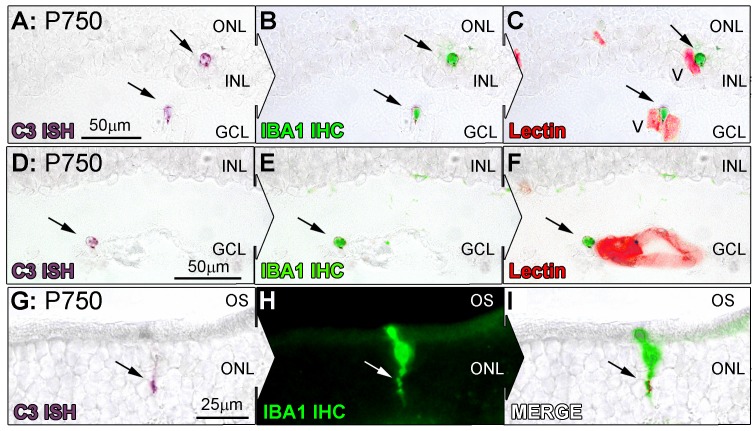
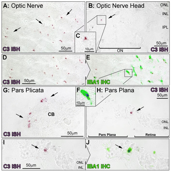
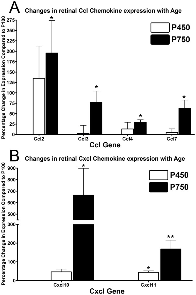
Results: C3 expression increased significantly at P750, and correlated with thinning of the ONL, at P750, and up-regulation of GFAP. In situ hybridization showed that C3 was expressed by microglia/monocytes, mainly from within the retinal vasculature, and occasionally the ONL. The number of C3-expressing microglia increased significantly by P750, and coincided spatiotemporally with thinning of the ONL, and up-regulation of Ccl- and Cxcl- chemokines.

Conclusions: Our data suggest that recruited microglia/monocytes contribute to activation of complement in the aging retina, through local expression of C3 mRNA. C3 expression coincides with age-related thinning of the ONL at P750, although it is unclear whether the C3-expressing monocytes are a cause or consequence. These findings provide evidence of activation of complement during natural aging, and may have relevance to cellular events underling the pathogenesis of age-related retinal diseases.


Conflict of interest statement

Competing Interests: The authors have declared that no competing interests exist.

Figures

Figure 1
Figure 1. Quantification of ONL thickness and GFAP expression with respect to age.
A: The thickness of the ONL decreased progressively throughout the aging time course, and was markedly reduced by P750 (P<0.05). B: Regional variation in ONL thickness was not observed at P100 and P450 age-groups across the vertical meridian (P>0.05). At P750, the greater thinning was observed in the superior retina than the inferior (<0.05). C: The expression of GFAP was found to increase at P450, however this was not significant compared to P100 (P>0.05). At P750 a 276% increase was observed in the expression of GFAP, compared to P100 (P<0.05). P100 n = 3, P450 n = 3, P750 n = 3; error bars represent SEM. ‘*’ denotes a significant change using ANOVA with Tukey’s post-test where P<0.05, ‘**’ denotes P<0.01.
Figure 2
Figure 2. Expression of C3 in the neural retina by qPCR over the aging timecourse.
No appreciable difference was observed in the expression of C3 between P100 and P450 age groups (P>0.05). By P750 there was a robust 264.3% increase in the expression of C3, compared to P100 and P450 (P<0.05). P100 n = 3, P450 n = 3, P750 n = 3; error bars represent SEM. ‘*’ denotes a significant change using ANOVA with Tukey’s post-test where P<0.05.
Figure 3
Figure 3. In situ hybridisation for C3 mRNA in the retina with respect to age.
A–F: Representative images show In situ hybridisation for C3 mRNA (purple) and vessels (lectin staining, green) in the retina. In retinas from P750, expression of C3 was apparent in cells (A, C, E, arrows) closely associated with vessels from the retinal vasculature stained with lectin (B, D, E). H: Staining for C3 was also occasionally observed among cells situated in the ONL (arrow). Histogram: Quantification of C3-expressing cells per retina showed no significant change at P450 compared to P100 (0.6 per retina, P>0.05). At P750, C3-expressing cells increased significantly (P<0.05) to 11.6 per retina compared to both P100 and P450 groups. P100 n = 3, P450 n = 3, P750 n = 3; error bars represent SEM. ‘*’ denotes a significant change using ANOVA with Tukey’s post-test where P<0.05, ‘**’ denotes P<0.01. V, retinal blood vessel; INL, inner nuclear layer; IPL, inner plexiform layer; GCL, ganglion cell layer; ONL, outer nuclear layer; OS, outer segments.
Figure 4
Figure 4. Distribution of In situ hybridisation for C3 mRNA in the over the aging timecourse.
A: C3-expressing cells were evenly distributed across superior and inferior retina P100 and P450 animals (P>0.05). At P750, C3-expressing cells were more numerous in the superior portion of the retina than the inferior (9.7 and 2.1 respectively, P<0.05). B: Increases in C3-expressing cells were predominately associated with the retinal vasculature at P750 (P<0.05), with more modest increases apparent in the ONL and subretinal space over the same period (P<0.05). P100 n = 3, P450 n = 3, P750 n = 3; error bars represent SEM. ‘*’ denotes a significant change using ANOVA with Tukey’s post-test where P<0.05, ‘**’ denotes P<0.01.
Figure 5
Figure 5. Co-immunolabeling of C3-expressing cells (purple) for IBA1 (green) at P750.
A–I: Representative images at P750 show In situ hybridisation for C3 mRNA, IBA1 immunoreactivity, and lectin staining (red) for retinal vasculature in the retina. C3 staining (A, D, arrows) was found to correlate strongly with IBA1-immunoreactive microglia/monocytes (B, E, arrows), which were in close association with both superficial and deep retinal vasculature (C, F, arrows). C3-expressing cells in the ONL (G, arrow) also showed specificity for IBA1-immunreactive microglia at the ONL/OS margin (H,I, arrows). V, retinal blood vessel; INL, inner nuclear layer; GCL, ganglion cell layer; ONL, outer nuclear layer; OS, outer segments.
Figure 6
Figure 6. In situ hybridisation for C3-expressing cells in the optic nerve and ciliary body.
Representative images demonstrate In situ hybridisation for C3 mRNA (purple) and IBA1-immunoreactive microglia/monocytes (green) at the optic nerve (A-F) and ciliary body (G-J), at P750. A–F: An abundance of C3-expressing cells were evident in section of the optic nerve tissue (A, arrows), and were immunoreactive for IBA1 (D–F). C3-expressing cells were also occasionally found emerging from the optic nerve head (B–C, arrow). G–J: At P750, C3-expressing cells were observed within the ciliary body, including the pars plicata (G, arrows) and the pars plana, particularly at the retinal margin (H, arrow). These cells were also immunoreactive for IBA1 (I–J). CB, ciliary body; V, retinal blood vessel; INL, inner nuclear layer; IPL, inner plexiform layer; ONL, outer nuclear layer.
Figure 7
Figure 7. Expression of Ccl- (A) and Cxcl- (B) chemokines in the neural retina by qPCR over the aging timecourse.
A: The expression of Ccl2, Ccl3, Ccl4, Ccl7 did not change appreciably in animals aged P450 (P>0.05), while a significant up-regulation was observed in P750 animals for all Ccl- genes (P<0.05), compared to P100; a large increase in Ccl2 expression was observed at P450, although this was not significant (P>0.05). B: At P450, there was a small increase in expression of Cxcl11 (P<0.05), but not Cxdc10 (P>0.05). By P750 there was a considerable increase in the expression of both Cxcl- genes compared to P100, particularly Cxcl10 (P<0.05). P100 n = 4, P450 n = 4, P750 n = 4; error bars represent SEM. ‘*’ denotes a significant change using ANOVA with Tukey’s post-test where P<0.05, ‘**’ denotes P<0.01.

Similar articles

Cited by

References

    1. Harman D (1981) The aging process. Proc Natl Acad Sci U S A 78: 7124–7128. - PMC - PubMed
    1. Bonnel S, Mohand-Said S, Sahel JA (2003) The aging of the retina. Exp Gerontol 38: 825–831. - PubMed
    1. Ambati J, Ambati BK, Yoo SH, Ianchulev S, Adamis AP (2003) Age-related macular degeneration: etiology, pathogenesis, and therapeutic strategies. Surv Ophthalmol 48: 257–293. - PubMed
    1. Ambati J (2011) Age-related macular degeneration and the other double helix. The Cogan Lecture. Invest Ophthalmol Vis Sci 52: 2165–2169. - PMC - PubMed
    1. Rein DB, Wittenborn JS, Zhang X, Honeycutt AA, Lesesne SB, et al. (2009) Forecasting age-related macular degeneration through the year 2050: the potential impact of new treatments. Arch Ophthalmol 127: 533–540. - PubMed

Publication types