. 2014 Jul;11(4):367-76.
doi: 10.1038/cmi.2014.16. Epub 2014 Apr 7.

Long-term exposure to decabrominated diphenyl ether impairs CD8 T-cell function in adult mice

Affiliations

Long-term exposure to decabrominated diphenyl ether impairs CD8 T-cell function in adult mice

Weihong Zeng et al. Cell Mol Immunol. 2014 Jul.

Abstract

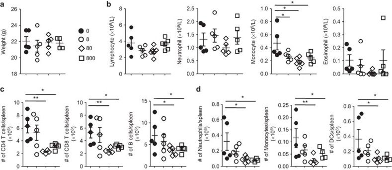
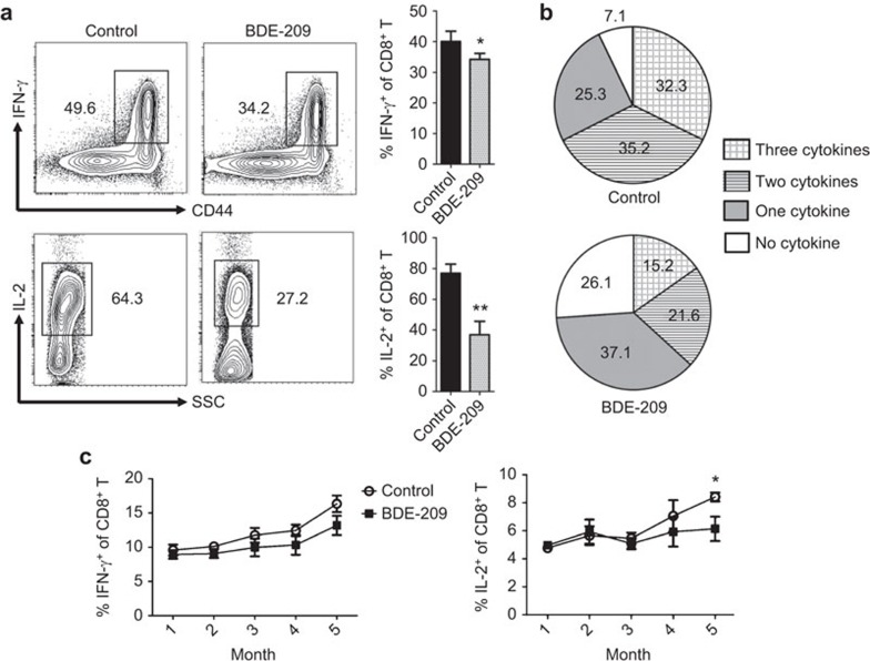
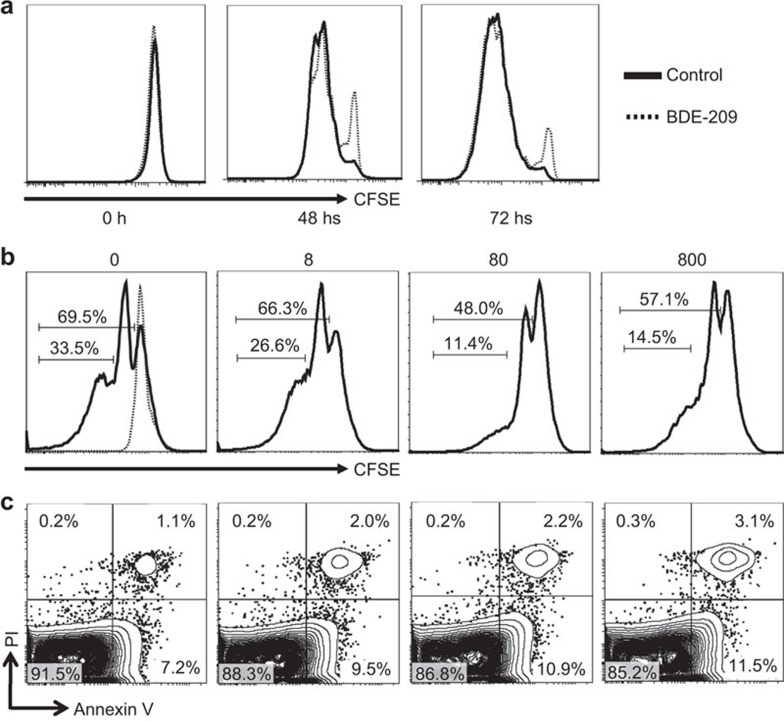
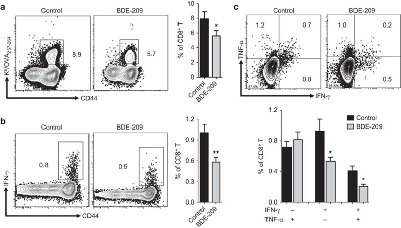
Polybrominated diphenyl ethers (PBDEs) are ubiquitous environmental pollutants that accumulate to high levels in human populations that are subject to occupational or regional industry exposure. PBDEs have been shown to affect human neuronal, endocrine and reproductive systems, but their effect on the immune system is not well understood. In this study, experimental adult mice were intragastrically administered 2,2',3,3',4,4',5,5',6,6'-decabromodiphenyl ether (BDE-209) at doses of 8, 80 or 800 mg/kg of body weight (bw) at 2-day intervals. Our results showed that continuous exposure to BDE-209 resulted in high levels of BDE-209 in the plasma that approached the levels found in people who work in professions with high risks of PDBE exposure. Reduced leukocytes, decreased cytokine (IFN-γ, IL-2 and TNF-α) production and lower CD8 T-cell proliferation were observed in the mice exposed to BDE-209. Additionally, mice with long-term BDE-209 exposure had lower numbers of antigen-specific CD8 T cells after immunization with recombinant Listeria monocytogenes expressing ovalbumin (rLm-OVA) and the OVA-specific CD8 T cells had reduced functionality. Taken together, our study demonstrates that continuous BDE-209 exposure causes adverse effects on the number and functionality of immune cells in adult mice.


Figures

Figure 1
Figure 1
Long-term BDE-209 exposure resulted in a high burden of BDE-209 in the plasma, retarded growth and reduced leukocytes in the peripheral blood. The BDE-209-exposed mice were intragastrically administered BDE-209 at a dose of 800 mg/kg bw at 2-day intervals. (a) The BDE-209 concentration in the plasma was determined by GC–MS under the NCI mode at day 7 and months 1, 2, 5 and 7 (n=10 mice per group, *P<0.05 and ***P<0.001 compared to the control group, two-way repeated-measures ANOVA with Bonferroni post-tests, the means±s.d. are depicted). (b) Body weight was monitored over a period of 7 months (n=10 mice per group, *P<0.05 compared to control group, two-way repeated-measures ANOVA with Bonferroni post-tests, the means±s.d. are depicted). (c) Peripheral blood was collected from control and BDE-209-exposed mice at month 7, and the hematological parameters, including leukocytes (WBC), red blood cells (RBC), platelets (PLT), hemoglobin (HGB), packed cell volume (PCV) and mean corpuscular volume (MCV), were detected by a CAD (**P<0.01, unpaired Student's t-test, the means±s.d. are depicted). BDE, brominated diphenyl ether; bw, body weight; CAD, Coulter AcT Diff hematology analyzer; GC–MS, gas chromatography-mass spectrometry; NCI, negative chemical ionization.
Figure 2
Figure 2
The effects of BDE-209 exposure on the body weight and the number of immune cells in the peripheral blood and spleen. BDE-209-exposed mice were intragastrically administered BDE-209 at doses of 8, 80 or 800 mg/kg bw at 2-day intervals. (a) The body weight was evaluated at month 3. (b) The immune cells, including lymphocytes, neutrophils, monocytes and eosinophils, in the peripheral blood were detected by a HEMAVET 950 multispecies hematology instrument (DREW Scientific Inc.) at month 1. (c, d) Splenocytes were collected from control mice and BDE-209-exposed mice at month 3, and the numbers of CD4 T (CD3+CD4+), CD8 T (CD3+CD8+) and B cells (B220+), as well as monocytes (CD11b+Ly6Chi), neutrophils (CD11b+Ly6Cmid) and dendritic cells (CD11b+CD11c+) in the spleen were calculated by flow cytometry through staining with anti-CD3e, anti-CD4, anti-CD8a, anti-B220, anti-CD11b, anti-Ly6C and anti-CD11c (*P<0.05, **P<0.01, one-way ANOVA followed by Tukey's post-tests, the means±s.d. are depicted). BDE, brominated diphenyl ether; bw, body weight.
Figure 3
Figure 3
Long-term exposure to BDE-209 at a dose of 800 mg/kg bw had adverse effects on the functionality of CD8 T cells in the peripheral blood. The cytokine production by CD8 T cells in the peripheral blood was measured by ICS following stimulation with PMA (50 ng/ml) and ionomycin (500 ng/ml) for 5 h in vitro. (a) Comparison of the IFN-γ and IL-2 expression by CD8+ T cells between the control mice and the BDE-209-exposed mice at month 7. As shown in the flow diagram (gated on CD8+ T cells), the percentages (%) indicate the IFN-γ- or IL-2-positive population of CD8+ T cells (left panels). The bar graphs display the corresponding statistical results of IFN-γ+ CD8+ T cells or IL-2+ CD8+ T cells between the control and BDE-209-exposed mice. (b) Polyfunctional CD8 T cells were detected by ICS with multicolor flow cytometry in control and BDE-209-exposed mice at month 7. The percentages of four combinations of IFN-γ, IL-2 and TNF-α expression of CD8+ T cells are shown in the pie charts (three cytokines: IFN-γ+TNF-α+IL-2+; two cytokines: IFN-γ+TNF-α+IL-2, IFN-γ+TNF-αIL-2+ and IFN-γTNF-α+IL-2+; one cytokine: IFN-γ+TNF-αIL-2, IFN-γTNF-αIL-2+ and IFN-γTNF-α+IL-2; no cytokines: IFN-γTNF-αIL-2). (c) Kinetics of the percentages of IFN-γ+CD8+ (left) or IL-2+CD8+ (right) T cells from months 1 to 5 in the control and BDE-209-exposed mice. The data in a were analyzed using an unpaired Student's t-test, and the data in c were assessed using two-way repeated-measures ANOVA (with Bonferroni post-tests) (n=10 per group, the means±s.d. are depicted, *P<0.05 and **P<0.01 compared to the control group). BDE, brominated diphenyl ether; bw, body weight; ICS, intracellular cytokine staining.
Figure 4
Figure 4
Continuous BDE-209 exposure impaired proliferation and induced apoptosis of CD8 T cells. (a) PBMCs from control mice and mice administered BDE-209 at a dose of 800 mg/kg bw were labeled with CFSE (5 µM) and stimulated with pre-bound anti-CD3e (2 µg/ml) plus soluble anti-CD28 (5 µg/ml) for 0, 48 or 72 h. The proliferation of CD8 T cells was visualized by FACS analysis of CFSE fluorescence in CD8+ cells (solid lines, control mice; dotted lines, BDE-209-exposed mice). (b, c) Mice were intragastrically administered BDE-209 at a dose of 8, 80 or 800 mg/kg bw at 2-day intervals, and splenic CD8 T cells were purified from the control mice and BDE-209-exposed mice using a FACS Aria II based on the surface expression of CD3 and CD8 (CD3+CD8+) at month 3. The purified splenic-CD8 T cells were labeled with CFSE (5 µM) and then stimulated with anti-CD3 and anti-CD28 mAbs coupled to magnetic beads at a cell-bead ratio of 4∶1 for 72 h (dotted lines, unstimulated splenic CD8+ T cells). The proliferation (b) and the proportions of early (Annexin V+PI) and late apoptotic or necrotic (Annexin V+PI+) cells (c) were evaluated by FACS analysis of the CD8+ cells. Each group included five mice; representative images are shown. BDE, brominated diphenyl ether; bw, body weight; PBMC, peripheral blood mononuclear cell.
Figure 5
Figure 5
Long-term BDE-209 exposure at a dose of 800 mg/kg bw decreased the abundance of memory-phenotype CD8 T cells in the peripheral blood. The percentages of central memory (TCM, CD44hiCD62Lhi) and effector memory (TEM, CD44hiCD62Llo) phenotype CD8 T cells were determined by flow cytometry. (a) Flow diagrams (left) and bar graphs (right) show the percentages of TCM and TEM CD8 T cells in control and BDE-209-exposed mice at month 7 (n=8 per group, *P<0.05, unpaired Student's t-test, the means±s.d.). (b) Kinetics of the percentages of TCM and TEM in CD8 T cells from months 1 to 7 in control and BDE-209-exposed mice (n=8 per group, *P<0.05 compared to the control group, two-way repeated-measures ANOVA with Bonferroni post-tests, the means±s.d. are depicted). BDE, brominated diphenyl ether; bw, body weight.
Figure 6
Figure 6
Long-term BDE-209 exposure impairs antigen-specific CD8 T-cell responses. At month 10, control mice and mice exposed to BDE-209 at a dose of 800 mg/kg bw were infected with rLm-OVA (intravenously) at a dose of 5×10⁴ CFU/mouse. Seven days later, OVA257–264-specific CD8 T cells in peripheral blood were detected by MHC/peptide tetramers or intracellular cytokine staining. (a, b) Detection of OVA257–264-specific CD8 T cells by MHC/peptide tetramers (Kb/OVA257–264, a) or intracellular IFN-γ (IFN-γ+/OVA257–264, b) staining. (c) Detection of OVA257–264-specific polyfunctional CD8 T cells by intracellular cytokine staining with multicolor flow cytometry. Polyfunctional CD8 T cells were considered the population of IFN-γ+TNF-α+ (double-positive) cells gated on CD8+ T cells. The data were assessed using an unpaired Student's t-test (n=8 per group, the means±s.d. are depicted, *P<0.05, **P<0.01). BDE, brominated diphenyl ether; bw, body weight.

Similar articles

Cited by

References

    1. Rahman F, Langford KH, Scrimshaw MD, Lester JN. Polybrominated diphenyl ether (PBDE) flame retardants. Sci Total Environ. 2001;275:1–17. - PubMed
    1. Costa LG, Giordano G. Is decabromodiphenyl ether (BDE-209) a developmental neurotoxicant. Neurotoxicology. 2011;32:9–24. - PMC - PubMed
    1. Petreas M, She J, Brown FR, Winkler J, Windham G, Rogers E, et al. High body burdens of 2,2′,4,4′-tetrabromodiphenyl ether (BDE-47) in California women. Environ Health Perspect. 2003;111:1175–1179. - PMC - PubMed
    1. Sjodin A, Jones RS, Focant JF, Lapeza C, Wang RY, McGahee EE, et al. Retrospective time-trend study of polybrominated diphenyl ether and polybrominated and polychlorinated biphenyl levels in human serum from the United States. Environ Health Perspect. 2004;112:654–658. - PMC - PubMed
    1. Frederiksen M, Vorkamp K, Thomsen M, Knudsen LE. Human internal and external exposure to PBDEs—a review of levels and sources. Int J Hyg Environ Health. 2009;212:109–134. - PubMed

Publication types

MeSH terms