. 2014 May;46(5):444-450.
doi: 10.1038/ng.2938. Epub 2014 Apr 6.

The genomic landscape of diffuse intrinsic pontine glioma and pediatric non-brainstem high-grade glioma

Affiliations

The genomic landscape of diffuse intrinsic pontine glioma and pediatric non-brainstem high-grade glioma

Gang Wu et al. Nat Genet. 2014 May.

Abstract

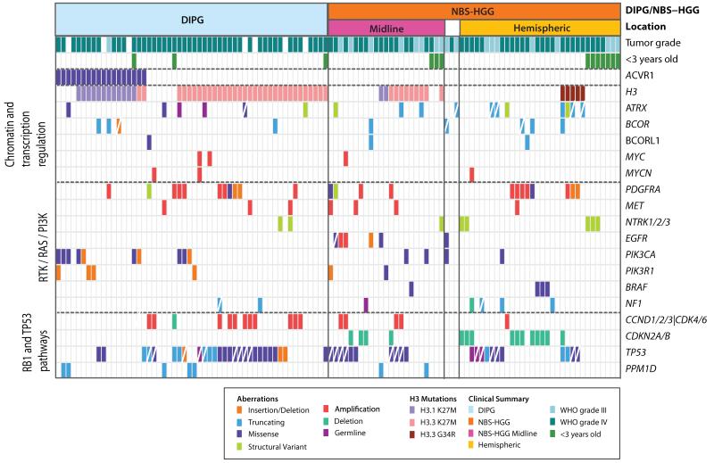
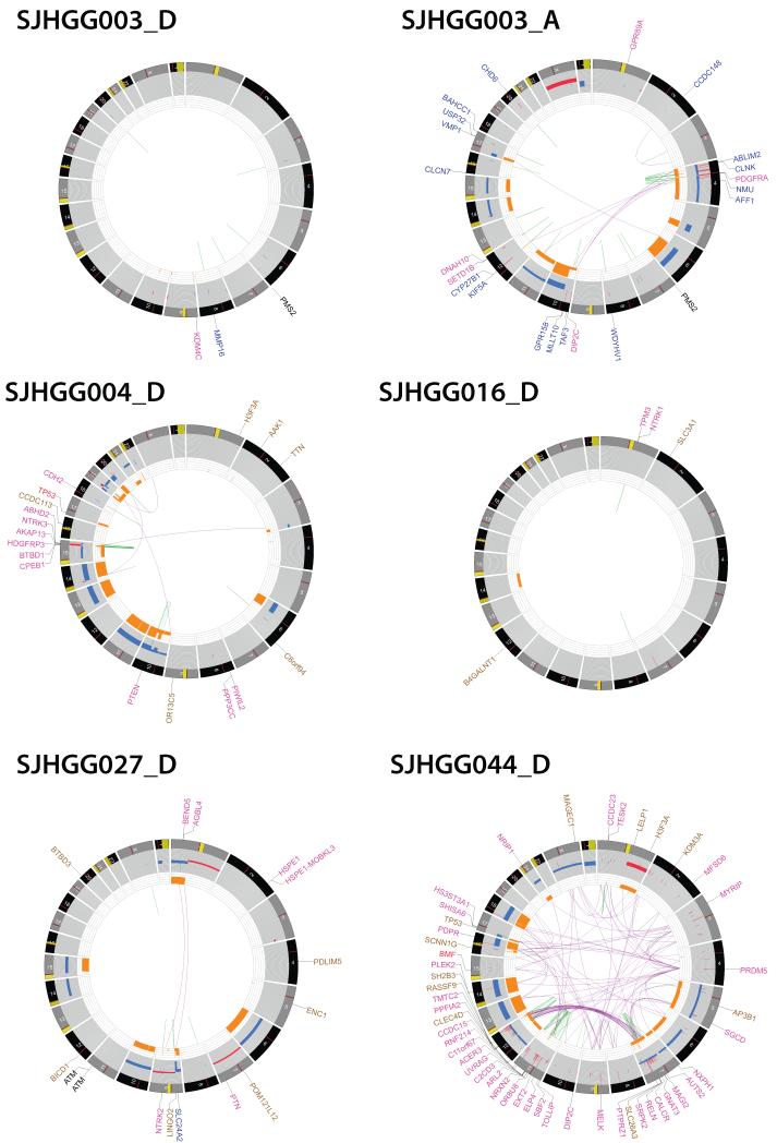
Pediatric high-grade glioma (HGG) is a devastating disease with a less than 20% survival rate 2 years after diagnosis. We analyzed 127 pediatric HGGs, including diffuse intrinsic pontine gliomas (DIPGs) and non-brainstem HGGs (NBS-HGGs), by whole-genome, whole-exome and/or transcriptome sequencing. We identified recurrent somatic mutations in ACVR1 exclusively in DIPGs (32%), in addition to previously reported frequent somatic mutations in histone H3 genes, TP53 and ATRX, in both DIPGs and NBS-HGGs. Structural variants generating fusion genes were found in 47% of DIPGs and NBS-HGGs, with recurrent fusions involving the neurotrophin receptor genes NTRK1, NTRK2 and NTRK3 in 40% of NBS-HGGs in infants. Mutations targeting receptor tyrosine kinase-RAS-PI3K signaling, histone modification or chromatin remodeling, and cell cycle regulation were found in 68%, 73% and 59% of pediatric HGGs, respectively, including in DIPGs and NBS-HGGs. This comprehensive analysis provides insights into the unique and shared pathways driving pediatric HGG within and outside the brainstem.


Figures

Fig. 1
Fig. 1. Recurrent genetic alterations in pediatric high-grade glioma
Genetic alterations detected in 19 genes, including ACVR1, and genes most recurrently mutated in the pathways indicated on the left, are displayed according to the color key shown below. Diagonal white line indicates loss of the wild-type allele, or male patient for ATRX, which is X-linked. H3F3A (H3.3) and HIST1H3B (H3.1) mutations are grouped together into the category H3. Structural variants involving NTRK1, NTRK2, or NTRK3, and copy number variants of components of the CyclinD1, D2, D3, or CDK4, CDK6 G1 checkpoint complex are grouped together as NTRK1/2/3 or CCND1/2/3/CDK4/6, respectively. Tumor subgroup (DIPG or NBS-HGG), location of NBS-HGGs (midline versus tumors in cerebral hemispheres), and tumor grade are indicated. White boxes for location or tumor grade indicates information not available. < 3 y.o denotes less than 3 years of age. Mutations for 112 HGGs are shown. Four hypermutator samples and 11 samples for which only RNA-seq data was available, were excluded from this summary. Data is shown in tabular form in Supplementary Table 9.
Fig. 2
Fig. 2. ACVR1 mutations in DIPG activate BMP signaling
a. Missense ACVR1 substitutions in DIPG were clustered in the glycine/serine rich domain (G/S) or kinase domain. Each red circle indicates a DIPG carrying the specified mutation, and an * indicates mutations previously found as germline mutations in individuals with FOP. The extracellular domain (EC) and transmembrane domain (TM) did not contain mutations. b. ACVR1 mutations ventralize zebrafish embryos. Graph shows the percentage of embryos exhibiting a dorsalized or ventralized phenotype. Embryos injected with wild-type ACVR1 mRNA (WT) showed a dorsalized phenotype, while embryos injected with mutant ACVR1 mRNA showed a ventralized phenotype (increasing severity from left to right). R258G had the least severe effect, resulting only in the V3-V4 ventralized phenotype, whereas G328V had the most severe effect with 90% of embryos showing the V5 ventralized phenotype. The number of embryos examined is shown on top. c. Representative phenotype images of zebrafish embryos injected with the indicated ACVR1 mRNA. Untreated mutants R258G, G328E, G328W, and R206H have little to no dorsal structures, and G356D and G328V are more severely affected. Treatment with LDN-193189 (LDN) reversed the ventralization effects in the ACVR1 mutants, as can be seen by the partial rescue of dorsal structures (i.e. head) for R258G (100%, n=20), G328E (83%, n=23), G328W (100%, n=20), R206H (100%, n=27), and the reduced severity of ventralization without the formation of dorsal structures for G356D and G328V. Scale bar is 200 μm. d. ACVR1 mutations drive increased levels of phospho-SMAD1/5 in primary astrocyte cultures. Western blots from lysates of primary astrocytes isolated from brainstem of neonatal Tp53 conditional knockout mice, transduced with retroviruses expressing FLAG-tagged ACVR1 wild-type, or indicated mutants, and serum starved for 2 hours. Quantitation of the ratio of phospho-SMAD/Total SMAD normalized to the empty vector control is shown below.
Fig. 3
Fig. 3. Structural variants generate oncogenic chimeric NTRK fusion proteins
a. All fusions included the C-terminal kinase domain from NTRK1, NTRK2 or NTRK3 (blue). N-terminal fusion partners include the tropomyosin domain (yellow) of TPM3, an actin-binding protein fused to NTRK1, the BTB/POZ dimerization domain (gray), and Kelch domain (orange) from the topoisomerase I-interacting protein BTBD1, or the pointed protein-protein interaction domain (purple), of the ETS transcription factor ETV6, fused to NTRK3. The N-terminus of the actin-binding protein Vinculin (light blue, VCL) was fused to NTRK2, and the N-terminus (green) of the ATP/GTP binding protein AGBL4 was fused to NTRK2. The functional carboxypeptidase domain of AGBL4 is not present in the fusion protein. For each fusion protein, the dotted red line shows the fusion point, with the amino acid of the N-terminal and C-terminal fusion partners breakpoint indicated. The full-length of the fusion protein is shown on the right end. b. NTRK fusion proteins induce high-grade astrocytomas. Tp53-null mouse primary astrocytes isolated from neonatal cortex or brainstem were transduced with FLAG-tagged TPM3-NTRK1 (top row) or BTBD1-NTRK3 (lower row) respectively, and implanted into mouse brain. Representative (of 7 independent mice per construct) H&E stains show pleomorphic tumor cells, many with features of astrocytic differentiation and high mitotic activity. Tumors induced by BTBD1-NTRK3 showed the frequent presence of giant cells reminiscent of giant cell glioblastoma. Immunohistochemical analysis showed expression of FLAG-tagged NTRK fusion proteins, and elevated phospho-Akt, and phospho-p42/44 Mapk in tumor relative to surrounding normal tissue. Scale bar=50μm.
Fig. 4
Fig. 4. Pediatric HGG mutations in histone modifiers or chromatin regulators
Mutations identified in NBS-HGG (pink) or DIPG (blue) are shown. Genetic alterations were identified in proteins that attach (writers, shown above) or remove (erasers, shown below) post-translational modifications of lysines (K4, K9, K18, K27, K36) in the tail of histone H3, as well as proteins involved in modifications of other histones, or chromatin remodelers. Notably, there were no mutations in writers or erasers of K27, which is directly mutated at high frequency in DIPG, and to a lesser degree in NBS-HGG.
Fig. 5
Fig. 5. CIRCOS plots showing the range of structural alterations in pediatric HGG
CIRCOS plots display the genome by chromosome in a circular plot, and depict structural genetic variants, including DNA copy number alterations, intra- and inter-chromosomal translocations, and non-sequence mutations. Loss of heterozygosity, orange; amplification, red; deletion, blue Sequence mutations in RefSeq genes: missense SNVs, brown; indels, red; splice site SNVs, blue, Genes at SV breakpoints: genes involved in in-frame fusions, pink; others, blue. Patient SJHGG003 carried a germline PMS2 mutation, and developed two independent tumors, first a hemispheric malignant glioneuronal tumor (SJHGG003_D), and 2 years later a DIPG (SJHGG003_A). The SNVs and indels for these two cases were too numerous to include on the plot for these two cases. The hypermutator tumor with more than 800,000 somatic mutations had an extremely stable genome (SJHGG003_D), while the second tumor with approximately 100-fold fewer SNVs, carried typical genomic copy number and structural abnormalities as seen in other HGGs (SJHGG003_A), thus demonstrating the broad range and complexity of mutations associated with germline PMS2 mutation. SJHGG016_D is an infant NBS-HGG with a TPM3-NTRK1 fusion and very stable genome. SJHGG027_D is an NBS-HGG from a patient with A-T, showing a relatively stable genome. This tumor sample was collected prior to radiotherapy. SJHGG004_D, is a DIPG with chromothripsis driving BTBD1-NTRK3 fusion, shown in more detail in Supplementary Fig. 8a. SJHGG044_D is an NBS-HGG showing dramatic chromothripsis. For the examples of chromothripsis, the names of genes disrupted by structural variants were too numerous to display.

Comment in

References

    1. Gottardo NG, Gajjar A. Chemotherapy for malignant brain tumors of childhood. J Child Neurol. 2008;23:1149–59. - PMC - PubMed
    1. Pollack IF, et al. Age and TP53 mutation frequency in childhood malignant gliomas: results in a multi-institutional cohort. Cancer Res. 2001;61:7404–7. - PubMed
    1. Schwartzentruber J, et al. Driver mutations in histone H3.3 and chromatin remodelling genes in paediatric glioblastoma. Nature. 2012;482:226–31. - PubMed
    1. Wu G, et al. Somatic histone H3 alterations in pediatric diffuse intrinsic pontine gliomas and non-brainstem glioblastomas. Nat Genet. 2012;44:251, 3. - PMC - PubMed
    1. Khuong-Quang DA, et al. K27M mutation in histone H3.3 defines clinically and biologically distinct subgroups of pediatric diffuse intrinsic pontine gliomas. Acta Neuropathol. 2012;124:439–47. - PMC - PubMed

Publication types

MeSH terms