. 2014 May;46(5):457-461.
doi: 10.1038/ng.2925. Epub 2014 Apr 6.

Recurrent activating ACVR1 mutations in diffuse intrinsic pontine glioma

Affiliations

Recurrent activating ACVR1 mutations in diffuse intrinsic pontine glioma

Kathryn R Taylor et al. Nat Genet. 2014 May.

Abstract

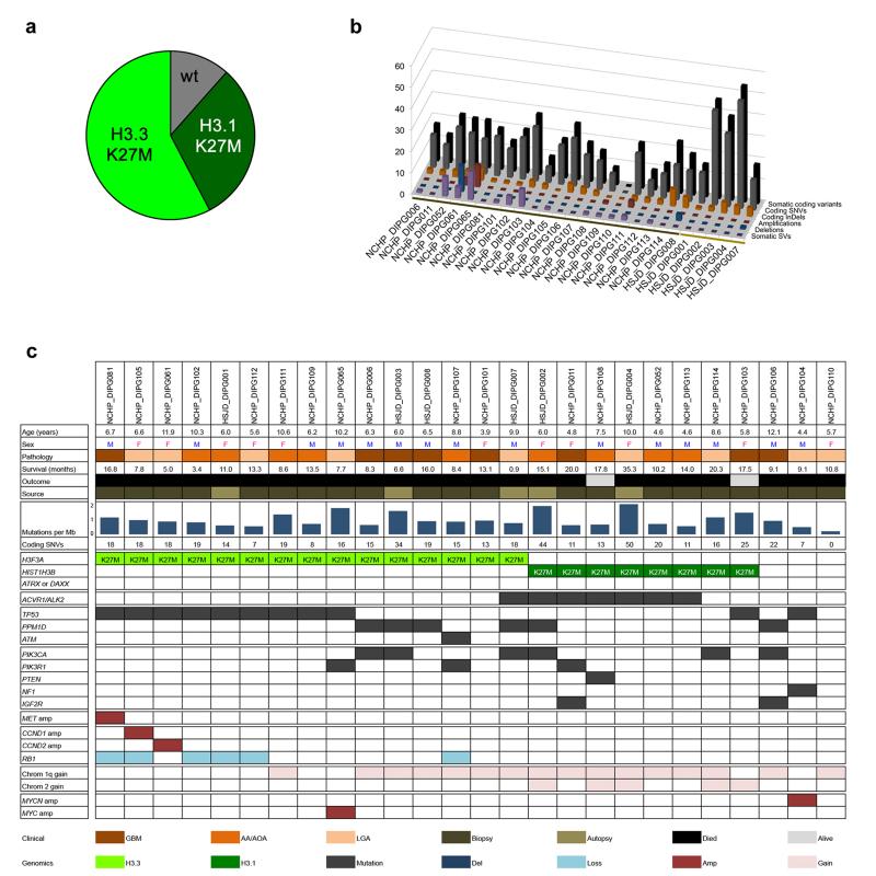
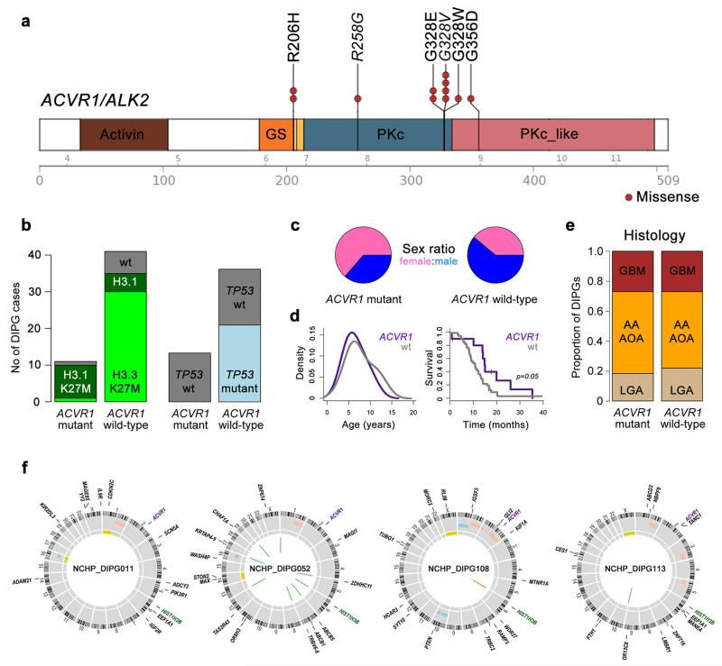
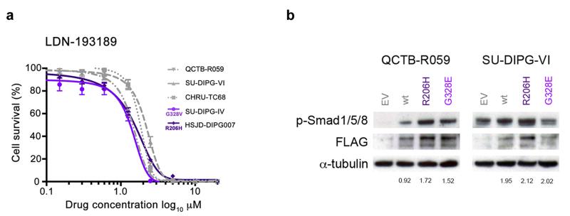
Diffuse intrinsic pontine gliomas (DIPGs) are highly infiltrative malignant glial neoplasms of the ventral pons that, due to their location within the brain, are unsuitable for surgical resection and consequently have a universally dismal clinical outcome. The median survival time is 9-12 months, with neither chemotherapeutic nor targeted agents showing substantial survival benefit in clinical trials in children with these tumors. We report the identification of recurrent activating mutations in the ACVR1 gene, which encodes a type I activin receptor serine/threonine kinase, in 21% of DIPG samples. Strikingly, these somatic mutations (encoding p.Arg206His, p.Arg258Gly, p.Gly328Glu, p.Gly328Val, p.Gly328Trp and p.Gly356Asp substitutions) have not been reported previously in cancer but are identical to mutations found in the germ line of individuals with the congenital childhood developmental disorder fibrodysplasia ossificans progressiva (FOP) and have been shown to constitutively activate the BMP-TGF-β signaling pathway. These mutations represent new targets for therapeutic intervention in this otherwise incurable disease.


Figures

Figure 1
Figure 1. The genomic landscape of DIPG
(a) Pie chart showing breakdown of histone H3 mutations in our series of 26 DIPG samples (H3F3A K27M – 15/26, 58%; HIST1H3B K27M – 8/26, 31%, wild-type 3/26, 11%). (b) The mutational spectrum of DIPG. Barchart showing total somatic coding variants (black), coding SNVs (grey) and InDels (orange), amplifications (red), deletions (blue) and SVs (purple) for each DIPG case. Number of events are plotted along the z axis. Biopsy cases are marked by the dark brown bar, autopsy cases by light brown. (c) Summary of major alterations found. Clinicopathological information of the 26 DIPG samples are provided along with mutation rate and number of somatic coding SNVs. Mutations, amplifications and deletions are noted for the histone H3 genes and ATRX/DAXX; ACVR1; ATM/TP53/PPM1D axis; members of the PI3K/MAPK signalling pathways; receptor tyrosine kinases; members of RB pathway, chromosome 1q and 2 single copy gains, and amplification of MYC/MYCN.
Figure 2
Figure 2. Recurrent ACVR1 mutations in DIPG
(a) Cartoon showing recurrent missense mutations in ACVR1, overlaid with functional protein domains and exon boundaries. In total, 11/52 (21%) of DIPG harboured somatic mutations at four residues, all of which have been previously described in the germline of patients with fibrodysplasia ossificans progressiva. The specific base changes which may be unique to DIPG are highlighted in italics. Activin: activin types I and II receptor domain; GS: TGF-β glycine-serine rich domain; PKc: protein kinase catalytic domain; PKc_like: protein kinase catalytic domain-like. (b) Bar graphs showing segregation of activating mutations in ACVR1 with HIST1H3B K27M mutations (p<0.0001, Fishers exact test) and wild-type TP53 (p=0.0103, Fishers exact test) in our extended series of 52 DIPG patients. (c) Sex distribution of patients with ACVR1 mutations, showing a strong predominance of females in mutant samples. (d) Age distribution (left) and overall survival (right) of DIPG patients with ACVR1 mutations (purple), compared with wild-type (grey). (e) Barplot representing histological breakdown of ACVR1 mutant and wild-type samples. GBM: glioblastoma multiforme; AA: anaplastic astrocytoma; AOA: anaplastic oligoastrocytoma; LGA: low grade astrocytoma. Brown=WHO grade 4, orange=grade 3, tan=grade 2. (f) Circos plots representing the whole genome sequences of the four cases of concurrent ACVR1 mutant / HIST1H3B K27M mutant / TP53 wild-type DIPGs. Outer ring contains chromosomal ideograms, annotated for somatic SNVs in coding genes. Inner ring plots copy number derived from coverage data, dark red=amplification, pink=gain, dark blue=deletion, light blue=loss. Innermost ring represents loss of heterozygosity (LOH, yellow). Inside the circle are drawn SVs, red=interchromosomal translocations, blue=intrachromosomal translocations, orange=deletion, purple=inversion.
Figure 3
Figure 3. ACVR1 mutations are weakly activating and responsive to targeted inhibition
(a) In vitro cytotoxicity of the ALK2 inhibitor LDN-193189. Primary cultures were treated with inhibitor for 72 hours and cell viability measured by CellTiter Glo. The cells used were HSJD-DIPG007 (DIPG, ACVR1 R206H, H3F3A K27M), SUDIPG-IV (DIPG, ACVR1 G328V, HIST1H3B K27M), CHRU-TC68 (DIPG, ACVR1 wt, H3F3A K27M), SU-DIPG-VI (DIPG, ACVR1 wt, H3F3A K27M), QCTB-R059 (thalamic paediatric GBM, ACVR1 wt, H3F3A K27M). (b) ACVR1 mutations confer increased signalling through phospho-Smad 1/5/8. QCTB-R059 and SU-DIPG-VI cells were transfected with FLAG-tagged ACVR1 R206H and G328E mutations, and assessed for phospho-Smad 1/5/8 by Western blot. EV: empty vector; wt: wild-type ACVR1. α-tubulin is included as a loading control. Figures are given for phospho-Smad 1/5/8 levels quantitated relative to FLAG expression.

Comment in

References

    1. Jones C, Perryman L, Hargrave D. Paediatric and adult malignant glioma: close relatives or distant cousins? Nat Rev Clin Oncol. 2012;9:400–13. - PubMed
    1. Katagiri T. Recent topics in fibrodysplasia ossificans progressiva. J Oral Biosciences. 2012;54:119–123.
    1. Wu G, et al. Somatic histone H3 alterations in pediatric diffuse intrinsic pontine gliomas and non-brainstem glioblastomas. Nat Genet. 2012;44:251–3. - PMC - PubMed
    1. Lewis PW, et al. Inhibition of PRC2 Activity by a Gain-of-Function H3 Mutation Found in Pediatric Glioblastoma. Science. 2013 - PMC - PubMed
    1. Roujeau T, et al. Stereotactic biopsy of diffuse pontine lesions in children. J Neurosurg. 2007;107:1–4. - PubMed

Publication types

Substances