. 2014 Jun;38(6):756-67.
doi: 10.1097/PAS.0000000000000208.

Proposed morphologic classification of prostate cancer with neuroendocrine differentiation

Affiliations

Proposed morphologic classification of prostate cancer with neuroendocrine differentiation

Jonathan I Epstein et al. Am J Surg Pathol. 2014 Jun.

Abstract

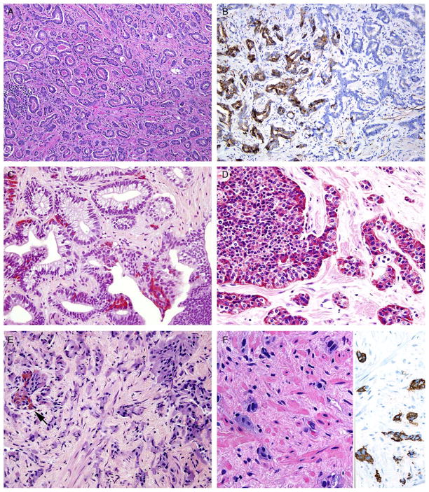
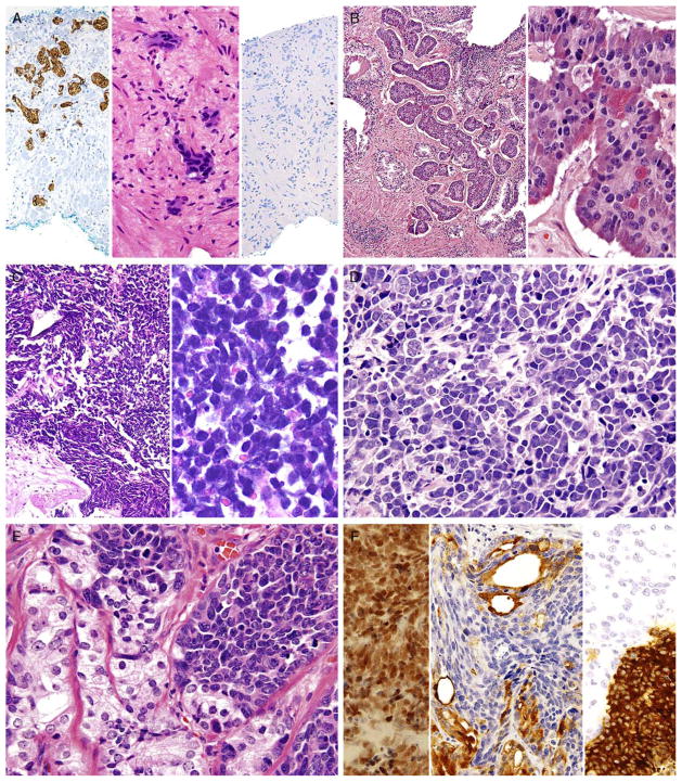
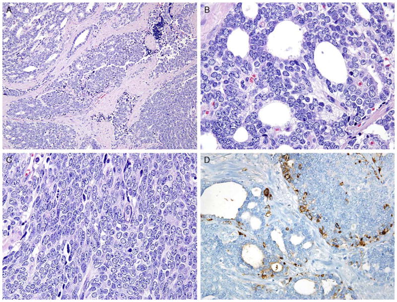
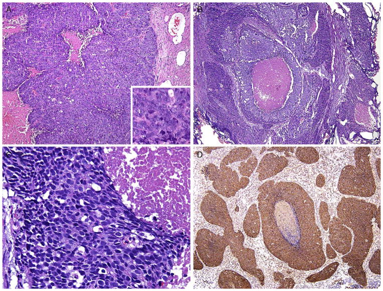
On July 31, 2013, the Prostate Cancer Foundation assembled a working committee on the molecular biology and pathologic classification of neuroendocrine (NE) differentiation in prostate cancer. New clinical and molecular data emerging from prostate cancers treated by contemporary androgen deprivation therapies, as well as primary lesions, have highlighted the need for refinement of diagnostic terminology to encompass the full spectrum of NE differentiation. The classification system consists of: Usual prostate adenocarcinoma with NE differentiation; 2) Adenocarcinoma with Paneth cell NE differentiation; 3) Carcinoid tumor; 4) Small cell carcinoma; 5) Large cell NE carcinoma; and 5) Mixed NE carcinoma - acinar adenocarcinoma. The article also highlights "prostate carcinoma with overlapping features of small cell carcinoma and acinar adenocarcinoma" and "castrate-resistant prostate cancer with small cell cancer-like clinical presentation". It is envisioned that specific criteria associated with the refined diagnostic terminology will lead to clinically relevant pathologic diagnoses that will stimulate further clinical and molecular investigation and identification of appropriate targeted therapies.


Conflict of interest statement

Conflicts of Interest and Source of Funding: The authors have disclosed that they have no significant relationships with, or financial interest in, any commercial companies pertaining to this article.

Figures

FIGURE 1
FIGURE 1
A, Adenocarcinoma of the prostate Gleason score 3+3 = 6. B, Same case as (A) with areas showing positivity for synaptophysin (left) that are indistinguishable in their glandular morphology from areas that are synaptophysin negative (right). C, Gleason score 6 adenocarcinoma and adjacent high-grade prostatic intraepithelial neoplasia with Paneth cell–like NE granules. D, Sheets and cords of prostatic adenocarcinoma with Paneth cell–like NE granules. E, Cords of adenocarcinoma of the prostate with amphophilic cytoplasm and scattered cells with eosinophilic granules (arrow). F, Cords of adenocarcinoma of the prostate with amphophilic cytoplasm (left) staining diffusely for synaptophysin (right).
FIGURE 2
FIGURE 2
A, Cords of adenocarcinoma of the prostate with amphophilic cytoplasm (center) staining diffusely for synaptophysin (left) with a low Ki67 proliferation rate (right). B, Carcinoid-like tumor with nests of cells (left). Higher magnification (right) shows uniform round nuclei with “salt and pepper” chromatin and scattered Paneth cell–like granules. The tumor was positive for NE markers and negative for PSA. Adjacent was usual adenocarcinoma (not shown). C, Low (left) and high (right) magnification of small cell carcinoma of the prostate. D, Intermediate cell type variant of small cell carcinoma of the prostate with slightly more open chromatin and occasional small nucleoli. Tumor was positive for NE markers (not shown). E, Mixed usual adenocarcinoma of the prostate (left) with small cell carcinoma (right). F, Same case as (E) with small cell carcinoma component positive for TTF-1 (left). Synaptophysin labeled the small cell carcinoma component (right, bottom) and not the adenocarcinoma component (right, top). PSA (center) was positive in the adenocarcinoma component but not in the small cell carcinoma.
FIGURE 3
FIGURE 3
A, Case with overlapping features between small cell—usual adenocarcinoma with gland formation (upper left) and sheets of cells (lower right). B, Higher magnification of Figure 4A. Despite showing lumen formation, there is a lack of apical cytoplasm typical of usual adenocarcinoma of the prostate. C, Higher magnification of Figure 4A with solid sheets of cells. Cells have a high nuclear to cytoplasmic ratio like small cell carcinoma yet lack its high mitotic/apoptotic rate. Nucleoli are intermediate between small cell and usual adenocarcinoma of the prostate. D, Same case as Figure 4A with positivity for synaptophysin in both solid and glandular areas.
FIGURE 4
FIGURE 4
A, LCNEC sheets of cells and geographic necrosis. Inset shows numerous prominent nucleoli. Tumor was diffusely positive for chromogranin. B, Another example of LCNEC with large nests of cells and geographic necrosis. C, Higher magnification of Figure 4A with cells having abundant cytoplasm. D, LCNEC (same case as Fig. 4A) with diffuse synaptophysin immunoreactivity.

References

    1. Abrahamsson PA. Neuroendocrine cells in tumour growth of the prostate. Endocr Relat Cancer. 1999;6:503–519. - PubMed
    1. Abrahamsson PA, Wadstrom LB, Alumets J, et al. Peptidehormone- and serotonin-immunoreactive tumour cells in carcinoma of the prostate. Pathol Res Pract. 1987;182:298–307. - PubMed
    1. Bonkhoff H, Stein U, Remberger K. Androgen receptor status in endocrine-paracrine cell types of the normal, hyperplastic, and neoplastic human prostate. Virchows Arch A Pathol Anat Histopathol. 1993;423:291–294. - PubMed
    1. Azzopardi JG, Evans DJ. Argentaffin cells in prostatic carcinoma: differentiation from lipofuscin and melanin in prostatic epithelium. J Pathol. 1971;104:247–251. - PubMed
    1. Kazzaz BA. Argentaffin and argyrophil cells in the prostate. J Pathol. 1974;112:189–193. - PubMed

MeSH terms

Substances