. 2014 Aug 15;23(16):4315-27.
doi: 10.1093/hmg/ddu148. Epub 2014 Apr 4.

Activating mutations in RRAS underlie a phenotype within the RASopathy spectrum and contribute to leukaemogenesis

Affiliations

Activating mutations in RRAS underlie a phenotype within the RASopathy spectrum and contribute to leukaemogenesis

Elisabetta Flex et al. Hum Mol Genet. .

Abstract

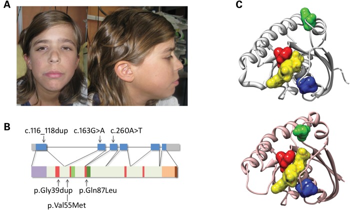
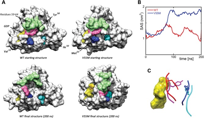
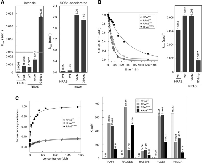
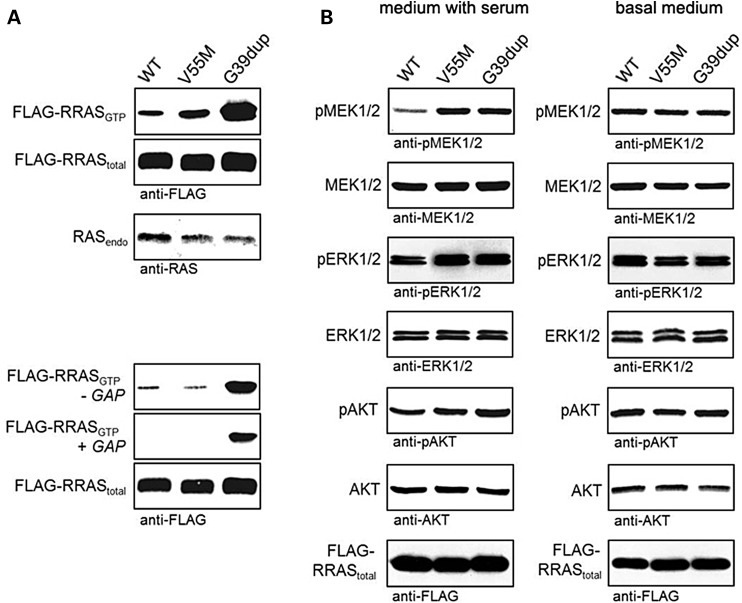
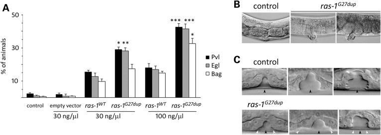
RASopathies, a family of disorders characterized by cardiac defects, defective growth, facial dysmorphism, variable cognitive deficits and predisposition to certain malignancies, are caused by constitutional dysregulation of RAS signalling predominantly through the RAF/MEK/ERK (MAPK) cascade. We report on two germline mutations (p.Gly39dup and p.Val55Met) in RRAS, a gene encoding a small monomeric GTPase controlling cell adhesion, spreading and migration, underlying a rare (2 subjects among 504 individuals analysed) and variable phenotype with features partially overlapping Noonan syndrome, the most common RASopathy. We also identified somatic RRAS mutations (p.Gly39dup and p.Gln87Leu) in 2 of 110 cases of non-syndromic juvenile myelomonocytic leukaemia, a childhood myeloproliferative/myelodysplastic disease caused by upregulated RAS signalling, defining an atypical form of this haematological disorder rapidly progressing to acute myeloid leukaemia. Two of the three identified mutations affected known oncogenic hotspots of RAS genes and conferred variably enhanced RRAS function and stimulus-dependent MAPK activation. Expression of an RRAS mutant homolog in Caenorhabditis elegans enhanced RAS signalling and engendered protruding vulva, a phenotype previously linked to the RASopathy-causing SHOC2(S2G) mutant. Overall, these findings provide evidence of a functional link between RRAS and MAPK signalling and reveal an unpredicted role of enhanced RRAS function in human disease.


Figures

Figure 1.
Figure 1.
RASopathy-causing and leukaemia-associated RRAS mutations. (A) Facial features of the affected subject (9802) heterozygous for the de novo germline c.116_118dup. (B) RRAS exon–intron arrangement with coding exons as blue boxes. RRAS functional motifs include the GTP/GDP binding domain (G1 to G5, starting from the N-terminus) (red), switch I (light green), switch II (dark green) and hypervariable region (light brown) with the C-terminal CAAX motif (dark brown). The unique N-terminal region is also shown (violet). Location of disease-associated mutations is reported. (C) Position of affected residues on the three-dimensional structure of RRAS in its GDP-bound, inactive state (PDB: 2FN4) (above) and that of non-hydrolysable GTP analogue (GppNHp)-bound, active HRAS (PDB: 5P21) (below). The red surface indicates Gly39 and Val40 (Gly13 and Val14, in HRAS), whereas Val55 (Val29) and Gln87 (Gln61) are shown in blue and green, respectively. GDP is reported as semi-transparent yellow surface.
Figure 2.
Figure 2.
Molecular dynamics (MD) simulations. (A) Structural perturbations promoted by the p.Val55Met substitution as obtained from MD simulations of the RRAS/GDP complex. The wild-type (WT) protein is also shown for comparison. Top panels report the protein structures at the beginning of simulations, whereas the final structures (200 ns) are shown at the bottom. The final structure of RRASV55M is well representative of the last 120 ns of the trajectory. The protein surface of RRAS is shown with GDP (yellow). The mutated residues and those forming a cluster in the simulation of mutated RRAS are coloured as follows: Val55/Met55 (blue), Tyr58 (pink) and Ile50 (cyan). Residues 59–64, which, together with Tyr58, form the switch I region, are coloured in green. (B) Solvent accessible surface of GDP in the MD simulations of wild-type (red) and mutant (blue) RRAS/GDP complexes. (C) Conformation of the loop comprised between Val55/Met55 and Asp59 in wild-type (red) and mutant (blue) RRAS/GDP complexes obtained from MD simulations. GDP is reported as semi-transparent yellow surface. Superimposed conformations of the corresponding loop (residues 29–33) in GDP-bound HRAS (violet) (PDB: 4Q21) and GDP-bound HRAS complexed with SOS1 (cyan) (PDB: 1BKD) are shown for comparison. The side chains of Tyr58 and the corresponding residue in HRAS, Tyr32, are displayed as sticks.
Figure 3.
Figure 3.
In vitro biochemical characterization of the RRASG39dup and RRASV55M mutants. (A) Intrinsic (left) and SOS1-accelerated (right) mantGDP nucleotide dissociation measured in the presence of 20-fold excess of non-labelled GDP. The decrease in mant fluorescence was fitted by single exponentials to obtain kobs values for nucleotide dissociation. RRASG39dup exhibited a 35-fold increased intrinsic dissociation of mantGDP, whereas SOS1-accelerated mantGDP dissociation was augmented by ∼30-fold for both mutants, compared with RRASWT and HRASWT. The Kobs values are an average of five to seven independent measurements. (B) Intrinsic GTP hydrolysis kinetics (left) and rate constants (right) of RRASG39dup and RRASV55M proteins, documenting the impaired catalytic activity in the former. The Kobs values are an average of five to seven independent measurements. (C) Binding of RRASWT, RRASV55M and RRASG39dup to the RAS-binding domain of RAF1 measured as variation in fluorescence polarization of each mantGppNHp-bound RRAS protein at increasing concentrations of RAF1-RBD (left), and dissociation constants (Kd) for the interaction of HRASWT and RRAS proteins to the RBDs of RAF1, RALGDS, PLCE1, PIK3CA and RASSF5 (right). Kd values were obtained by quadratic fitting of the concentration-dependent binding curves from fluorescence polarization measurement as exemplified for RAF1-RBD. Of note, RRASWT binds to RAF1, RALGDS, RASSF5 and PLCE1 less efficiently than HRAS, whereas an increased binding affinity to PIK3CA is observed.
Figure 4.
Figure 4.
RRASG39dup and RRASV55M signalling activities in cells. (A) Determination of GTP-bound RRAS levels in COS-7 cells transiently expressing wild-type or mutant FLAG-tagged RRAS proteins. Assays were performed in the presence of serum (above), and in serum-free conditions (−GAP) or in the presence of the neurofibromin GAP domain (+GAP) (below). RRASG39dup was predominantly present in the active GTP-bound form and was resistant to GAP stimulation, whereas a slightly increased level of GTP-bound RRASV55M was observed in the presence of serum. Representative blots of three performed experiments are shown. (B) Determination of MEK, ERK and AKT phosphorylation levels (pMEK, pERK and pAKT) in transiently transfected COS-7 cells cultured in medium with serum (left) or basal medium (right). Expression of each RRAS mutant resulted in variably enhanced MEK, ERK and also partially AKT phosphorylation after stimulation. Total MEK, ERK and AKT in cell lysates are shown for equal protein expression and loading. Expression levels of exogenous, FLAG-tagged RRAS in cell lysates are shown for each experiment. Representative blots of three performed experiments are shown.
Figure 5.
Figure 5.
Consequences of ras-1G27dup expression on C. elegans vulval development. (A) Heat-shock-driven expression of ras-1WT and ras-1G27dup at early L3 stage results in protruding vulva (Pvl), egg laying defective (Egl) and bag-of-worms (Bag) phenotypes. Isogenic animals that had lost the transgene (control group) and worms expressing the heat shock-inducible vector (empty vector) were subjected to heat shock and scored in parallel for comparison. The dose at which the transgene has been injected is reported at the bottom. Error bars indicate SD of three independent experiments. Asterisks indicate significant differences compared with ras-1WT at the corresponding dose of injection (*P < 0.05; **P < 0.005; ***P < 0.0005; Fisher's Exact Test). (B) A proper vulva develops in heat-shocked control animals (left), whereas a protruding vulva is observed in heat-shocked ras-1G27dup young adults (middle) and adult worms (right). (C) Nomarski images of vulval precursor cells in late L3 (left), early L4 (middle) and mid-late L4 (right) stages from synchronized animals heat-shocked at early L3. In control animals (N = 48), only P6.p descendants invaginate (upper panel), whereas in 10 of 30 analysed ras-1G27dup-expressing worms, P5.p and/or P7.p descendants also detach from the cuticle, generating asymmetric invaginations (lower panel). Black arrowheads point to P6.p descendant invagination, whereas white arrowheads point to P5.p and P7.p descendant invagination. Anterior is to the left and dorsal is up, in all images.

References

    1. Mitin N., Rossman K.L., Der C.J. Signaling interplay in Ras superfamily function. Curr. Biol. 2005;15:R563–R574. - PubMed
    1. Mendoza M.C., Er E.E., Blenis J. The Ras-ERK and PI3K-mTOR pathways: cross-talk and compensation. Trends Biochem. Sci. 2011;36:320–328. - PMC - PubMed
    1. Harris T.J.R., McCormick F. The molecular pathology of cancer. Nat. Rev. Clin. Oncol. 2010;7:251–265. - PubMed
    1. Pylayeva-Gupta Y., Grabocka E., Bar-Sagi D. RAS oncogenes: weaving a tumorigenic web. Nat. Rev. Cancer. 2011;11:761–774. - PMC - PubMed
    1. Roberts A.E., Allanson J.E., Tartaglia M., Gelb B.D. Noonan syndrome. Lancet. 2013;381:333–342. - PMC - PubMed

Publication types

MeSH terms