. 2014 Jun;63(6):2063-72.
doi: 10.2337/db13-1279. Epub 2014 Apr 4.

Epidermal growth factor receptor inhibition slows progression of diabetic nephropathy in association with a decrease in endoplasmic reticulum stress and an increase in autophagy

Affiliations

Epidermal growth factor receptor inhibition slows progression of diabetic nephropathy in association with a decrease in endoplasmic reticulum stress and an increase in autophagy

Ming-Zhi Zhang et al. Diabetes. 2014 Jun.

Abstract

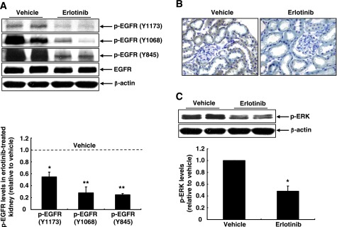
Previous studies by us and others have reported renal epidermal growth factor receptors (EGFRs) are activated in models of diabetic nephropathy. In the present study, we examined the effect of treatment with erlotinib, an inhibitor of EGFR tyrosine kinase activity, on the progression of diabetic nephropathy in a type 1 diabetic mouse model. Inhibition of renal EGFR activation by erlotinib was confirmed by decreased phosphorylation of EGFR and extracellular signal-related kinase 1/2. Increased albumin/creatinine ratio in diabetic mice was markedly attenuated by erlotinib treatment. Erlotinib-treated animals had less histological glomerular injury as well as decreased renal expression of connective tissue growth factor and collagens I and IV. Autophagy plays an important role in the pathophysiology of diabetes mellitus, and impaired autophagy may lead to increased endoplasmic reticulum (ER) stress and subsequent tissue injury. In diabetic mice, erlotinib-treated mice had evidence of increased renal autophagy, as indicated by altered expression and activity of ATG12, beclin, p62, and LC3A II, hallmarks of autophagy, and had decreased ER stress, as indicated by decreased expression of C/EBP homologous protein, binding immunoglobulin protein, and protein kinase RNA-like ER kinase. The mammalian target of rapamycin (mTOR) pathway, a key factor in the development of diabetic nephropathy and an inhibitor of autophagy, is inhibited by AMP-activated protein kinase (AMPK) activation. Erlotinib-treated mice had activated AMPK and inhibition of the mTOR pathway, as evidenced by decreased phosphorylation of raptor and mTOR and the downstream targets S6 kinase and eukaryotic initiation factor 4B. Erlotinib also led to AMPK-dependent phosphorylation of Ulk1, an initiator of mammalian autophagy. These studies demonstrate that inhibition of EGFR with erlotinib attenuates the development of diabetic nephropathy in type 1 diabetes, which is mediated at least in part by inhibition of mTOR and activation of AMPK, with increased autophagy and inhibition of ER stress.


Figures

Figure 1
Figure 1
EGFR inhibition with erlotinib attenuated progression of diabetic nephropathy. Albuminuria, measured by 24-h urinary albumin/creatinine ratio (ACR), was markedly attenuated by erlotinib treatment in both STZ–wild-type (A) and STZ-eNOS−/− mice (B). C: Periodic acid-Schiff staining indicated that mesangial expansion in STZ–wild-type mice and mesangial expansion, mesangiolysis, and glomerulosclerosis in STZ-eNOS−/− mice were markedly attenuated with erlotinib treatment (original magnification ×400). *P < 0.05 vs. corresponding nondiabetic mice; †P < 0.05 vs. corresponding STZ + vehicle group; n = 4–6.
Figure 2
Figure 2
A: Erlotinib treatment markedly inhibited kidney EGFR phosphorylation at the indicated tyrosine residues in STZ-eNOS−/− mice. B: Immunostaining of p-EGFR (Y1068) was primarily restricted to tubular epithelial cells in STZ-eNOS−/− mice and reduced by erlotinib treatment (original magnification ×250). C: Erlotinib also marked inhibited kidney ERK1/2 phosphorylation in STZ-eNOS−/− mice. *P < 0.05; **P < 0.01 vs. vehicle group; n = 3 in vehicle group and n= 4 in erlotinib group.
Figure 3
Figure 3
A: Erlotinib treatment markedly reduced renal expression of CTGF, collagen I, and collagen IV in STZ-eNOS−/− mice. Original magnification: CTGF, ×250; collagen I and collagen IV, ×400. B: Erlotinib treatment also reduced kidney macrophage infiltration (indicated by F4/80 immunoexpression) and oxidative stress (indicated by nitrotyrosine immunostaining) in STZ-eNOS−/− mice. Original magnification: nitrotyrosine, ×160; F4/80, ×250. **P < 0.01 vs. vehicle group; n = 4. hpf, high-power field.
Figure 4
Figure 4
Erlotinib reduced kidney ER stress but stimulated the autophagic pathway in STZ-eNOS−/− mice. A: Erlotinib inhibited kidney CHOP expression in STZ-eNOS−/− mice. *P < 0.05 vs. vehicle group; n = 3 in vehicle group and n = 4 in erlotinib group. B: Erlotinib increased expression of ATG12 and beclin and decreased expression of p62. Erlotinib-induced activation of autophagic pathway was indicated by increased expression levels of LC3A II, a membrane-bound form of LC3A produced during formation of autophagosomes. **P < 0.01 vs. vehicle group; n = 3–5. C: Erlotinib treatment increased Ulk1 phosphorylation on the AMPK phosphorylation site Ser317, but decreased Ulk1 phosphorylation on the mTOR-dependent phosphorylation site Ser757. **P < 0.01 vs. vehicle group; n = 3 in vehicle group and n = 4 in erlotinib group.
Figure 5
Figure 5
A: Erlotinib treatment decreased kidney ER stress, as indicated by decreased glomerular and tubule epithelial expression of CHOP, PERK, and BIP in STZ-eNOS−/− mice. B: LC3A immunostaining was detected in tubular epithelial cells, but not in glomerulus, in vehicle-treated STZ-eNOS−/− mouse kidneys. With erlotinib treatment, LC3A expression was detectable in glomerulus and was markedly increased in tubular epithelial cells. Original magnification: CHOP and BIP, ×250; PERK and LC3A, ×400.
Figure 6
Figure 6
EGFR inhibition with erlotinib inhibited the kidney mTOR pathway but stimulated AMPK activation in STZ-eNOS−/− mice. A: Erlotinib inhibited phosphorylation of mTOR, raptor, p70 S6K, and eIF-4B. B: Erlotinib stimulated phosphorylation of AMPKα and AMPKβ. C: Erlotinib treatment increased kidney AMPKα activity in both epithelia and glomerulus (original magnification ×400). **P < 0.01 vs. vehicle group; n = 3–5.
Figure 7
Figure 7
EGFR inhibition stimulated AMPK activity but inhibited S6K activity in mesangial cells. A: AG1478 (300 nmol/L) effectively inhibited EGFR phosphorylation in mesangial cells cultured in high-glucose medium (25 mmol/L). B: AG1478 treatment for 6 h led to inhibition of S6K activity and stimulation of AMPK activity. *P < 0.05; **P < 0.01 vs. control group; n = 3.

References

    1. Kanwar YS, Wada J, Sun L, et al. Diabetic nephropathy: mechanisms of renal disease progression. Exp Biol Med (Maywood) 2008;233:4–11 - PubMed
    1. Chen J, Chen JK, Nagai K, et al. EGFR signaling promotes TGFβ-dependent renal fibrosis. J Am Soc Nephrol 2012;23:215–224 - PMC - PubMed
    1. Chen X, Abair TD, Ibanez MR, et al. Integrin alpha1beta1 controls reactive oxygen species synthesis by negatively regulating epidermal growth factor receptor-mediated Rac activation. Mol Cell Biol 2007;27:3313–3326 - PMC - PubMed
    1. Zhang MZ, Yao B, Cheng HF, Wang SW, Inagami T, Harris RC. Renal cortical cyclooxygenase 2 expression is differentially regulated by angiotensin II AT(1) and AT(2) receptors. Proc Natl Acad Sci U S A 2006;103:16045–16050 - PMC - PubMed
    1. Zhang MZ, Yao B, Wang S, et al. Intrarenal dopamine deficiency leads to hypertension and decreased longevity in mice. J Clin Invest 2011;121:2845–2854 - PMC - PubMed

Publication types

MeSH terms