. 2014 May;10(5):386-91.
doi: 10.1038/nchembio.1498. Epub 2014 Apr 6.

Design of activated serine-containing catalytic triads with atomic-level accuracy

Affiliations

Design of activated serine-containing catalytic triads with atomic-level accuracy

Sridharan Rajagopalan et al. Nat Chem Biol. 2014 May.

Abstract

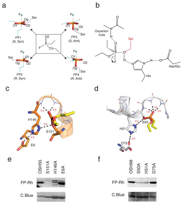
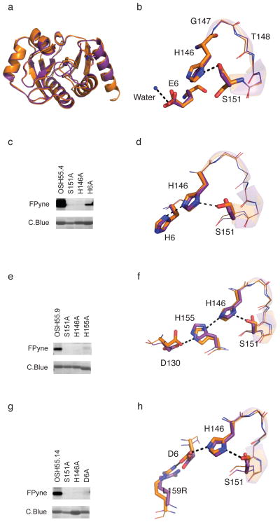
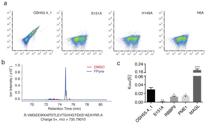
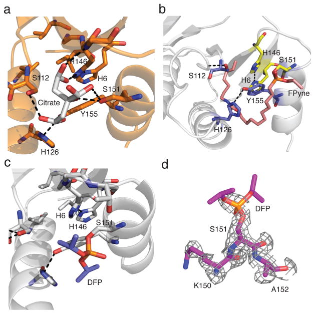
A challenge in the computational design of enzymes is that multiple properties, including substrate binding, transition state stabilization and product release, must be simultaneously optimized, and this has limited the absolute activity of successful designs. Here, we focus on a single critical property of many enzymes: the nucleophilicity of an active site residue that initiates catalysis. We design proteins with idealized serine-containing catalytic triads and assess their nucleophilicity directly in native biological systems using activity-based organophosphate probes. Crystal structures of the most successful designs show unprecedented agreement with computational models, including extensive hydrogen bonding networks between the catalytic triad (or quartet) residues, and mutagenesis experiments demonstrate that these networks are critical for serine activation and organophosphate reactivity. Following optimization by yeast display, the designs react with organophosphate probes at rates comparable to natural serine hydrolases. Co-crystal structures with diisopropyl fluorophosphate bound to the serine nucleophile suggest that the designs could provide the basis for a new class of organophosphate capture agents.


Conflict of interest statement

Competing financial interests: The authors declare no financial competing interests.

Figures

Figure 1
Figure 1. Design strategy
(a) Transition-state (TS) models. The fluorophosphonate (FP) ligand (ethyl ethylphosphonofluoridate) used in design calculations is in the middle panel flanked by TS models corresponding to syn-and anti- attack on the two isomers (R & S) of the ligand with the leaving atom fluorine (F) in blue-spheres. The serine nucleophile is shown in purple. (b) Theozyme geometry. Geometric parameters were derived from the computed ideal active site geometries and native hydrolase statistics previously reported. Theozymes with the histidine hydrogen bonding reversed were also considered during RosettaMatch (See Methods). (c.d) Design models of OSH55 (c) and OSH98 (d). Active-site residues of OSH55 and OSH98 are shown in orange and white sticks respectively. The modeled fluorophosphonate ligand is shown in yellow sticks. In both designs, multiple backbone NHs form the oxyanion hole to stabilize the transition state. (e,f) Selective FP-Rh labeling of OSH55 (e) and OSH98 (f) but not the active site serine knockouts. Serine reactivity was assessed by gel-based ABPP (top, in-gel fluorescence; bottom, coomassie staining, see Supplementary Fig. 15 for full-size gel images. The experiments were performed in duplicates with consistent results).
Figure 2
Figure 2. Characterization of OSH55 derived designs
(a) Structural superposition of design (brown) and crystal structure (purple) of OSH55 with overall backbone RMSD of 0.6 Å. The all-atom RMSD over the binding pocket residues is 0.34 Å. (b) A zoom-in view of the active site of OSH55 showing the orientation of Ser151 and His146 as designed and Glu6 coordinating a water molecule. (c,e,g) Selective fluorophosphonate probe labeling of OSH55.4 (c), OSH55.9 (e) and OSH55.14 (g) but not the active-site knockout mutants. Serine reactivity was assessed by gel-based ABPP (top, in-gel fluorescence; bottom, coomassie staining, see Supplementary Fig. 16 for full-size gel images. The experiments were performed in duplicates with consistent results). There is much less labeling in the catalytic triad knockouts but no decrease in the amount of protein. (d,f,h) Superimposition of the designed catalytic triad active sites (brown) overlaid on the experimentally determined crystal structures (purple) for OSH55.4 (d), OSH55.9 (f) and OSH55.14 (h); the designed hydrogen bond networks are recapitulated with very high accuracy.
Figure 3
Figure 3. Organophosphate reactivity of OSH55.4_1 is comparable to native enzymes
(a) Flow cytometry analysis of OSH55.4_1 and active site knockouts displayed on yeast following incubation with FP-biotin. Signal along x-axis indicates extent of yeast surface display; signal along y-axis; extent of probe binding. The active site residue knockouts abolish binding to FP-biotin. (b) MS1 chromatographic traces of the OSH55.4_1 active-site peptide adducted with FPyne probe. LC-MS/MS experiments (See Methods) identified a fully tryptic peptide (m/z = 735.79010 and z = 5+) with probe modification on the catalytic Ser151 (denoted with an asterisk). The extracted MS1 chromatographic traces (±15 p.p.m.) showed a distinct peak present only in the FPyne-treated (blue) but not in the DMSO-treated (red) sample. (c) Comparison of rates of FP-Rh labeling of the OSH55.4_1 design to those of a representative set of native serine hydrolases by fluorescence polarization. The kobs/[E] (mean ± s.e.m.) value for OSH55.4_1 is greater than those of RBBP9 and PME1. The S151A knockout mutation reduces the reaction rate to near background level (*p<0.05, **p<0.01, ***p<0.001, t-test, comparison with OSH55.4_1).
Figure 4
Figure 4. Crystal structures of OSH55.4_1 bound to Diispropyl fluoroposphate (DFP)
(a) Crystal Structure of the apo form of OSH55.4_1 with a citrate molecule (white) from the crystallization buffer cocktail making extensive interactions with active site residues. (b) Crystal structure of OSH55.4_1 covalently modified at Ser151 with FPyne (pink). (c) Crystal structure of OSH55.4_1 covalently modified at Ser151 with DFP (red and blue). (d) Simulated annealing omit map around Ser151 (fo-fc). Density is clearly evident on the serine but the isopropyl groups of DFP are not distinguishable probably due to free rotation about the P-C bond.

References

    1. Botos I, Wlodawer A. The expanding diversity of serine hydrolases. Curr Opin Struct Biol. 2007;17:683–690. - PMC - PubMed
    1. Hedstrom L. Serine protease mechanism and specificity. Chem Rev. 2002;102:4501–4524. - PubMed
    1. Ekici OD, Paetzel M, Dalbey RE. Unconventional serine proteases: variations on the catalytic Ser/His/Asp triad configuration. Protein Sci. 2008;17:2023–2037. - PMC - PubMed
    1. Carter P, Wells JA. Dissecting the catalytic triad of a serine protease. Nature. 1988;332:564–568. - PubMed
    1. Corey DR, Craik CS. An Investigation into the Minimum Requirements for Peptide Hydrolysis by Mutation of the Catalytic Traid of Trypsin. J Am Chem Soc. 1992;114:1784–1790.

Publication types

LinkOut - more resources