. 2014 Jul;57(7):1476-84.
doi: 10.1007/s00125-014-3227-7. Epub 2014 Apr 6.

Doc2b enrichment enhances glucose homeostasis in mice via potentiation of insulin secretion and peripheral insulin sensitivity

Affiliations

Doc2b enrichment enhances glucose homeostasis in mice via potentiation of insulin secretion and peripheral insulin sensitivity

Latha Ramalingam et al. Diabetologia. 2014 Jul.

Abstract

Aims/hypothesis: Insulin secretion from pancreatic beta cells and insulin-stimulated glucose uptake into skeletal muscle are processes regulated by similar isoforms of the soluble N-ethylmaleimide-sensitive factor-attachment protein receptor (SNARE) and mammalian homologue of unc-18 (Munc18) protein families. Double C2 domain β (Doc2b), a SNARE- and Munc18-interacting protein, is implicated as a crucial effector of glycaemic control. However, whether Doc2b is naturally limiting for these processes, and whether Doc2b enrichment might exert a beneficial effect upon glycaemia in vivo, remains undetermined.

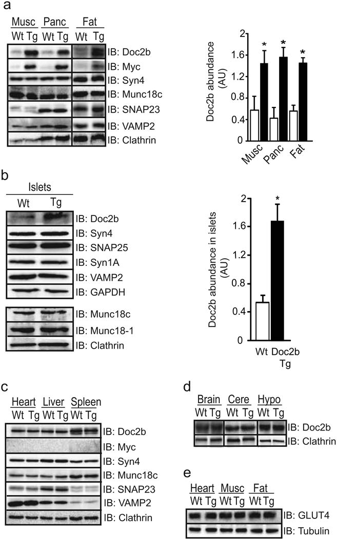
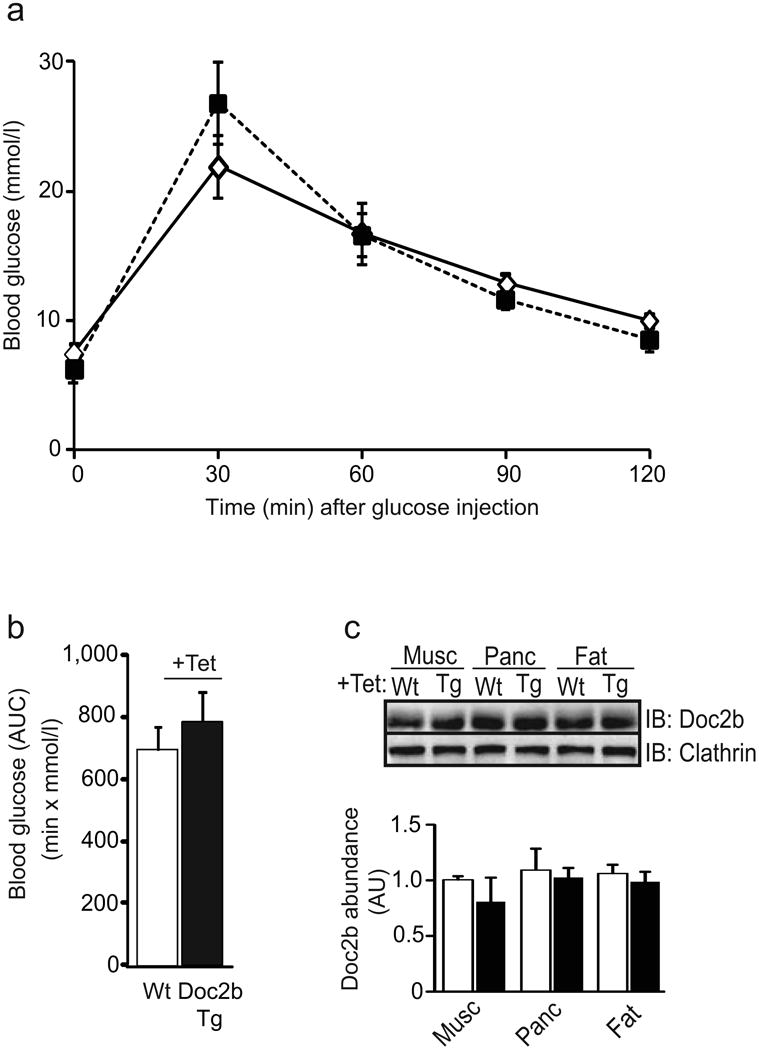
Methods: Tetracycline-repressible transgenic (Tg) mice engineered to overexpress Doc2b simultaneously in the pancreas, skeletal muscle and adipose tissues were compared with wild-type (Wt) littermate mice regarding glucose and insulin tolerance, islet function in vivo and ex vivo, and skeletal muscle GLUT4 accumulation in transverse tubule/sarcolemmal surface membranes. SNARE complex formation was further assessed using Doc2b overexpressing L6-GLUT4-myc myoblasts to derive mechanisms relatable to physiological in vivo analyses.

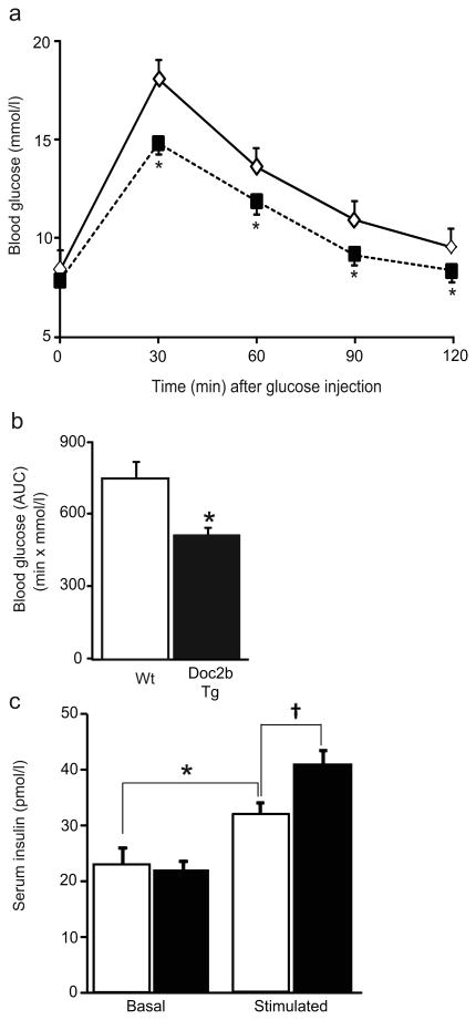
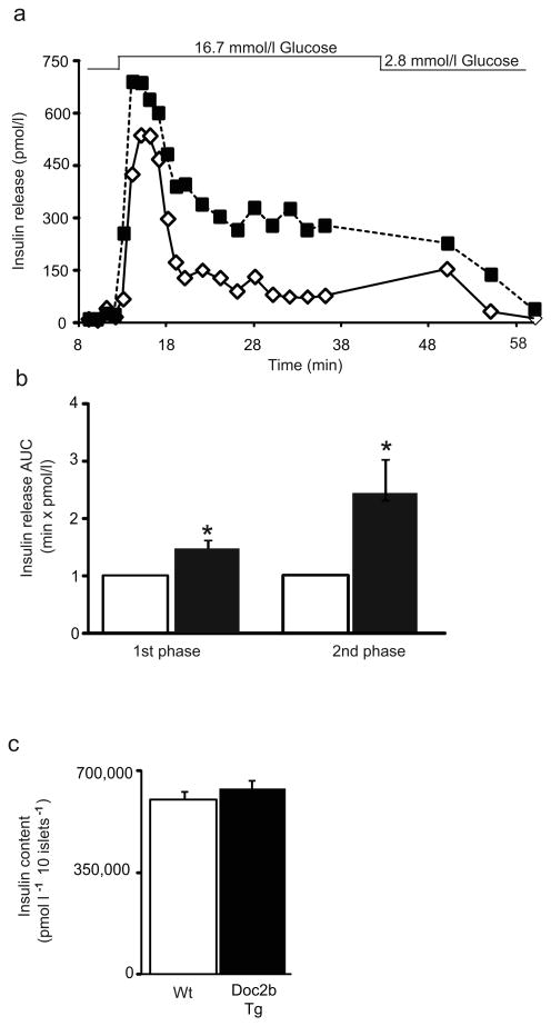
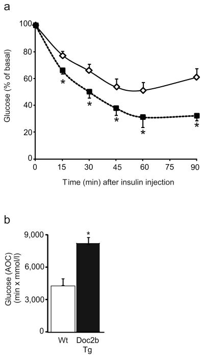
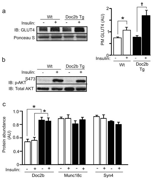
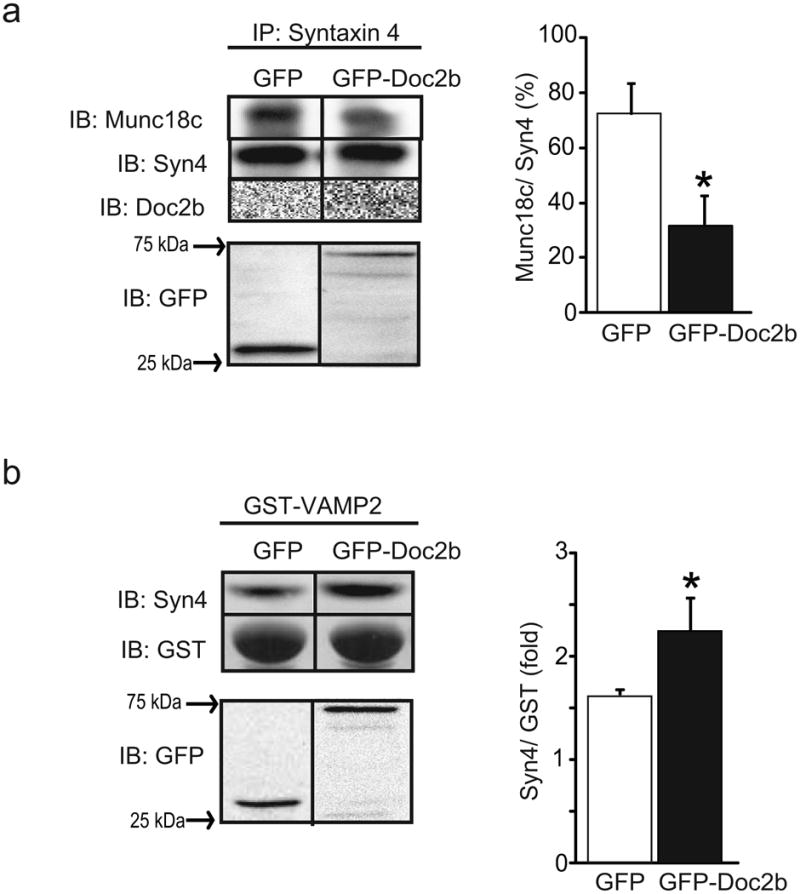
Results: Doc2b Tg mice cleared glucose substantially faster than Wt mice, correlated with enhancements in both phases of insulin secretion and peripheral insulin sensitivity. Heightened peripheral insulin sensitivity correlated with elevated insulin-stimulated GLUT4 vesicle accumulation in cell surface membranes of Doc2b Tg mouse skeletal muscle. Mechanistic studies demonstrated Doc2b enrichment to enhance syntaxin-4-SNARE complex formation in skeletal muscle cells.

Conclusions/interpretation: Doc2b is a limiting factor in SNARE exocytosis events pertinent to glycaemic regulation in vivo. Doc2b enrichment may provide a novel means to simultaneously boost islet and skeletal muscle function in vivo in the treatment and/or prevention of diabetes.


Conflict of interest statement

Duality of interest: The authors declare that there is no duality of interest associated with this manuscript.

Figures

Fig. 1
Fig. 1
Protein expression in tissues of Doc2b transgenic mice. (a) Gastrocnemius skeletal muscle (Musc), pancreas (Panc) and epigonadal fat were isolated from three to five pairs of Doc2b Tg mice (black bars) and Wt littermates (white bars) and immunoblotted (IB) for detection of SNARE and SNARE accessory proteins. Doc2b abundances were normalised to clathrin to account for minor variations in protein loading; *p<0.05 vs Wt. (b) Isolated islets were assessed for Doc2b and SNARE protein expression as described in (a) above. (c, d) Heart, liver and spleen were similarly assessed for Doc2b levels (c), as was whole-brain lysate, cerebellum (Cere) and hypothalamus (Hypo) (d). (e) GLUT4 protein abundance was assessed in heart, skeletal muscle and fat from mice from panel (a). Data are representative of three to five pairs of mice. AU, arbitrary units
Fig. 2
Fig. 2
Doc2b Tg mice have enhanced glucose tolerance. (a) IPGTT in Doc2b Tg mice (black squares) and Wt littermates (white diamonds) was performed in 4- to 6-month-old female mice fasted for 6 h. (b) AUC data are shown as the average ± SE from seven pairs of mice; *p<0.05, vs Wt. (c) Insulin content present in serum taken before (Basal) and 10 min post injection of glucose (Stimulated) during the IPGTT in Doc2b Tg (black bars) and Wt mice (white bars). Data represent the average ± SE from six pairs of mice; *p<0.05 vs Wt basal; p<0.05 vs Wt glucose-stimulated
Fig. 3
Fig. 3
Tet-mediated repression of the Doc2b transgene reduces glucose tolerance compared with that of the Wt mice. (a) Doc2b Tg (black squares) and Wt (white diamonds) female mice assessed in Fig. 2 assays were subsequently administered tet (1 mg/ml) in the drinking water for 1 week and the IPGTT was re-performed. (b) AUC analysis is shown as the average ± SE from seven pairs of mice. (c) Tissue extracts were immunoblotted and quantified as described in Fig. 1 for Doc2b Tg (black bars) and Wt mice (white bars). Data represent the average ± SE of three independent sets of tissues
Fig. 4
Fig. 4
Islets from Doc2b Tg mice exhibit potentiated biphasic insulin release. (a) Islets isolated from Doc2b Tg mice (black squares) and Wt littermates (white diamonds) were perifused in parallel at 2.8 mmol/l glucose for 10 min followed by 16.7 mmol/l glucose for 35 min and then returned to low glucose for 20 min. Eluted fractions were collected and insulin secretion was determined by RIA, as depicted in this representative pair of traces. (b) AUC for first (11–17 min) and second (18–45 min) phases of insulin secretion was quantified in islets, normalised to baseline, from Doc2b Tg (black bars) and Wt (white bars) mice. Data represent the average ± SE from three independent sets of perifused islets; *p<0.05 vs Wt (Wt set equal to 1.0 and Tg normalised thereto for each phase per set). (c) Average insulin content per 10 islets from Doc2b Tg mice and Wt littermates
Fig. 5
Fig. 5
Doc2b Tg mice exhibit enhanced insulin sensitivity. (a) Insulin tolerance testing of seven pairs of female Doc2b Tg mice (black squares) and Wt littermates (white diamonds) fasted for 6 h. Data are shown as the mean percentage of starting basal blood glucose concentrations ± SE; *p<0.05 vs Wt. (b) Area over the curve (AOC) data are shown as the average ± SE from seven pairs of mice; *p<0.05, vs Wt
Fig. 6
Fig. 6
Doc2b Tg mice show increased insulin-stimulated GLUT4 accumulation at the sarcolemma/transverse tubule PMs of skeletal muscle. (a) GLUT4 abundance in the PM fractions was detected by immunoblot (Ponceau S shows protein loading). Quantification of GLUT4 accumulation in PM fractions is shown in the adjacent bar graph where data are shown as the average ± SE for three sets of mice; *p<0.05 vs Wt basal, p<0.05 vs Tg basal. (b) Whole skeletal muscle detergent homogenates from mice stimulated with insulin were immunoblotted for activated Akt (p-AktS473). Blots were stripped and reprobed for total Akt content. Data are representative of three independent sets of tissue homogenates. (c) PM fractions prepared from Doc2b Tg mice (black bars) and Wt littermates (white bars) from panel (a) were immunoblotted for Doc2b, Munc18c and Syn4. Data are shown as the average ± SE for three sets of mice. While Doc2b was significantly elevated in Tg vs Wt fractions (*p<0.05), no statistical differences in either Syn4 or Munc18c abundances were observed. AU, arbitrary units
Fig. 7
Fig. 7
Overexpression of Doc2b coordinately decreases Munc18c–Syn4 binding while increasing Syn4 activation in L6 GLUT4-myc myoblasts. Detergent lysates prepared from L6 GLUT4-myc myoblasts transfected to express GFP-tagged Doc2b or GFP alone and stimulated with insulin for 5 min were used in anti-Syn4 immunoprecipitation reactions and co-precipitated Munc18c or Doc2b proteins were detected by immunoblotting (a), or in GST-VAMP2 interaction assays for detection of the Syn4 present in lysates that is accessible to the exogenous GST–VAMP2 probe (b). Proteins were immunoblotted for Syn4, GST and GFP or GFP–Doc2b (∼75 kDa). Quantification is represented in the adjacent bar graphs. Data are representative of the average ± SE of three independent experiments of the ratio of Munc18c/Syn4 (%), and Syn4/GST–VAMP2 (fold), respectively; *p<0.05 vs GFP

References

    1. Ohara-Imaizumi M, Fujiwara T, Nakamichi Y, et al. Imaging analysis reveals mechanistic differences between first- and second-phase insulin exocytosis. J Cell Biol. 2007;177:695–705. - PMC - PubMed
    1. Zhu D, Koo E, Kwan E, et al. Syntaxin-3 regulates newcomer insulin granule exocytosis and compound fusion in pancreatic beta cells. Diabetologia. 2013;56:359–369. - PubMed
    1. Spurlin BA, Thurmond DC. Syntaxin 4 facilitates biphasic glucose-stimulated insulin secretion from pancreatic beta-cells. Mol Endocrinol. 2006;20:183–193. - PubMed
    1. Regazzi R, Wollheim CB, Lang J, et al. VAMP-2 and cellubrevin are expressed in pancreatic beta-cells and are essential for Ca(2+)-but not for GTP gamma S-induced insulin secretion. EMBO J. 1995;14:2723–2730. - PMC - PubMed
    1. Sadoul K, Berger A, Niemann H, et al. SNAP-23 is not cleaved by botulinum neurotoxin E and can replace SNAP-25 in the process of insulin secretion. J Biol Chem. 1997;272:33023–33027. - PubMed

Publication types

LinkOut - more resources