. 2014 May;6(5):569-73.
doi: 10.1002/emmm.201404055.

Diffusion MRI: what water tells us about the brain

Affiliations

Diffusion MRI: what water tells us about the brain

Denis Le Bihan. EMBO Mol Med. 2014 May.

Abstract

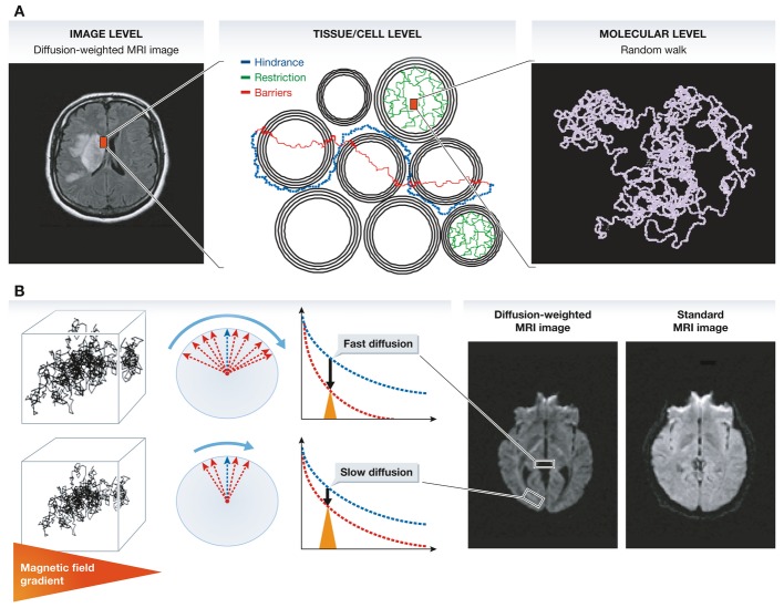
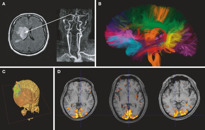
Diffusion MRI has been used worldwide to produce images of brain tissue structure and connectivity, in the normal and diseased brain. Diffusion MRI has revolutionized the management of acute brain ischemia (stroke), saving life of many patients and sparing them significant disabilities. In addition to stroke, diffusion MRI is now widely used for the detection of cancers and metastases (breast, prostate, liver). Another major field of application of diffusion MRI regards the wiring of the brain. Diffusion MRI is now used to map the circuitry of the human brain with incredible accuracy, opening up new lines of inquiry for human neuroscience and for the understanding of brain illnesses or mental disorders. Here, as a pioneer of the field, I provide a personal account on the historical development of these concepts over the last 30 years.


Figures

Figure 1
Figure 1
(A) Contrast and signal levels in the diffusion-weighted image (left) reflect water diffusion behavior (random walk) (right). Diffusion behavior is modulated by tissue structure at the cellular level (middle): For instance, diffusion can be restricted within cells, water may escape when cell membranes are permeable and might then experience a tortuous pathway in the extracellular space (hindrance). (B) In the presence of a magnetic field gradient (variation of the magnetic field along one spatial direction), magnetized water molecule hydrogen atoms are dephased. The amount of dephasing is directly related to the diffusion distance (a few micrometers) covered by water molecules during measurement (a few tens of milliseconds). Given the great many water molecules experiencing individual random walk displacements, the overall effect of this dephasing is an interference, which reduces MRI signal amplitudes. In areas with fast water diffusion (e.g. within ventricules), the signal is deeply reduced, while in areas of slow water diffusion (e.g. white matter bundles), the signal is only slightly reduced. This differential effect results in a contrast in the diffusion-weighted MRI images, which is not visible in standard MRI images.
Figure 2
Figure 2
(A) Acute stroke. The angiogram (right) shows an occlusion of the right middle cerebral artery. The diffusion-weighted image (left) clearly shows a bright signal corresponding to a drop in water diffusion resulting from cell swelling (cytotoxic edema) in the tissue undergoing acute ischemia. (B) Brain connectivity. Water diffusion in white matter fibers is anisotropic, faster in the direction of the fibers. By measuring water diffusion in many directions, the orientations of the whiter matter bundles can be retrieved at each brain location. Algorithms are then used to identify bundles, which are represented with arbitrary colors (courtesy CONNECT/NeuroSpin). (C) Cancer. Water diffusion in the glioblastoma has been imaged and measured after the patient underwent a chemotherapy treatment. Areas within the tumor where the treatment has been efficient appear in green (return of water diffusion to baseline values), while areas in red highlight parts of the lesion where diffusion still remains low (courtesy B. Ross, University of Chicago). (D) Functional brain imaging. Areas in color represent parts of the brain (visual cortex) where water diffusion has slightly decreased during activation (visual presentation of a flickering checkerboard for 10 s).

References

    1. Aso T, Urayama S, Poupon C, Sawamoto N, Fukuyama H, Le Bihan D. An intrinsic diffusion response function for analyzing diffusion functional MRI time series. Neuroimage. 2009;47:1487–1495. - PubMed
    1. Assaf Y, Alexander DC, Jones DK, Bizzi A, Behrens TE, Clark CA, Cohen Y, Dyrby TB, Huppi PS, Knoesche TR, et al. The CONNECT project: combining macro- and micro-structure. Neuroimage. 2013;80:273–829. - PubMed
    1. Basser PJ, Mattiello J, Le Bihan D. MR diffusion tensor spectroscopy and imaging. Biophys J. 1994;66:259–267. - PMC - PubMed
    1. Crick F. Do dentritic spines twitch? Tins. 1982;5:44–47.
    1. Douek P, Turner R, Pekar J, Patronas NJ, Le Bihan D. MR color mapping of myelin fiber orientation. J Comput Assist Tomogr. 1991;15:923–929. - PubMed

Publication types