

Deep anterior lamellar keratoplasty in case of Hurler-Scheie syndrome
- PMID: 24706723
- PMCID: PMC3987521
- DOI: 10.1136/bcr-2013-202730
Deep anterior lamellar keratoplasty in case of Hurler-Scheie syndrome
Abstract
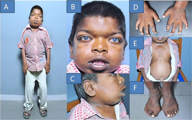
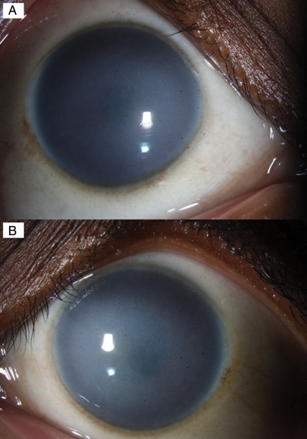
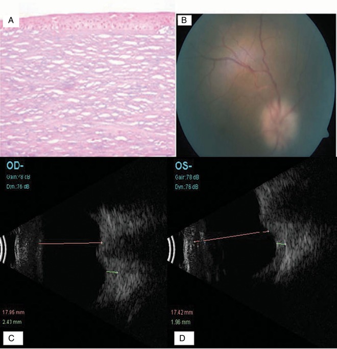
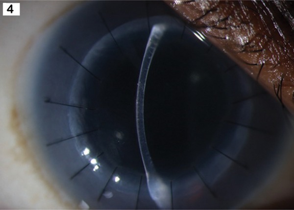
A 12-year-old boy with Hurler-Scheie syndrome (H/S syndrome) reported with reduced vision in both the eyes for past few years. Deep anterior lamellar keratoplasty (DALK) was performed for visual rehabilitation in his left eye. During surgery, the predescemet's plane was reached by meticulously dissecting the lamellar fibres using a manual technique. Histopathology of the dissected cornea showed the presence of numerous alcian blue positive deposits corroborating with the diagnosis of mucopolysaccharidosis (MPS). Postoperative course was uneventful. One year following surgery, the graft was clear and had a visual acuity of 20/50 with +1.00 170° - 0.75 refractive correction. Endothelial cell count, as measured by non-contact specular microscopy, was 2473.4 cells/mm(2). This case report highlights the application of DALK in a case of MPS-related corneal stromal opacification.
Figures
Similar articles
-
Multimodal imaging of Hurler syndrome-related keratopathy treated with deep anterior lamellar keratoplasty.BMC Ophthalmol. 2020 Oct 31;20(1):433. doi: 10.1186/s12886-020-01689-2. BMC Ophthalmol. 2020. PMID: 33129306 Free PMC article.
-
Deep Anterior Lamellar Keratoplasty in a Case of Hurler-Scheie Syndrome Undergoing Enzyme Replacement Therapy.Cornea. 2019 Mar;38(3):376-378. doi: 10.1097/ICO.0000000000001840. Cornea. 2019. PMID: 30575621
-
Deep anterior lamellar keratoplasty for Maroteaux-Lamy syndrome.Cornea. 2010 Dec;29(12):1459-61. doi: 10.1097/ICO.0b013e3181d927d6. Cornea. 2010. PMID: 20856106
-
Long-Term Outcomes of Big Bubble Deep Anterior Lamellar Keratoplasty in Mucopolysaccharidoses: A Retrospective Case Series and Review of the Literature.Cornea. 2022 Jul 1;41(7):809-814. doi: 10.1097/ICO.0000000000003041. Epub 2022 Apr 13. Cornea. 2022. PMID: 35439776 Review.
-
Surgical Corneal Anatomy in Deep Anterior Lamellar Keratoplasty: Suggestion of New Acronyms.Cornea. 2019 Apr;38(4):515-522. doi: 10.1097/ICO.0000000000001845. Cornea. 2019. PMID: 30681518 Review.
Cited by
-
Keratoplasty: are children missing out on the lamellar revolution-the 2023 Bowman Club, David L. Easty Lecture.BMJ Open Ophthalmol. 2024 Oct 24;9(1):e001804. doi: 10.1136/bmjophth-2024-001804. BMJ Open Ophthalmol. 2024. PMID: 39455068 Free PMC article.
-
Multimodal imaging of Hurler syndrome-related keratopathy treated with deep anterior lamellar keratoplasty.BMC Ophthalmol. 2020 Oct 31;20(1):433. doi: 10.1186/s12886-020-01689-2. BMC Ophthalmol. 2020. PMID: 33129306 Free PMC article.
-
Objectively measuring anterior segment alterations in the eyes of mucopolysaccharidoses: Its utility in early diagnosis of glaucoma.Indian J Ophthalmol. 2022 Dec;70(12):4180-4185. doi: 10.4103/ijo.IJO_1300_22. Indian J Ophthalmol. 2022. PMID: 36453310 Free PMC article.
References
-
- Ashar JN, Pahuja S, Ramappa M, et al. Deep anterior lamellar keratoplasty in children. Am J Ophthalmol 2013;155:570–4 - PubMed
-
- Ramappa M, Ashar J, Vaddavalli PK, et al. Endothelial keratoplasty in children: surgical challenges and early outcomes. Br J Ophthalmol 2012;96:1149–51 - PubMed
-
- Harding SA, Nischal KK, Upponi-Patil A, et al. Indications and outcomes of deep anterior lamellar keratoplasty in children. Ophthalmology 2010;117:2191–5 - PubMed
-
- Buzzonetti L, Petrocelli G, Valente P. Big-bubble deep anterior lamellar keratoplasty assisted by femtosecond laser in children. Cornea 2012;31:1083–6 - PubMed
-
- Alldredge OC KJ. Clinical types of corneal transplant rejection: their manifestations, frequency, preoperative correlates, and treatment. Arch Ophthalmol 1981;99:599–604 - PubMed
Publication types
MeSH terms
LinkOut - more resources
Full Text Sources
Other Literature Sources
Medical
Miscellaneous