Comparative Study
. 2014 Apr 15;111(15):5723-8.
doi: 10.1073/pnas.1323842111. Epub 2014 Mar 31.

Microtubule binding distinguishes dystrophin from utrophin

Affiliations
Comparative Study

Microtubule binding distinguishes dystrophin from utrophin

Joseph J Belanto et al. Proc Natl Acad Sci U S A. .

Abstract

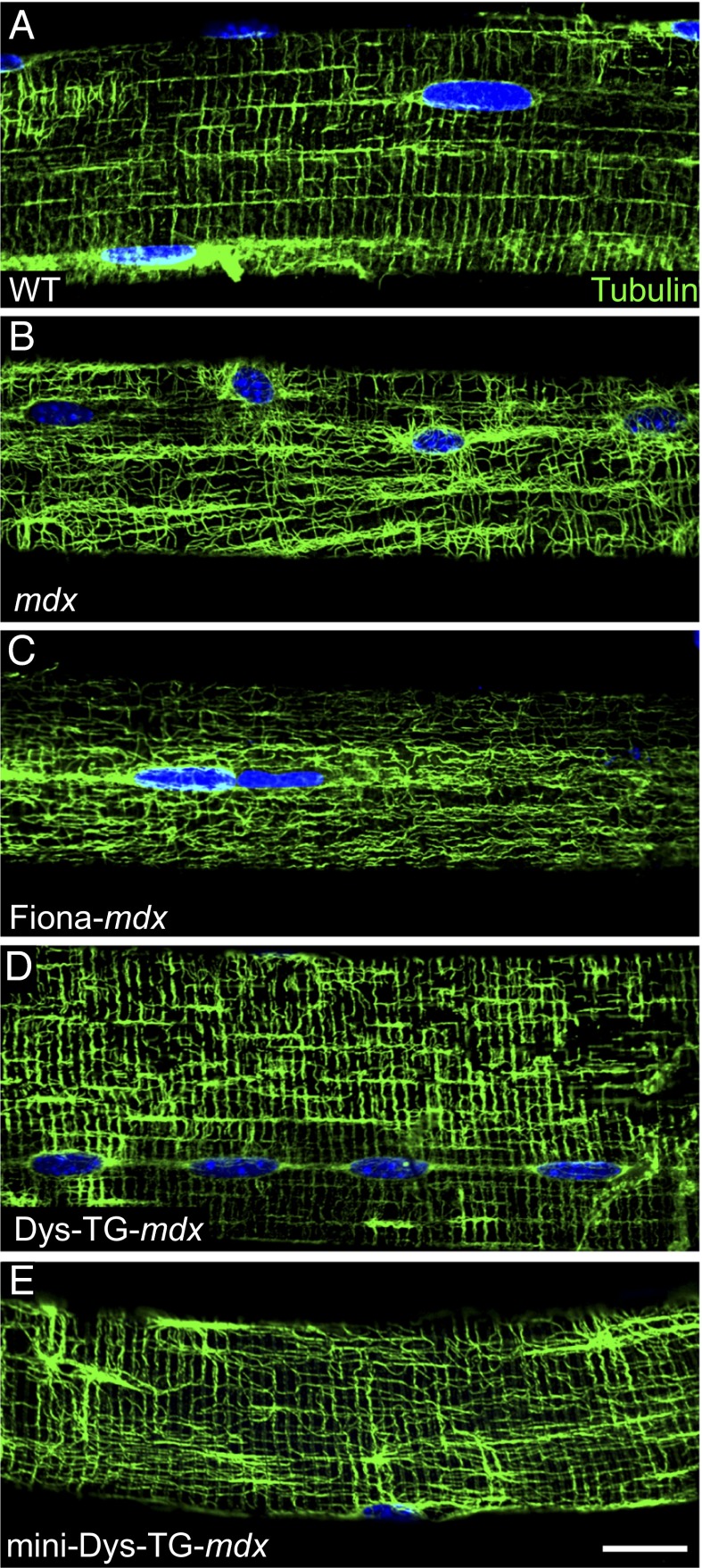
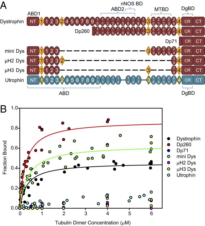
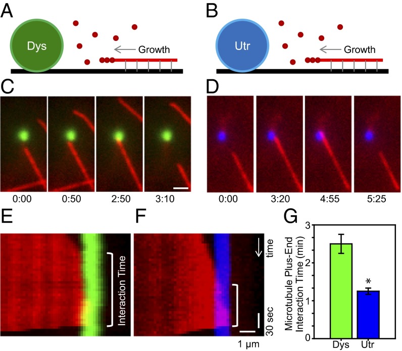
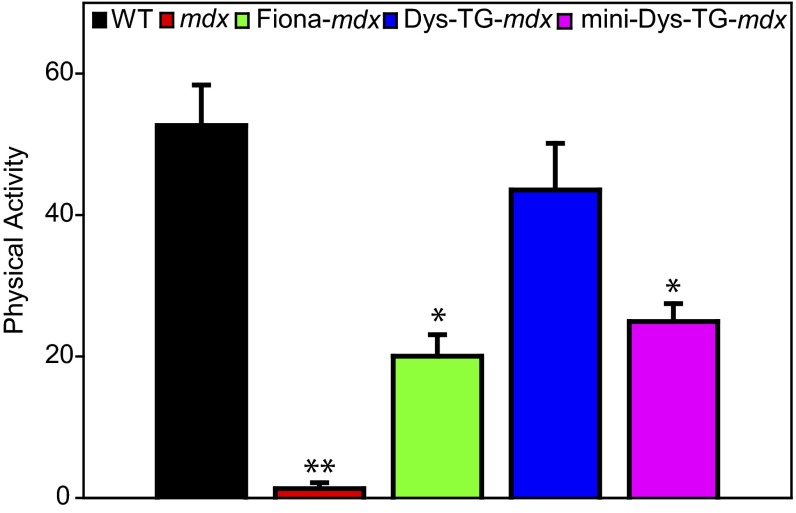
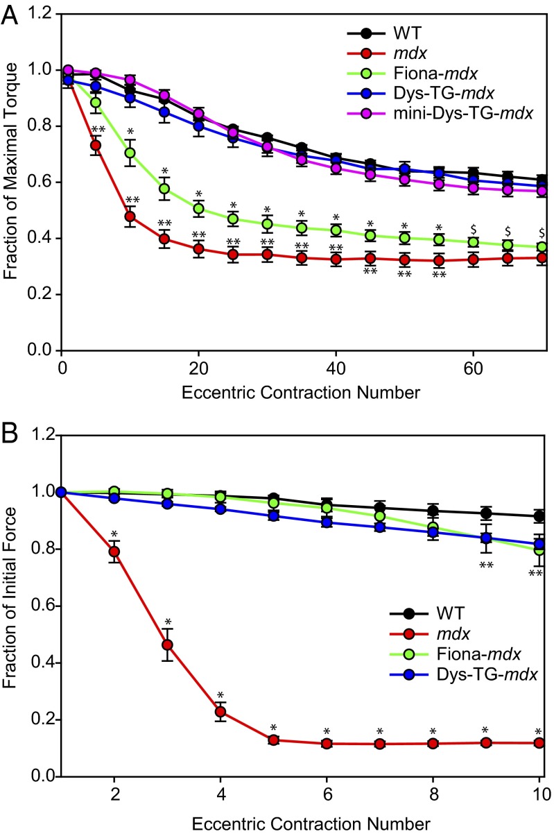
Dystrophin and utrophin are highly similar proteins that both link cortical actin filaments with a complex of sarcolemmal glycoproteins, yet localize to different subcellular domains within normal muscle cells. In mdx mice and Duchenne muscular dystrophy patients, dystrophin is lacking and utrophin is consequently up-regulated and redistributed to locations normally occupied by dystrophin. Transgenic overexpression of utrophin has been shown to significantly improve aspects of the disease phenotype in the mdx mouse; therefore, utrophin up-regulation is under intense investigation as a potential therapy for Duchenne muscular dystrophy. Here we biochemically compared the previously documented microtubule binding activity of dystrophin with utrophin and analyzed several transgenic mouse models to identify phenotypes of the mdx mouse that remain despite transgenic utrophin overexpression. Our in vitro analyses revealed that dystrophin binds microtubules with high affinity and pauses microtubule polymerization, whereas utrophin has no activity in either assay. We also found that transgenic utrophin overexpression does not correct subsarcolemmal microtubule lattice disorganization, loss of torque production after in vivo eccentric contractions, or physical inactivity after mild exercise. Finally, our data suggest that exercise-induced inactivity correlates with loss of sarcolemmal neuronal NOS localization in mdx muscle, whereas loss of in vivo torque production after eccentric contraction-induced injury is associated with microtubule lattice disorganization.


Conflict of interest statement

The authors declare no conflict of interest.

Figures

Fig. 1.
Fig. 1.
Microtubule organization in 6-mo-old mouse EDL muscle fibers. (A) When dystrophin is present (WT), microtubules are organized into a rectilinear lattice beneath the sarcolemma. (B) In the absence of dystrophin (mdx), the microtubule lattice becomes disordered. (C) Transgenic overexpression of utrophin in the absence of dystrophin (Fiona-mdx) does not rescue the disorganized microtubule lattice. (D) Transgenic overexpression of nearly full-length dystrophin (Dys-TG-mdx) or (E) mini-dystrophin (mini-Dys-TG-mdx) in the absence of endogenous dystrophin rescues the microtubule lattice. Images are representative of those obtained for n ≥ 10 fibers from each of n ≥ 3 mice per genotype. (Scale bar, 20 µm.)
Fig. 2.
Fig. 2.
Domain structure and microtubule binding properties of dystrophin and utrophin. (A) Schematic representation of dystrophin and utrophin protein constructs. Ovals represent spectrin-like repeats, diamonds represent hinge regions. Homologous repeats are aligned. Dystrophin repeats 15 and 19 do not have homologous repeats in utrophin. ABD, actin binding domain; nNOS BD, nNOS binding domain; MTBD, microtubule binding domain; DgBD, β-dystroglycan binding domain; NT, N terminus; CT, C terminus; CR, cysteine rich domain. (B) Microtubule binding curves of constructs assayed. Dystrophin binds microtubules with high affinity (KD = 0.33 µM), as do Dp260 (KD = 0.26 µM) and mini-dystrophin (KD = 0.38 µM), whereas all other dystrophin constructs and utrophin display no specific microtubule binding.
Fig. 3.
Fig. 3.
Effect of dystrophin and utrophin on microtubule localization. (A and B) Schematic representation of assay performed. Dystrophin (Dys, green) or utrophin (Utr, blue). (C) Time-lapse images of a direct dystrophin/microtubule interaction. (Scale bar, 2 µm.) (D) Time-lapse images of a direct utrophin/microtubule interaction. (E) Kymograph of the data presented in C. (F) Kymograph of the data presented in D. (G) The dystrophin/microtubule (n = 6) interaction time is significantly greater than the utrophin/microtubule (n = 8) interaction time. Data are presented as means ± SE. *P < 0.001.
Fig. 4.
Fig. 4.
Physical activity of mice after mild treadmill exercise. WT mice exhibited ∼50% of their initial cage activity after mild exercise, whereas postexercise activity in mdx mice dropped more than 95% of their initial activity. Transgenic overexpression of dystrophin in mdx mice (Dys-TG-mdx) restored postexercise activity levels similar to that in WT, whereas transgenic overexpression of utrophin (Fiona-mdx) or mini-dystrophin (mini-Dys-TG-mdx) yielded intermediate (∼25%) restoration of activity. *Statistically different from WT, Dys-TG-mdx, and mdx; **statistically different from all other lines.
Fig. 5.
Fig. 5.
In vivo and ex vivo contraction-induced injury loss by anterior crural muscles. (A) Compared with WT mice, mdx mice showed drastic loss of torque during a series of eccentric contractions. Dys-TG-mdx and mini-Dys-TG-mdx mice showed much less loss of torque, analogous to that of WT mice. Conversely, the Fiona-mdx mice showed intermediate loss of torque, comparable to the intermediate loss of physical activity seen in Fig. 4. *Statistically different from WT, Dys-TG-mdx, mini-Dys-TG-mdx, and mdx; **statistically different from WT, Dys-TG-mdx, Fiona-mdx, and mini-Dys-TG-mdx; $statistically different from WT, Dys-TG-mdx, and mini-Dys-TG-mdx but not mdx. (B) Significant loss of force by mdx muscles at contractions 2 through 10 compared with WT (P < 0.001). EDL muscles from Fiona-mdx and Dys-TG-mdx mice were protected from force loss and were not different from WT at contractions 2 through 8 (P > 0.05). At contractions 9 and 10, Fiona-mdx (P = 0.031 and P = 0.002 for contractions 9 and 10, respectively) and Dys-TG-mdx (P = 0.014 and P = 0.005 for contractions 9 and 10, respectively) began to show differences from WT but were not different from each other (P = 0.945 and P = 0.532 for contractions 9 and 10, respectively).

References

    1. Mendell JR, et al. Evidence-based path to newborn screening for Duchenne muscular dystrophy. Ann Neurol. 2012;71(3):304–313. - PubMed
    1. Hoffman EP, Brown RH, Jr, Kunkel LM. Dystrophin: The protein product of the Duchenne muscular dystrophy locus. Cell. 1987;51(6):919–928. - PubMed
    1. Koenig M, Monaco AP, Kunkel LM. The complete sequence of dystrophin predicts a rod-shaped cytoskeletal protein. Cell. 1988;53(2):219–228. - PubMed
    1. Koenig M, Kunkel LM. Detailed analysis of the repeat domain of dystrophin reveals four potential hinge segments that may confer flexibility. J Biol Chem. 1990;265(8):4560–4566. - PubMed
    1. Way M, Pope B, Cross RA, Kendrick-Jones J, Weeds AG. Expression of the N-terminal domain of dystrophin in E. coli and demonstration of binding to F-actin. FEBS Lett. 1992;301(3):243–245. - PubMed

Publication types