. 2014 Apr 15;111(15):5706-11.
doi: 10.1073/pnas.1402215111. Epub 2014 Mar 31.

Differential ubiquitination and degradation of huntingtin fragments modulated by ubiquitin-protein ligase E3A

Affiliations

Differential ubiquitination and degradation of huntingtin fragments modulated by ubiquitin-protein ligase E3A

Kavita P Bhat et al. Proc Natl Acad Sci U S A. .

Abstract

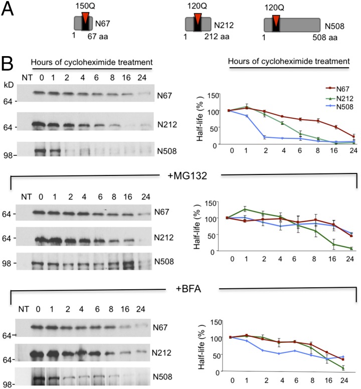
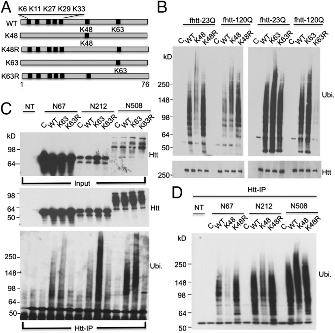
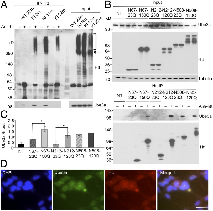
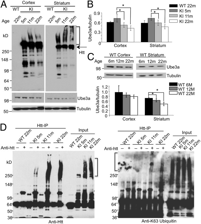
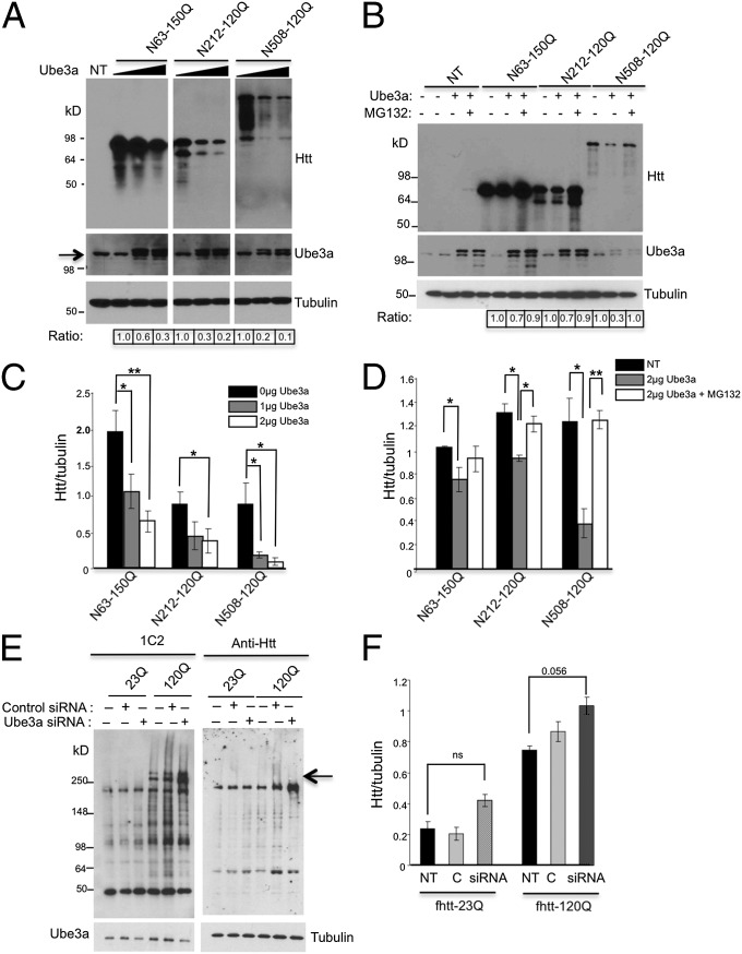
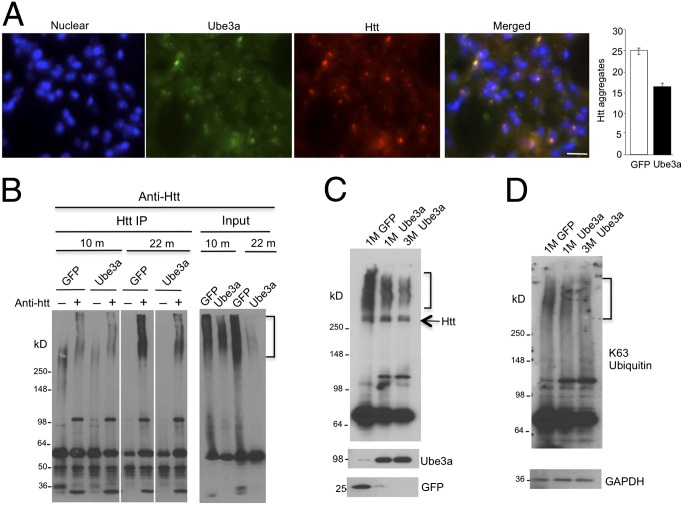
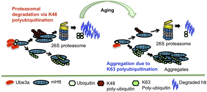
Ubiquitination of misfolded proteins, a common feature of many neurodegenerative diseases, is mediated by different lysine (K) residues in ubiquitin and alters the levels of toxic proteins. In Huntington disease, polyglutamine expansion causes N-terminal huntingtin (Htt) to misfold, inducing neurodegeneration. Here we report that shorter N-terminal Htt fragments are more stable than longer fragments and find differential ubiquitination via K63 of ubiquitin. Aging decreases proteasome-mediated Htt degradation, at the same time increasing K63-mediated ubiquitination and subsequent Htt aggregation in HD knock-in mice. The association of Htt with the K48-specific E3 ligase, Ube3a, is decreased in aged mouse brain. Overexpression of Ube3a in HD mouse brain reduces K63-mediated ubiquitination and Htt aggregation, enhancing its degradation via the K48 ubiquitin-proteasome system. Our findings suggest that aging-dependent Ube3a levels result in differential ubiquitination and degradation of Htt fragments, thereby contributing to the age-related neurotoxicity of mutant Htt.

Keywords: misfolding; proteolysis.


Conflict of interest statement

The authors declare no conflict of interest.

Figures

Fig. 1.
Fig. 1.
Shorter N-terminal Htt fragments have longer half-lives in aging HD KI mice. (A) Schematic representation of N-terminal Htt fragments used in this study. (B) Cycloheximide chase (100 μM) assay of HEK293 cells to determine the t1/2 of transfected mHtt fragments (N-67-150Q, 212-120Q, and N508-150Q). The transfected cells were untreated or treated with MG132 (10 mM) or bafilomycin A (100 nM). The relative protein levels were assessed by densitometric analysis of the protein bands (Right), and the value at 0 h was considered 100%. All Western blots are representative of results from at least three independent experiments, and data are presented as mean ± SEM. NT, nontransfected cells.
Fig. 2.
Fig. 2.
Polyubiquitination of full-length and N-terminal fragments of mHtt preferentially uses lysine 63 (K63) of ubiquitin. (A) Schematic representation of ubiquitin lysine mutants used for in vitro ubiquitination assay. (B) In vitro ubiquitination assay using ubiquitin lysine mutants to determine ubiquitination topology of mHtt in full-length Htt (i.e., fHtt-23Q and fHtt-120Q) expressed in stably transfected cell lines. Various lysine mutants of HA-ubiquitin were expressed in the fHtt-23Q and fHtt-120Q cell lines, and mHtt was immunoprecipitated and detected by anti-HA for ubiquitin (Ubi.; Upper) and anti-Htt (Lower). (C and D) In vitro ubiquitination assay of HEK293 cells to determine ubiquitination topology of N-terminal mHtt fragments (N67-150Q, N212-120Q, and N508-1200Q) by using ubiquitin lysine mutant K63R (C) or K48R (D). All Western blots are representative of results from at least three independent experiments. C, Htt transfected cells without ubiquitin contransfection; NT, nontransfected cells.
Fig. 3.
Fig. 3.
Ube3a interacts with mHtt fragments in vivo and in vitro. (A) Coimmunoprecipitation assay by using Htt-specific antibody to immunoprecipitate Htt (Upper) from the striatum of 5-, 11-, and 22-mo-old HD KI mice and 22-mo-old WT mice to determine Ube3a (Lower) binding to Htt in vivo. Bracket indicates aggregated mHtt, and arrow indicates full-length mHtt. (B) Coimmunoprecipitation assay in HEK293 cells using anti-Htt to immunoprecipitate Ube3a and N-terminal Htt fragments (N67-23Q/150Q, N212-23Q/120Q, and N508-23Q/120Q; two samples for each Htt fragment). Inputs are shown (Top) along with the precipitated Ube3a (Middle) and Htt (Bottom). (C) Densitometric analysis of the amount of immunoprecipitated Ube3a (Ube3a/input) in each lane, which is normalized to the total amount of Ube3a in input. All Western blots are representative of results from at least three independent experiments, and data are mean ± SEM (D) Fluorescent immunocytochemistry showing the colocalization of Ube3a with mHtt aggregate (arrow) in fHtt-120Q cell lines. NT, nontransfected cells. (Scale bar: 10 μm.)
Fig. 4.
Fig. 4.
Age-dependent decrease in Ube3a in HD KI mice. (A) Western blot analysis of mouse brain cortex and striatum from 5-, 11-, and 22 mo-old HD KI mice and 22-mo-old WT mouse. Arrow indicates full-length mHtt. (B) The relative levels of Ube3a in HD KI mouse brains at different ages. (C) Western blotting of the brain cortex and striatum in normal mice at 6, 12, and 22 mo of age. Densitometric analysis of the relative levels of Ube3a (the ratio of Ube3a to tubulin) is shown beneath the blots (*P < 0.05). (D) Coimmunoprecipitation assay using anti-Htt to immunoprecipitate Htt (Left) from HD KI mouse brain striatum at 5, 11, and 22 mo of age to determine in vivo Htt-specific K63 ubiquitination (Right). All Western blots are representative of results from at least three independent experiments, and data are mean ± SEM. Bracket indicates aggregated mHtt (Left) and K63 ubiquitinated mHtt (Right).
Fig. 5.
Fig. 5.
Overexpressing Ube3a reduces mHtt stability and t1/2, whereas knocking down Ube3a has the opposite effect. (A) Western blot analysis of the effect of increasing transfected Ube3a (0, 1, and 2 μg DNA) on the protein stability and ubiquitination levels of N-terminal mHtt fragments (N63-150Q, N212-120Q, and N508-150Q) in HEK293 cells. Because anti-Htt (mEM48) reacts more intensively with shorter mHtt fragments with longer polyQ domain, the individual panels containing N63-150Q, N212-120Q, or N508-150Q are different exposures of Western blots from the same experiment to clearly show Ube3a-mediated differences at their levels. (B) Western blot analysis of the effects of MG132 (10 μM) and Ube3a (2 μg) on the protein stability of N-terminal mHtt fragments (N63-150Q, N212-120Q, and N508-120Q) in HEK293 cells. In A and B, ratios of mHtt to tubulin are normalized by the control without Ube3a and are shown beneath the blot probed with anti-tubulin. (C and D) Densitometric analysis of the relative levels of mHtt (ratio of mHtt to tubulin) in A (C) and B (D). *P < 0.05 and **P < 0.01. (E) Knockdown assay to determine the effects of Ube3a siRNA on the protein stability of mHtt fragments in fHtt-23Q and fHtt-120Q cell lines. The blots were probed with 1C2 antibody that reacts with expanded polyQ repeats and mEM48 antibody for Htt. Arrow indicates a high molecular weight form of mHtt recognized by 1C2. (F) Densitometric analysis of the results in C quantifying the levels of full-length mHtt that are normalized to the corresponding tubulin levels. All Western blots are representative of results from at least three independent experiments, and data are presented as mean ± SEM (*P < 0.05). C, control siRNA transfected cells; NT, nontransfected cells; siRNA, Ube3a siRNA transfected cells.
Fig. 6.
Fig. 6.
Ube3a overexpression reduces mHtt aggregation and K63 ubiquitination in the striatum of HD KI mice. (A) Fluorescent immunohistochemistry staining of 24-mo-old HD KI mouse brain injected with AAV9-Myc-Ube3a. Three months after injection, mouse brain sections were stained with anti-Htt (mEM48, red) and anti-myc for the injected viral Ube3a (green). Relative amounts of Htt aggregates quantified as fluorescent signals per image are shown (Right; n = 15 images). (B) Coimmunoprecipitation assay using anti-Htt to immunoprecipitate Htt (Left) from the striatum from 10- and 22-mo-old HD KI mice that had been injected with AAV-GFP or AAV-Ube3a. Bracket indicates aggregated mHtt. (C and D) Western blot analysis of 10-mo-old HD KI mouse striatum injected with AAV-GFP or AAV-Ube3a for 1 or 3 mo to detect levels of mHtt aggregation (C) and K63 ubiquitination (D). Bracket indicates aggregated mHtt (B and C) and K63 specific ubiquitination (D). Arrow indicates full-length mHtt. (Scale bar: 20 μm.)
Fig. 7.
Fig. 7.
A proposed model for age-dependent mHtt toxicity. In young HD KI mice, Ube3a is abundant and ubiquitinates mHtt via lysine 48 of ubiquitin and targets it for proteasomal degradation. With aging, the level of Ube3a decreases, reducing K48-mediated mHtt ubiquitination as well as its targeting to the proteasome for degradation and promoting mHtt ubiquitination via K63. As a result, more K63 polyubiquitinated mHtt fragments accumulate and form aggregates.

Similar articles

Cited by

References

    1. Gusella JF, MacDonald ME, Ambrose CM, Duyao MP. Molecular genetics of Huntington’s disease. Arch Neurol. 1993;50(11):1157–1163. - PubMed
    1. Snell RG, et al. Relationship between trinucleotide repeat expansion and phenotypic variation in Huntington’s disease. Nat Genet. 1993;4(4):393–397. - PubMed
    1. Kuemmerle S, et al. Huntington aggregates may not predict neuronal death in Huntington’s disease. Ann Neurol. 1999;46(6):842–849. - PubMed
    1. Vonsattel JP, DiFiglia M. Huntington disease. J Neuropathol Exp Neurol. 1998;57(5):369–384. - PubMed
    1. DiFiglia M, et al. Aggregation of huntingtin in neuronal intranuclear inclusions and dystrophic neurites in brain. Science. 1997;277(5334):1990–1993. - PubMed

Publication types

LinkOut - more resources