. 2014 Apr 15;111(15):E1491-500.
doi: 10.1073/pnas.1400568111. Epub 2014 Mar 31.

Acentriolar mitosis activates a p53-dependent apoptosis pathway in the mouse embryo

Affiliations

Acentriolar mitosis activates a p53-dependent apoptosis pathway in the mouse embryo

Hisham Bazzi et al. Proc Natl Acad Sci U S A. .

Abstract

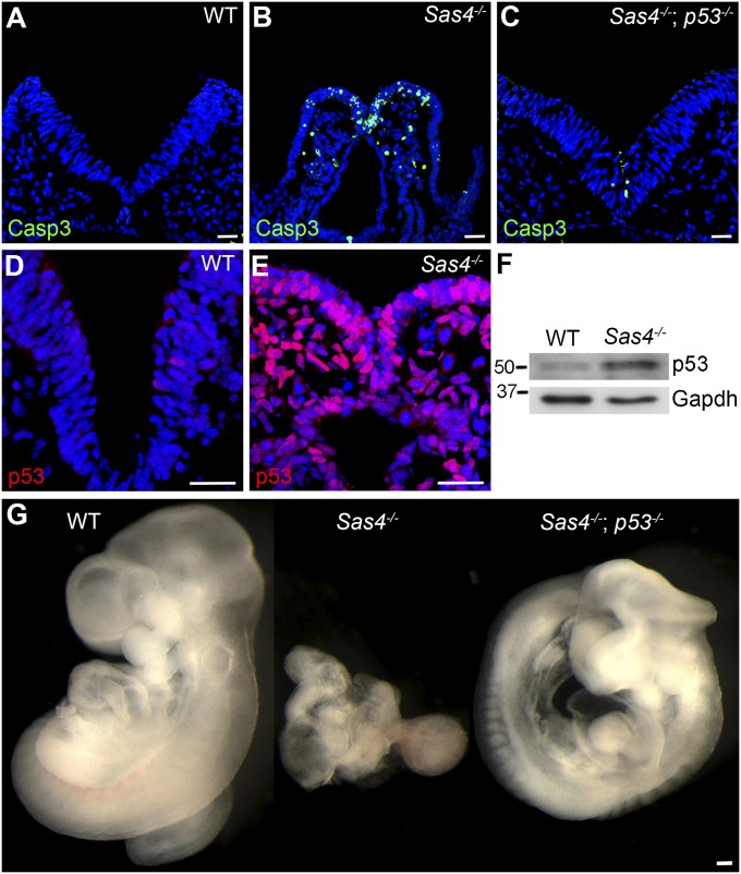
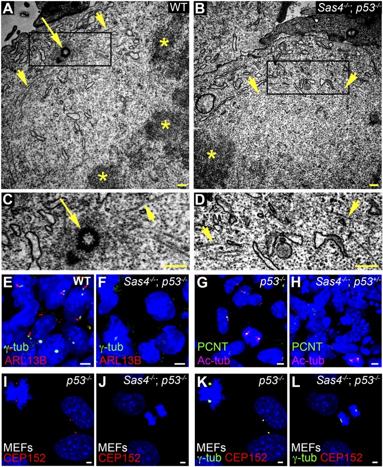
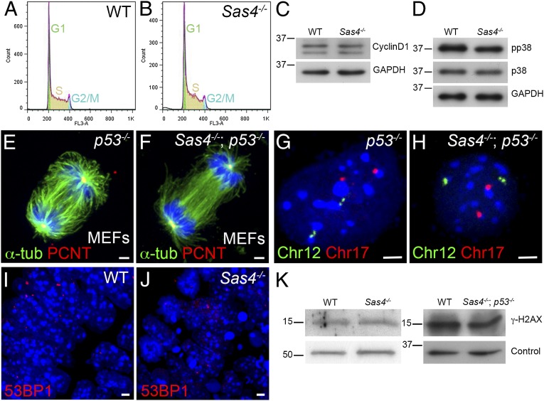
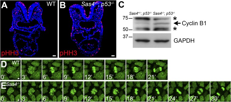
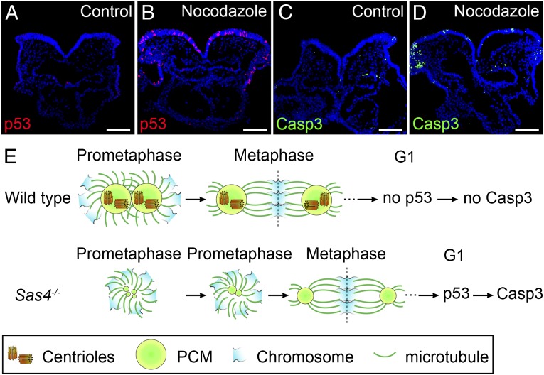
Centrosomes are the microtubule-organizing centers of animal cells that organize interphase microtubules and mitotic spindles. Centrioles are the microtubule-based structures that organize centrosomes, and a defined set of proteins, including spindle assembly defective-4 (SAS4) (CPAP/CENPJ), is required for centriole biogenesis. The biological functions of centrioles and centrosomes vary among animals, and the functions of mammalian centrosomes have not been genetically defined. Here we use a null mutation in mouse Sas4 to define the cellular and developmental functions of mammalian centrioles in vivo. Sas4-null embryos lack centrosomes but survive until midgestation. As expected, Sas4(-/-) mutants lack primary cilia and therefore cannot respond to Hedgehog signals, but other developmental signaling pathways are normal in the mutants. Unlike mutants that lack cilia, Sas4(-/-) embryos show widespread apoptosis associated with global elevated expression of p53. Cell death is rescued in Sas4(-/-) p53(-/-) double-mutant embryos, demonstrating that mammalian centrioles prevent activation of a p53-dependent apoptotic pathway. Expression of p53 is not activated by abnormalities in bipolar spindle organization, chromosome segregation, cell-cycle profile, or DNA damage response, which are normal in Sas4(-/-) mutants. Instead, live imaging shows that the duration of prometaphase is prolonged in the mutants while two acentriolar spindle poles are assembled. Independent experiments show that prolonging spindle assembly is sufficient to trigger p53-dependent apoptosis. We conclude that a short delay in the prometaphase caused by the absence of centrioles activates a previously undescribed p53-dependent cell death pathway in the rapidly dividing cells of the mouse embryo.


Conflict of interest statement

The authors declare no conflict of interest.

Figures

Fig. 1.
Fig. 1.
Sas4−/− mouse embryos arrest at mid-gestation. (A) Dorsal view of wild-type and Sas4−/− embryos at E8.5. (B) Wild-type and Sas4−/− embryos at E9.5. (C and D) Cross-sections of wild-type and Sox2-Cre; Sas4fl/- embryos at E8.5 stained for E-cadherin (red). Arrows indicate the neural plate (ectoderm), arrowheads indicate the mesenchyme (mesoderm), and asterisks indicate the gut (endoderm). (Scale bars: 300 µm.)
Fig. 2.
Fig. 2.
Cell death and early lethality in Sas4−/− embryos are rescued by removal of p53. (AC) Cleaved Casp3 (green) expression in cross-sections of wild-type embryos (0.6 ± 0.4%, n = 3,073 from two embryos) (A), Sas4−/− embryos (23 ± 4%, n = 3,150 from three embryos, P = 0.0048) (B), and Sas4−/− p53−/− embryos (2.7 ± 0.5%, n = 3,454 from two embryos, P = 0.0063) (C) at E8.5. (D and E) p53 (red) is rare in wild-type embryos (0.4 ± 0.1%, n = 3,534 from two embryos) but is strongly expressed in Sas4−/− mutant embryos (91 ± 1%, n = 2,629 from three embryos, P < 0.0001). (F) Western blot analysis of p53 in E8.5 embryo extracts. (G) Wild-type, Sas4−/−, and Sas4−/− p53−/− embryos at E9.5. P values are relative to wild type in each case. (Scale bars: 30 µm in AE, 300 µm in G.)
Fig. 3.
Fig. 3.
Sas4−/− embryos lack centrioles, centrosomes, and cilia. (AD) TEM of E8.5 neural folds. Microtubules (arrowheads) converge at metaphase spindle poles, where there are centrioles (arrow) in wild-type but not Sas4−/− p53−/− embryos. Asterisks mark the condensed chromosomes in A and B. C and D are magnifications of the boxed regions at the spindle poles in A and B, respectively. (EH) Expression of the centrosomal and cilia markers in the mesenchymal cells in E8.5 wild-type and Sas4−/− embryos stained for γ-tubulin (γ-tub) or PCNT (green) and ARL13B (red) or Ac-tub (magenta). Cilia are absent, and focal staining of γ-tubulin and PCNT is detected only during mitosis in Sas4−/− mutants. (IL) Expression of the PCM proteins CEP152 (red) and γ-tubulin (green) in p53−/− MEFs [45 ± 5 arbitrary units (A.U.)] and Sas4−/− p53−/− MEFs (25 ± 9 A.U., P < 0.0001, n = 14 poles each). CEP152 is not detected in either interphase or mitotic cells in Sas4−/− mutants. (Scale bars: 0.3 µm in AD, 3 µm in EL.)
Fig. 4.
Fig. 4.
Hh, but not Wnt, signaling is disrupted in Sas4−/− mutants. (AD) Hh-dependent ventral cell types in the neural tube of E9.5 wild-type and Sas4−/− p53−/− embryos. In Sas4−/− p53−/− embryos floor plate cells (FOXA2, red) and V3 interneurons (NKX2.2, green) are absent (compare A and B), and PAX6+ interneurons (green) occupy the ventral neural tube (compare C and D). (E and F) The activity of the canonical Wnt reporter TOPGAL (blue) is normal in E8.5 Sas4−/− mutants. (GL) The PCP noncanonical Wnt signaling protein VANGL2 (green) localizes to the anterior/posterior (horizontal) faces of the cells in the embryonic node of both wild-type (GI) and Sas4−/− p53−/− embryos (JL) at E7.75. Rhodamine-conjugated phalloidin (red) marks F-actin at cell borders. (Scale bars: 300 µm in AF, 3 µm in GL.)
Fig. 5.
Fig. 5.
Cell-cycle profile, chromosome segregation, and the DNA damage response are normal in Sas4−/− mutants. (A and B) Cell-cycle profiles of cells isolated from E8.5 wild-type (A) and Sas4−/− (B) embryos. (C) Western blots show similar levels of cyclin D1 in wild-type and mutant E8.5 embryo extracts. (D) Phospho-p38 (pp38, T180/Y182) is not elevated in E8.5 Sas4−/− embryo extracts. (E and F) Mitotic spindles from p53−/− (control) and Sas4−/− p53−/− MEFs stained with α-tubulin (green) and PCNT (red). (G and H) Sample FISH signals from dissociated cells of p53−/− control and Sas4−/− p53−/− embryos at E8.5 with two copies of chromosome 12 (green) and two copies of chromosome 17 (red). (I and J) Wild-type (I) and Sas4−/− (J) embryo sections at E8.5 (neural plate) stained with the DNA damage marker 53BP1. (K) Western blots of embryo extracts show that γ-H2AX is not elevated in Sas4−/− or Sas4−/− p53−/− cells at E8.5 relative to the respective wild types. For loading controls, α-tubulin was used for Sas4−/− and GAPDH for Sas4−/− p53−/−. (Scale bars: 3 µm.)
Fig. 6.
Fig. 6.
Prolonged mitosis in Sas4−/− embryos. (A and B) Cross-sections of E8.5 embryos show an increase in the number of mitotic cells (pHH3+; red) in Sas4−/− p53−/− embryos. (C) Western blots show an increase in cyclin B1 (arrow) in E8.5 Sas4−/− p53−/− embryos. Asterisks mark nonspecific bands. (D and E) Snapshots from time-lapse imaging of dividing cells in cultured E7.5 wild-type (D) and Sas4−/− (E) H2B-GFP+ embryos. (Scale bars: 30 µm in A and B, 3 µm in D and E.)
Fig. 7.
Fig. 7.
Prolonged prometaphase activates p53 and apoptosis in the wild-type mouse embryo. (AD) Cross-sections of cultured wild-type E8.5 embryos treated with vehicle (DMSO) (A and C) or 1 µM nocodazole for 1 h (B and D) and harvested 4 h after nocodazole removal. Approximately 11% of the cells in nocodazole-treated embryos are p53+ (red) (B). The number of Casp3+ (green) cells also was elevated in nocodazole-treated embryos (D), indicating increased apoptotic cell death. (E) Model for the activation of p53 and apoptosis by acentriolar mitosis. (Upper) Mitotic cells in wild-type embryos use centrioles to recruit more PCM and form mature centrosomes that organize microtubules and establish the bipolar mitotic spindle during prometaphase. The chromosomes are aligned quickly and efficiently at the metaphase plate of a well-structured bipolar spindle. Mitosis proceeds normally, and p53 and Casp3 are not up-regulated in the daughter cells. (Lower) Mitotic cells in Sas4−/− embryos lack centrioles and rely primarily on a chromatin-based pathway to organize the mitotic spindle. The establishment of bipolar spindle poles is slower in the absence of centrioles, but acentriolar cells eventually assemble a bipolar spindle and align the chromosomes at the metaphase plate, followed by proper chromosome segregation. However, the daughter cells of the acentriolar division retain the memory of the prolonged prometaphase and up-regulate p53, which increases the probability of cell death (marked by Casp3 expression). (Scale bars: 100 µm.)

References

    1. Boveri T. Concerning the origin of malignant tumours by Theodor Boveri. Translated and annotated by Henry Harris. J Cell Sci. 2008;121(Suppl 1):1–84. - PubMed
    1. Bettencourt-Dias M, Glover DM. Centrosome biogenesis and function: Centrosomics brings new understanding. Nat Rev Mol Cell Biol. 2007;8(6):451–463. - PubMed
    1. Kirkham M, Müller-Reichert T, Oegema K, Grill S, Hyman AA. SAS-4 is a C. elegans centriolar protein that controls centrosome size. Cell. 2003;112(4):575–587. - PubMed
    1. Leidel S, Gönczy P. SAS-4 is essential for centrosome duplication in C elegans and is recruited to daughter centrioles once per cell cycle. Dev Cell. 2003;4(3):431–439. - PubMed
    1. Basto R, et al. Flies without centrioles. Cell. 2006;125(7):1375–1386. - PubMed

Publication types

MeSH terms

Substances