. 2014 Apr 8;111(14):5242-7.
doi: 10.1073/pnas.1320953111. Epub 2014 Mar 24.

Nkx6.1 regulates islet β-cell proliferation via Nr4a1 and Nr4a3 nuclear receptors

Affiliations

Nkx6.1 regulates islet β-cell proliferation via Nr4a1 and Nr4a3 nuclear receptors

Jeffery S Tessem et al. Proc Natl Acad Sci U S A. .

Abstract

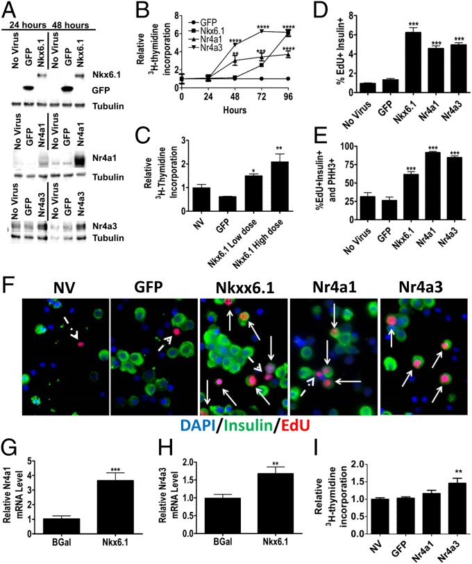
Loss of functional β-cell mass is a hallmark of type 1 and type 2 diabetes, and methods for restoring these cells are needed. We have previously reported that overexpression of the homeodomain transcription factor NK6 homeobox 1 (Nkx6.1) in rat pancreatic islets induces β-cell proliferation and enhances glucose-stimulated insulin secretion, but the pathway by which Nkx6.1 activates β-cell expansion has not been defined. Here, we demonstrate that Nkx6.1 induces expression of the nuclear receptor subfamily 4, group A, members 1 and 3 (Nr4a1 and Nr4a3) orphan nuclear receptors, and that these factors are both necessary and sufficient for Nkx6.1-mediated β-cell proliferation. Consistent with this finding, global knockout of Nr4a1 results in a decrease in β-cell area in neonatal and young mice. Overexpression of Nkx6.1 and the Nr4a receptors results in increased expression of key cell cycle inducers E2F transcription factor 1 and cyclin E1. Furthermore, Nkx6.1 and Nr4a receptors induce components of the anaphase-promoting complex, including ubiquitin-conjugating enzyme E2C, resulting in degradation of the cell cycle inhibitor p21. These studies identify a unique bipartite pathway for activation of β-cell proliferation, suggesting several unique targets for expansion of functional β-cell mass.


Conflict of interest statement

The authors declare no conflict of interest.

Figures

Fig. 1.
Fig. 1.
Overexpression of Nkx6.1 induces expression of Nr4a1 and Nr4a3. Rat islets were treated with AdCMV-GFP or AdCMV-Nkx6.1. (A and B) Nr4a1 (A) and Nr4a3 (B) mRNA levels. (C) Immunoblot analysis of whole-islet lysates 48 h after viral transduction. Data in A--C represent the means ± SEM of three independent experiments. (D and E) ChIP analysis of endogenous Nkx6.1 binding to the Nr4a1 (D) and Nr4a3 (E) promoter regions relative to a nonbound amplicon in INS-1–derived 832/13 cells. Data represent means ± SEM of six independent experiments. *P ≤ 0.05; **P ≤ 0.01; ***P ≤ 0.001. P values represent the comparison between Nkx6.1- and GFP-treated islets for RT-PCR and immunoblots and the comparison between anti-Nkx6.1 and IgG-precipitated samples in ChIP analyses.
Fig. 2.
Fig. 2.
Overexpression of Nr4a1 or Nr4a3 is sufficient to induce β-cell proliferation. (A) Immunoblot analysis demonstrating adenovirus-mediated overexpression of Nkx6.1, Nr4a1, or Nr4a3. (B) Incorporation of [3H-methyl]thymidine in rat islets. (C) Islets were treated with lentiviruses expressing GFP or Nkx6.1 or left untreated, and [3H-methyl]thymidine incorporation was measured after 136 h. (D) Percentage of EdU+insulin+ islet cells cultured with EdU for 96 h. (E) Percentage of EdU+insulin+ islet cells that are also PHH3+. (F) Representative images (100× magnification) of islets labeled with EdU. Solid arrows indicate insulin+EdU+ nuclei, and broken arrows indicate insulinEdU+ nuclei. DAPI is blue, insulin is green, and EdU is pink. (G and H) Treatment of human islets with AdCMV-Nkx6.1 increases Nr4a1 (G) and Nr4a3 (H) mRNA levels. (I) [3H-methyl]thymidine incorporation into human islets measured 96 h after indicated treatments. Data represent means ± SEM of a minimum of three independent experiments; human islet data represent eight independent experiments. *P ≤ 0.05; **P ≤ 0.01; ***P ≤ 0.001; ****P ≤ 0.0001. All comparisons with AdCMV-GFP–treated islets.
Fig. 3.
Fig. 3.
Nr4a1 and Nr4a3 are necessary for Nkx6.1-mediated proliferation. Rat islets were treated with AdCMV-Nkx6.1 and either Ad-siNr4a1, Ad-siNr4a3, or Ad-siControl (siCTRL) for 96 h. (A) Immunoblot assay of whole-islet lysates. (B) [3H-methyl]thymidine incorporation. Rat islets were treated with adenoviruses expressing GFP, Nkx6.1, or the dominant-negative Nr4a construct Nr4a1-M1 (M1), followed by immunoblot assay of whole-islet lysates (C). (D) [3H-methyl]thymidine incorporation. Data represent means ± SEM of a minimum of three independent experiments. **P ≤ 0.01; ***P ≤ 0.001 compared with AdCMV-Nkx6.1 plus Ad-siControl (B) or Nkx6.1-treated islets (D).
Fig. 4.
Fig. 4.
Mice deficient for Nr4a1 have decreased β-cell area and replication. (A) β-Cell area in pancreas sections from 11-d-old wild-type (WT) and Nr4a1 knockout (KO) mice, expressed as β-cell area relative to total pancreas area. (B) β-Cell area in pancreas sections from 2-mo-old WT or Nr4a1 KO mice. (C) Representative images at 20× magnification of pancreata from WT or Nr4a1 KO mice at 11 d of age. Insulin is red, amylase is green, and DAPI is blue. The percentage of Ki-67+insulin+ (D) and PHH3+insulin+ (E) cells was calculated at 11 d of age. *P < 0.05; **P < 0.01; ***P < 0.005.
Fig. 5.
Fig. 5.
Nkx6.1 up-regulates APC components, leading to degradation of p21. (A) Overexpression of Nr4a1 or Nr4a3 up-regulates expression of Ube2c mRNA at 48 h. (B) Nkx6.1-mediated up-regulation of Ube2c at 96 h is dependent on the expression of Nr4a1 and Nr4a3. (C) Overexpression of Nkx6.1 induces expression of p21 mRNA. (D) Immunoblot analyses demonstrate that p21 protein levels decrease between 48 and 72 h after Nkx6.1 overexpression. Data represent means ± SEM of three independent experiments. For A and D, P values represent the comparison with GFP-treated islets. For B, P values represent comparison with AdCMV-Nkx6.1 plus Ad-siControl islets. *P ≤ 0.05; **P ≤ 0.01; ***P ≤ 0.001; ****P ≤ 0.0001.
Fig. 6.
Fig. 6.
Loss of p21 stimulates β-cell proliferation (AE) and summary model (F). (A) p21 protein levels are maintained at baseline levels 48 h after Nr4a1 or Nr4a3 overexpression. (B) Overexpression of Ube2c for 72 h in INS-1 832/13 cells decreases p21 protein levels. (C) Treatment of islets with Ad-siUbe2c decreases [3H-methyl]thymidine incorporation in response to Nkx6.1 overexpression. (D) Percentage of insulin+Edu+ and insulinEdu+ islet cells in the presence and absence of p21 knockdown. (E) Percentage of EdU+insulin+ cells that are also PHH3+. Data represent means ± SEM of a minimum of three independent experiments. For A, B, and E, P values represent comparison with GFP-treated islets. For C, the P value is for the comparison with AdCMV-Nkx6.1 plus Ad-siControl islets. *P ≤ 0.05; **P ≤ 0.01; ***P ≤ 0.001; ****P ≤ 0.0001. (F) Schematic summary of bipartite mechanism of Nkx6.1-activated β-cell proliferation.

References

    1. Weir GC, Bonner-Weir S. Five stages of evolving beta-cell dysfunction during progression to diabetes. Diabetes. 2004;53(Suppl 3):S16–S21. - PubMed
    1. Muoio DM, Newgard CB. Mechanisms of disease: Molecular and metabolic mechanisms of insulin resistance and beta-cell failure in type 2 diabetes. Nat Rev Mol Cell Biol. 2008;9(3):193–205. - PubMed
    1. Butler AE, et al. Adaptive changes in pancreatic beta cell fractional area and beta cell turnover in human pregnancy. Diabetologia. 2010;53(10):2167–2176. - PMC - PubMed
    1. Tarabra E, Pelengaris S, Khan M. A simple matter of life and death-the trials of postnatal Beta-cell mass regulation. Int J Endocrinol. 2012;2012:516718. - PMC - PubMed
    1. Sander M, et al. Homeobox gene Nkx6.1 lies downstream of Nkx2.2 in the major pathway of beta-cell formation in the pancreas. Development. 2000;127(24):5533–5540. - PubMed

Publication types

Associated data