. 2014 Apr 8;111(14):5177-82.
doi: 10.1073/pnas.1403770111. Epub 2014 Mar 25.

Integrated strategy reveals the protein interface between cancer targets Bcl-2 and NAF-1

Affiliations

Integrated strategy reveals the protein interface between cancer targets Bcl-2 and NAF-1

Sagi Tamir et al. Proc Natl Acad Sci U S A. .

Abstract

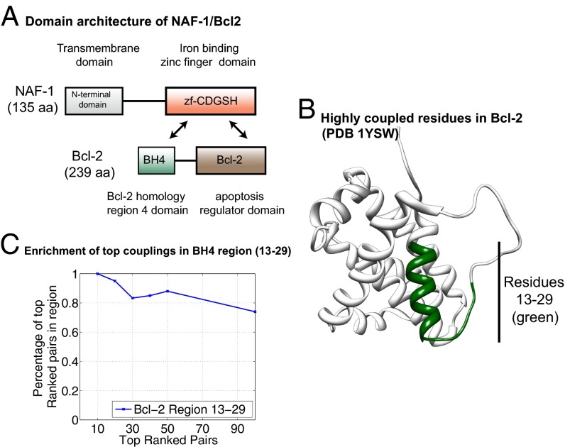
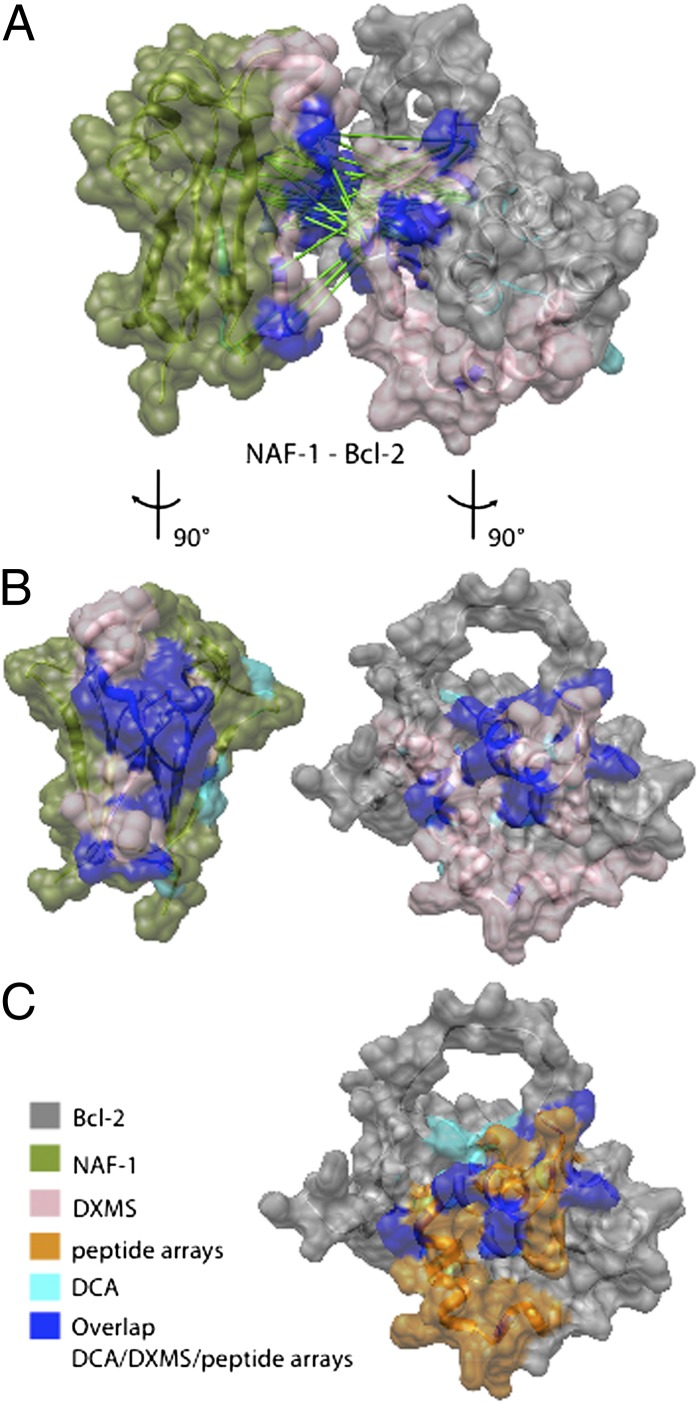
Life requires orchestrated control of cell proliferation, cell maintenance, and cell death. Involved in these decisions are protein complexes that assimilate a variety of inputs that report on the status of the cell and lead to an output response. Among the proteins involved in this response are nutrient-deprivation autophagy factor-1 (NAF-1)- and Bcl-2. NAF-1 is a homodimeric member of the novel Fe-S protein NEET family, which binds two 2Fe-2S clusters. NAF-1 is an important partner for Bcl-2 at the endoplasmic reticulum to functionally antagonize Beclin 1-dependent autophagy [Chang NC, Nguyen M, Germain M, Shore GC (2010) EMBO J 29(3):606-618]. We used an integrated approach involving peptide array, deuterium exchange mass spectrometry (DXMS), and functional studies aided by the power of sufficient constraints from direct coupling analysis (DCA) to determine the dominant docked conformation of the NAF-1-Bcl-2 complex. NAF-1 binds to both the pro- and antiapoptotic regions (BH3 and BH4) of Bcl-2, as demonstrated by a nested protein fragment analysis in a peptide array and DXMS analysis. A combination of the solution studies together with a new application of DCA to the eukaryotic proteins NAF-1 and Bcl-2 provided sufficient constraints at amino acid resolution to predict the interaction surfaces and orientation of the protein-protein interactions involved in the docked structure. The specific integrated approach described in this paper provides the first structural information, to our knowledge, for future targeting of the NAF-1-Bcl-2 complex in the regulation of apoptosis/autophagy in cancer biology.

Keywords: CDGSH; Cisd1; Cisd2; Miner1; mitoNEET.


Conflict of interest statement

The authors declare no conflict of interest.

Figures

Fig. 1.
Fig. 1.
Mapping the binding sites of the peptides interacting with NAF-1 on the Bcl-2 protein. (A) An array consisting of partly overlapping peptides derived from Bcl-2 was screened for binding to NAF-1. Each dark spot represents binding of NAF-1 to a specific peptide (Table 1). (B) The binding sites of the peptides discovered in the peptide array screening (described in A) are colored on the 3D structure of Bcl-2. The interaction surface involves regions previously shown to be involved in interactions with both pro- and antiapoptotic proteins.
Fig. 2.
Fig. 2.
Bcl-2 16–30 peptide destabilizes the 2Fe-2S cluster of NAF-1. UV-vis spectroscopy was used to observe the 2Fe-2S cluster stability of NAF-1 in the absence and presence of stoichiometric amounts of the Bcl-2 16–30 peptide. The Bcl-2 peptide interaction accelerates the cluster loss by a factor of two. All UV-vis spectra were measured from 250 to 800 nm on a Cary 50 spectrophotometer (Varian). Assay conditions were 100 µM NAF-1 with or without 100 µM Bcl-2 16–30 peptide in 50 mM bis-Tris buffer (pH 6.0) and 100 mM NaCl.
Fig. 3.
Fig. 3.
NAF-1’s 2Fe-2S cluster transfer is enhanced by the Bcl-2 16–30 peptide. NAF-1 was incubated with apo-Fd for 20 min at room temperature under different conditions, and the products were run on a native gel. Holo-Fd was run as a reference (line 4). Prereduction of the acceptor Cys ligand residues of apo-Fd with 5 mM DTT ensures transfer from NAF-1 to apo-Fd. Cluster transfer was enhanced by the addition of Bcl-2 16–30 peptide (line 3). (Upper) The gel is not stained (the red is from the 2Fe-2S in the cluster). (Lower) Coomassie blue staining of the gel verifying similar protein levels in all lanes. NAF-1 and Fd are indicated.
Fig. 4.
Fig. 4.
Bcl-2 16–30 peptide remains bound to NAF-1 following 2Fe-2S cluster transfer. Lane 1, apo-Fd; lane 2, holo-Fd; lane 3, NAF-1; lane 4, fluorescein-labeled Bcl-2 16–30 peptide; lane 5, mixture of NAF-1, holo-Fd, and peptide; lane 6, mixture of NAF-1, apo-Fd, and peptide under conditions to promote cluster transfer (as observed by the presence of the red band at the Fd position). These results show that the Bcl-2 peptide binds to NAF-1 and remains bound following 2Fe-2S cluster transfer to apo-Fd. (Upper) The gel is not stained (the red is from the 2Fe-2S in the cluster, and the yellow is from the fluorescein-labeled peptide). (Lower) Stained with Coomassie.
Fig. 5.
Fig. 5.
Direct coupling analysis for protein–protein recognition in the NAF-1–Bcl-2 system. (A) NAF-1 has a domain architecture including an N-terminal (PF10660) transmembrane domain and an iron binding zinc finger domain, zf-CDGSH (PF09360). Bcl-2 has a two-domain architecture as well as other unstructured regions. The domain BH4 (Bcl-2 homology region 4; PF02180) is found in several proteins analogous to Bcl-2 as well as its apoptosis regulator domain Bcl-2 (PF00452). Interaction between the zinc finger domain (zf-CDGSH) and the Bcl-2 domains (BH4 and Bcl-2) was evaluated by pairing and analyzing the genomic sequences of these domain families. (B) Bcl-2 structure (PDB ID code 1YSW). The green section shows the region with the DCA highest couplings for the domain pair zf-CDGSH–BH4. (C) Residues in region 13–29 of Bcl-2 constitute the highest percentage of residues in the top-ranked pairs. This suggests that this region has a higher probability of forming interfacial contacts between the iron binding zinc finger domain of NAF-1 and the BH4 domain of Bcl-2.
Fig. 6.
Fig. 6.
Effects of NAF-1–Bcl-2 protein complex formation on time-dependent solvent deuterium incorporation into Bcl-2 peptide probes. (A) Peptides 12–27, 48–85, and 71–102 in Bcl-2 show increased protection from deuterium incorporation (blue) upon complex formation. Residues 49–88 are missing from the structures available. (B) The peptides that undergo protection from solvent exchange (blue) are mapped onto the structure of Bcl-2 and are in good agreement with those identified in the peptide array and DCA experiments (Figs. 1 and 4). (Center) A representation of the helical Bcl-2 structure with the long loop that is protected from exchange upon complex formation.
Fig. 7.
Fig. 7.
Predicted interacting surface of NAF-1 and Bcl-2. (A) A putative interacting surface determined from DCA constraints (green links) is shown in dark blue and cyan. The DXMS results showing all protected residues are highlighted in pink and dark blue. Dark blue is the overlap between DXMS and DCA. The DCA residues are the top 30 DI pairs from each of the family pairings zf-CDGSH–BH4 (Table S1) and zf-CDGSH–Bcl-2 (Table S2). (B) NAF-1 and Bcl-2 are rotated by 90°. (C) Bcl-2 with interacting peptides from the array is shown in orange, and DCA residues are in cyan. Dark blue is the overlap between the peptide array and DCA.

References

    1. Pattingre S, et al. Bcl-2 antiapoptotic proteins inhibit Beclin 1-dependent autophagy. Cell. 2005;122(6):927–939. - PubMed
    1. Youle RJ, Strasser A. The BCL-2 protein family: Opposing activities that mediate cell death. Nat Rev Mol Cell Biol. 2008;9(1):47–59. - PubMed
    1. Chang NC, et al. Bcl-2-associated autophagy regulator Naf-1 required for maintenance of skeletal muscle. Hum Mol Genet. 2012;21(10):2277–2287. - PubMed
    1. Chang NC, Nguyen M, Germain M, Shore GC. Antagonism of Beclin 1-dependent autophagy by BCL-2 at the endoplasmic reticulum requires NAF-1. EMBO J. 2010;29(3):606–618. - PMC - PubMed
    1. Sohn YS, et al. NAF-1 and mitoNEET are central to human breast cancer proliferation by maintaining mitochondrial homeostasis and promoting tumor growth. Proc Natl Acad Sci USA. 2013;110(36):14676–14681. - PMC - PubMed

Publication types