. 2014 Apr 15;111(15):5514-9.
doi: 10.1073/pnas.1404545111. Epub 2014 Apr 2.

The docking protein FRS2α is a critical regulator of VEGF receptors signaling

Affiliations

The docking protein FRS2α is a critical regulator of VEGF receptors signaling

Pei-Yu Chen et al. Proc Natl Acad Sci U S A. .

Abstract

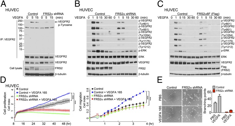
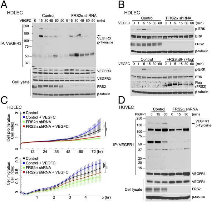
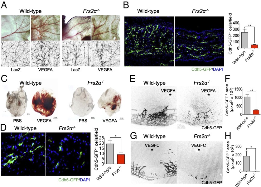
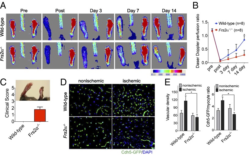
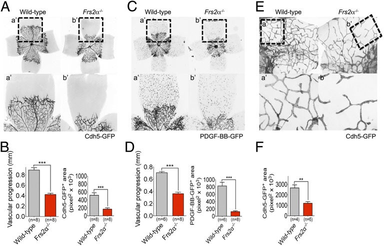
Vascular endothelial growth factors (VEGFs) signal via their cognate receptor tyrosine kinases designated VEGFR1-3. We report that the docking protein fibroblast growth factor receptor substrate 2 (FRS2α) plays a critical role in cell signaling via these receptors. In vitro FRS2α regulates VEGF-A and VEGF-C-dependent activation of extracellular signal-regulated receptor kinase signaling and blood and lymphatic endothelial cells migration and proliferation. In vivo endothelial-specific deletion of FRS2α results in the profound impairment of postnatal vascular development and adult angiogenesis, lymphangiogenesis, and arteriogenesis. We conclude that FRS2α is a previously unidentified component of VEGF receptors signaling.

Keywords: FGF receptor; MAP kinase; phosphorylation; receptor kinase inhibition; signal transduction.


Conflict of interest statement

The authors declare no conflict of interest.

Figures

Fig. 1.
Fig. 1.
FRS2α knockdown in HUVEC inhibits VEGF-A165–dependent signaling. (A) Control and FRS2α knockdown HUVEC were serum starved overnight and treated with VEGF-A165 (50 ng/mL) as indicated. Cell lysates were immunoprecipitated (IP) with an anti-VEGFR2 antibody and immunoblotted with anti–p-Tyrosine antibody. The same blot was stripped and blotted with anti-VEGFR2. Input lysates were blotted with anti-VEGFR2 and anti-FRS2α antibodies. (B) Control and FRS2α knockdown HUVEC were serum starved overnight and treated with VEGF-A165 (50 ng/mL) as indicated. Cell lysates were blotted with p-VEGFR2, VEGFR2, p-ERK, ERK, and FRS2α antibodies. (C) Control and FRS2α-6F (Flag) overexpressed HUVEC were serum starved overnight and treated with VEGF-A165 (50 ng/mL). Cell lysates were blotted with p-VEGFR2, VEGFR2, p-ERK, ERK, and Flag (FRS2α) antibodies. (D) Control and FRS2α knockdown HUVEC were serum starved overnight. Cell proliferation (Left) and cell migration (Right) in response to VEGF-A165 (50 ng/mL) profiles are shown as detected by xCELLigence. Approximately 1000 cells (for cell proliferation) or 25,000 cells (for cell migration) were loaded per well in duplicate (***P < 0.01 compared with control). (E) In vitro Matrigel: The extent of cords branching was assessed in control and FRS2α knockdown HUVEC placed on growth factor-depleted Matrigel and exposed to VEGF-A165 (50 ng/mL). Data in AC and E are based three independent experiments; data in D are based on two independent experiments.
Fig. 2.
Fig. 2.
FRS2α knockdown in HDLEC inhibits VEGF-C–dependent signaling. (A) Control and FRS2α knockdown HDLEC were serum starved overnight and treated with VEGF-C (50 ng/mL) for the indicated times. Cell lysates were immunoprecipitated (IP) with an anti-VEGFR3 antibody and immunoblotted with anti–p-Tyrosine antibody. The same blot was stripped and blotted with anti-VEGFR3. Input lysates were blotted with anti-VEGFR3 and anti-FRS2α antibodies. (B Upper) Control and FRS2α knockdown HDLEC were serum starved overnight and treated with VEGF-C (50 ng/mL). Cell lysates were blotted with p-ERK, ERK, and FRS2α antibodies. (B Lower) Control and FRS2α6F (Flag) overexpressed HDLEC were serum starved overnight and treated with VEGF-C (50 ng/mL). Cell lysates were blotted with p-ERK, ERK, and FRS2α antibodies. (C) Control and FRS2α knockdown HDLEC were serum starved overnight. Cell proliferation (Upper) and cell migration (Lower) in response to VEGF-C (50 ng/mL) profiles, detected by xCELLigence. Approximately 1,000 cells (for cell proliferation) or 25,000 cells (for cell migration) were loaded per well in duplicate (***P < 0.01 compared with control). (D) Control and FRS2α knockdown HUVEC were serum starved overnight and treated with PlGF-1 (50 ng/mL). Cell lysates were immunoprecipitated (IP) with an anti-VEGFR1 antibody and immunoblotted with anti–p-Tyrosine antibody. The same blot was stripped and blotted with anti-VEGFR1. Input lysates were blotted with anti-VEGFR1 and anti-FRS2α antibodies. Data shown in A, B, and D are based on three independent experiments; data in C are based on two independent experiments.
Fig. 3.
Fig. 3.
Impaired angiogenesis in Frs2α−/− mice. (A) Wild-type and Frs2α−/− mice were treated with 1 × 10⁹ pfu of Ad-LacZ or Ad-VEGF-A164 virus. VEGF-A–induced angiogenesis was recorded at day 7 by using a stereomicroscope and fluorescent scope. (B) Mouse ears were sectioned, and the number of vessels was counted (**P < 0.01 compared with control) (n = 4 mice per group). (C and D) Matrigel mixed with either PBS or VEGF-A165 (50 ng/mL) were placed s.c. in wild-type or Frs2α−/− mice. On day 7, matrigel plugs were sectioned, and the number of vessels was counted (*P < 0.05 compared with control) (n = 6 mice per group). (EH) Hydron pellet containing VEGF-A164 or VEGF-C was implanted into the cornea of wild-type and Frs2α−/− mice. Angiogenesis was assessed by stereo microscopy at day 7 following implantation (E and G) (asterisk marks the position of the implanted pellet). Vascular density was quantified in F and H (*P < 0.05; **P < 0.01 compared with control) (n = 6 mice per group).
Fig. 4.
Fig. 4.
Impaired arteriogenesis in Frs2α−/− mice. (A) Laser Doppler images showing blood flow before and after the induction of ischemia to the left hindlimb in wild-type and Frs2α−/− mice. (B) Laser-Doppler analysis of blood flow recovery in the left foot, expressed as a ratio of blood flow in left to right foot (L/R). *P < 0.05, wild-type vs. Frs2α−/− (n = 8 mice per group). (C) In Frs2α−/− mice, clinical score indicated a severe phenotype, leading to necrosis of limb. (D) Representative sections from nonischemic and ischemic groups of wild-type and Frs2α−/− on day 14 after ischemia. Quantification of capillary density (number/mm2 muscle area) and ratio of CD31/myocyte are shown in E. Data are mean ± SD from 10 fields per section (3 sections per mouse; n = 4 for each strain). *P < 0.05, wild-type vs. Frs2α−/−.
Fig. 5.
Fig. 5.
Postnatal angiogenesis and lymphangiogenesis defects in Frs2α−/− mice. (AD) P6 retinas from wild-type and Frs2α−/− littermate mice. (E and F) P6 diaphragms from wild-type and Frs2α−/− littermate mice. a’ and b’ panels show higher magnification of the angiogenic front (AD) and diaphragm lymphatic vessels (E and F).

References

    1. Carmeliet P, Jain RK. Molecular mechanisms and clinical applications of angiogenesis. Nature. 2011;473(7347):298–307. - PMC - PubMed
    1. Lohela M, Bry M, Tammela T, Alitalo K. VEGFs and receptors involved in angiogenesis versus lymphangiogenesis. Curr Opin Cell Biol. 2009;21(2):154–165. - PubMed
    1. Benedito R, et al. Notch-dependent VEGFR3 upregulation allows angiogenesis without VEGF-VEGFR2 signalling. Nature. 2012;484(7392):110–114. - PubMed
    1. Wang Y, et al. Ephrin-B2 controls VEGF-induced angiogenesis and lymphangiogenesis. Nature. 2010;465(7297):483–486. - PubMed
    1. Sawamiphak S, et al. Ephrin-B2 regulates VEGFR2 function in developmental and tumour angiogenesis. Nature. 2010;465(7297):487–491. - PubMed

Publication types

MeSH terms

Substances

LinkOut - more resources