. 2013 Aug 1;35(2):109-120.
doi: 10.1016/j.ppedcard.2013.09.002.

Transesophageal Echocardiography in Healthy Young Adult Male Baboons (Papio hamadryas anubis): Normal Cardiac Anatomy and Function in Subhuman Primates Compared to Humans

Affiliations

Transesophageal Echocardiography in Healthy Young Adult Male Baboons (Papio hamadryas anubis): Normal Cardiac Anatomy and Function in Subhuman Primates Compared to Humans

Arthur A Bert et al. Prog Pediatr Cardiol. .

Abstract

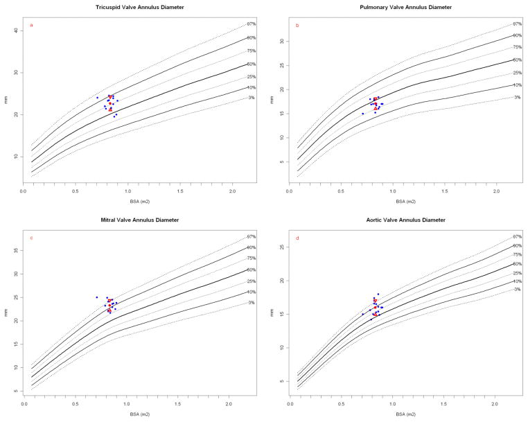
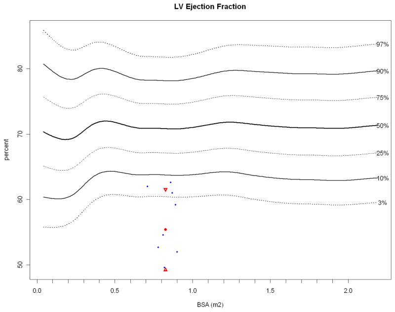
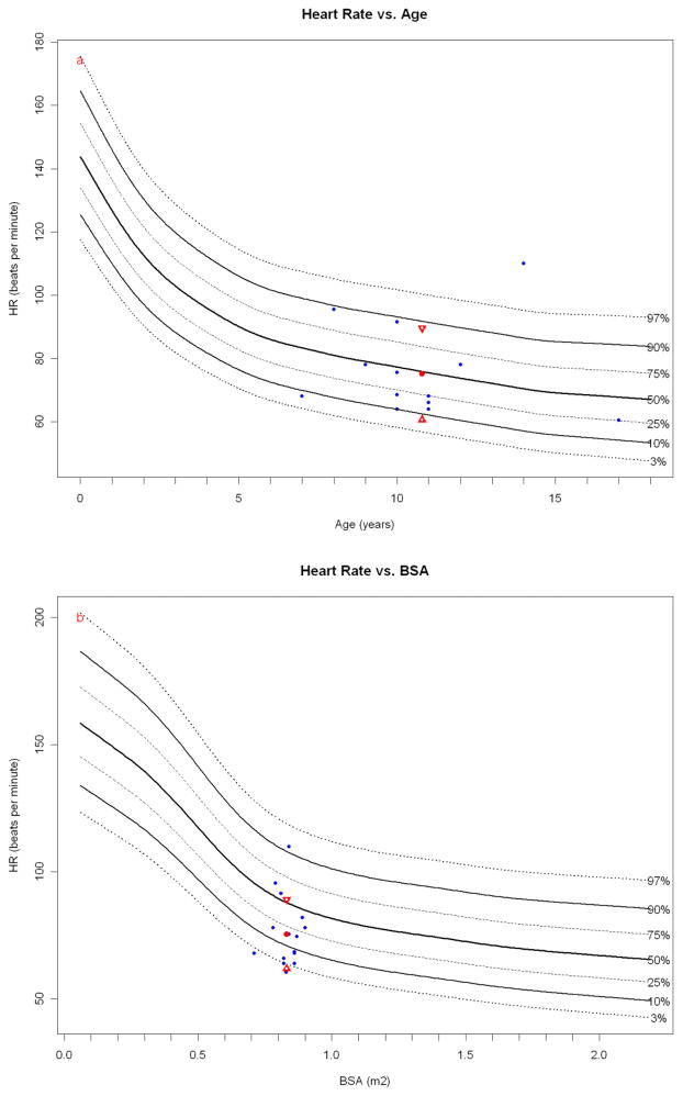
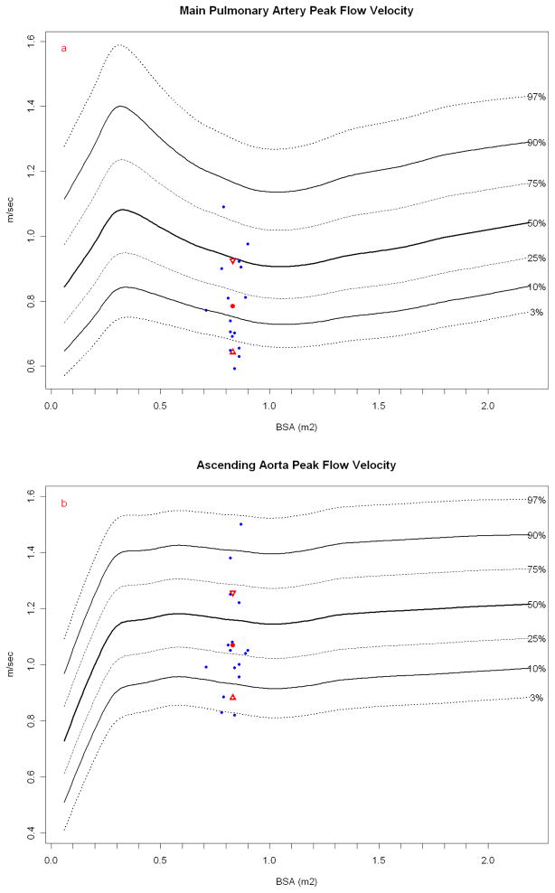
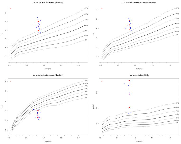
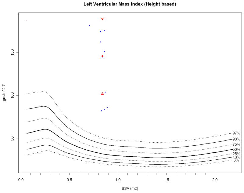
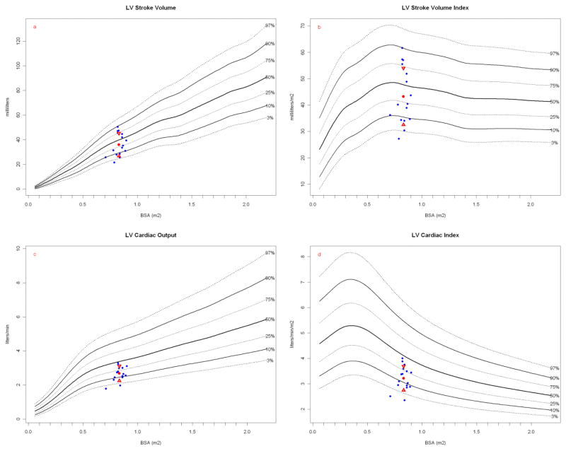
Implantable, viable tissue engineered cardiovascular constructs are rapidly approaching clinical translation. Species typically utilized as preclinical large animal models are food stock ungulates for which cross species biological and genomic differences with humans are great. Multiple authorities have recommended developing subhuman primate models for testing regenerative surgical strategies to mitigate xenotransplant inflammation. However, there is a lack of specific quantitative cardiac imaging comparisons between humans and the genomically similar baboons (Papio hamadryas anubis). This study was undertaken to translate to baboons transesophageal echocardiographic functional and dimensional criteria defined as necessary for defining cardiac anatomy and function in the perioperative setting. Seventeen young, healthy baboons (approximately 30 kg, similar to 5 year old children) were studied to determine whether the requisite 11 views and 52 measurement parameters could be reliably acquired by transesophageal echocardiography (TEE). The obtained measurements were compared to human adult normative literature values and to a large relational database of pediatric "normal heart" echo measurements. Comparisons to humans, when normalized to BSA, revealed a trend in baboons toward larger mitral and aortic valve effective orifice areas and much larger left ventricular muscle mass and wall thickness, but similar pulmonary and tricuspid valves. By modifying probe positioning relative to human techniques, all recommended TEE views except transgastric could be replicated. To supplement, two transthoracic apical views were discovered that in baboons could reliably replace the transgastric TEE view. Thus, all requisite echo views could be obtained for a complete cardiac evaluation in Papio hamadryas anubis to noninvasively quantify cardiac structural anatomy, physiology, and dimensions. Despite similarities between the species, there are subtle and important physiologic and anatomic differences when compared to human.

Keywords: Animal; Disease Models; Echocardiography; Heart; Heart Hypertrophy; Heart Valve Prosthesis; Papio; Primate; Transesophageal.


Figures

Figure 1
Figure 1
Valve diameters at the annulus. Papio measurements (blue dots) are plotted against a measured normal human cohort model. Papio mean (red dot) and standard deviation (red triangles) are shown. The number of normal human measurements used in the reference model is shown as “N=”. (a) Papio tricuspid valve annulus plotted on human normal model (N = 1353) and (b) Papio PV annulus plotted on normal human model (N = 1097). Both the tricuspid valve and PV measurements are within the expected range for humans of similar BSA. Both the (c) Papio MV annulus plotted on human normal model (N = 1164) and (d) Papio AV annulus plotted on human normal model (N = 1210) dimensions are consistently at the upper normal range for human, with the Papio mean around the 90th percentile for humans for both valves.
Figure 2
Figure 2
Papio LVEF (blue dots, obtained while under anesthesia) plotted against normal resting human LVEF (N = 14,261) obtained during transthoracic echocardiography, with the Papio mean (red dot) and standard deviation (red triangles). As with heart rate, general anesthesia may have depressed LV contractility.
Figure 3
Figure 3
Papio heart rate (obtained while under anesthesia) plotted against resting human heart rate normals obtained during transthoracic echocardiography (blue dots). The Papio mean (red dot) and standard deviation (red triangles) are shown. While the heart rates are obtained in slightly different physiologic states in each species, it is interesting to note that the age-based Papio heart rate compares more favorably to the human than the BSA-based heart rate.
Figure 4
Figure 4
Papio peak flow velocity (blue dots) in the (a) main PA and (b) ascending aorta plotted against resting human normals (ascending aorta, N = 13,284; PA, N = 13,634) obtained during resting transthoracic echocardiography with the Papio mean (red dot) and standard deviation (red triangles). Papio have similar flow velocities in the great arteries as humans, particularly considering these measurements are again done in anesthetized Papio.
Figure 5
Figure 5
LV wall thickness, short axis diastolic dimension, and calculated LV mass index. Papio measurements (blue dots) are plotted against a measured normal human cohort model. Papio mean (red dot) and standard deviation (red triangles) are shown. The number of normal human measurements used in the reference model is shown as “N=”. (a) Papio LV septal wall thickness plotted on human normal model (N = 14,489) and (b) Papio LV posterior wall thickness plotted on human normal model (N = 14,486). Both wall measurements are significantly thicker than the normal range for humans of similar BSA. The Papio short axis diastolic chamber dimension (c) plotted on human normal model (N = 14,161) is significantly smaller than that predicted for humans of a similar height. The resulting calculated Papio LV mass indexed to body surface area (d) is significantly higher than the predicted normal range for humans of similar BSA. Calculated LV mass was obtained using the Troy formula [61] and then indexed to calculated BSA (all diameters are calculated in diastole): LVmass=1.05([LVinternaldiameter+posteriorwallthickness+intraventricularseptumthickness)3-(LVinternaldiameter)3])
Figure 6
Figure 6
LV mass index (height based). Papio height-based LV mass index (blue dots, ref below) plotted against height based human LV mass index (N = 13,941), with the Papio mean (red dot) and standard deviation (red triangles). Papio have significantly more LV myocardial mass than humans at similar height [51].
Figure 7
Figure 7
LVSV and stroke index, LVCO and cardiac index. Papio measurements (blue dots) are plotted against a measured normal human cohort model. Papio mean (red dot) and standard deviation (red triangles) are shown. The number of normal human measurements used in the reference model is shown as “N=”. (a) Papio LVSV plotted on human normal model and (b) Papio LVSV indexed to BSA plotted on human normal model (N = 14,013 for both). Papio LVCO (c) and cardiac index (d) are plotted on human normal model (N = 1,860 for both). Papio stroke volume was calculated using the Continuity Equation (see text), whereas the human stroke volume was calculated from m-mode measurements using the Teichholz method [53, 31]. Note that baboon “height” is measured as crown to rump length.

References

    1. Barnhart GR, Jones M, Ishihara T, Chavez AM, Rose DM, Ferrans VJ. Bioprosthetic valvular failure. Clinical and pathological observations in an experimental animal model. J Thorac Cardiovasc Surg. 1982;83:618–31. - PubMed
    1. Gallegos RP, Nockel PJ, Rivard AL, Bianco RW. The current state of in-vivo pre-clinical animal models for heart valve evaluation. J Heart Valve Dis. 2005;14:423–32. - PubMed
    1. Grehan JF, Casagrande I, Oliveira EL, et al. A juvenile sheep model for the long-term evaluation of stentless bioprostheses implanted as aortic root replacements. J Heart Valve Dis. 2001;10:505–12. - PubMed
    1. Salerno CT, Droel J, Bianco RW. Current state of in vivo preclinical heart valve evaluation. J Heart Valve Dis. 1998;7:158–62. - PubMed
    1. Neethling W, Hodge A, Glancy R. Kangaroo versus Freestyle® stentless bioprostheses in a juvenile sheep model: hemodynamic performance and calcification behavior. J Card Surg. 2005;20:29–34. - PubMed

LinkOut - more resources