

Spontaneous resolution of vitreomacular traction in two patients with diabetic macular edema
- PMID: 24707275
- PMCID: PMC3975209
- DOI: 10.1159/000360219
Spontaneous resolution of vitreomacular traction in two patients with diabetic macular edema
Abstract
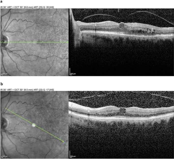
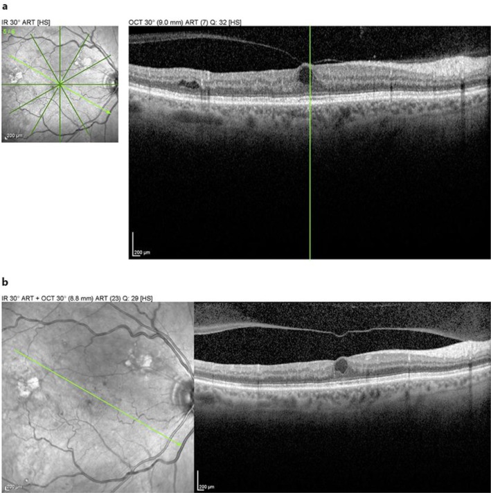
The nature of the vitreoretinal interface in conditions like diabetic macular edema (DME) remains incompletely understood. Furthermore, it is not clear what the role of pharmacological enzymatic vitreolysis will play in cases of vitreomacular traction (VMT) associated with macular disease like DME. We describe the spontaneous resolution of VMT in 2 patients with DME. As both surgical and pharmacologic interventions have been suggested to treat DME in the setting of VMT, we feel that a clarification of the nomenclature and reporting of these cases of natural history may be useful in more fully understanding the complex decision-making involved when determining whether to treat this subset of patients.
Keywords: Diabetic macular edema; Vitreomacular adhesion; Vitreomacular traction.
Figures
References
-
- Jackson TL, Nicod E, Simpson A, Angelis A, Grimaccia F, Kanavos P. Symptomatic vitreomacular adhesion. Retina. 2013;33:1503–1511. - PubMed
-
- Russell SR, Hageman GS. Optic disc, foveal, and extrafoveal damage due to surgical separation of the vitreous. Arch Ophthalmol. 2001;119:1653–1658. - PubMed
-
- Jackson TL, Nicod E, Angelis A, Grimaccia F, Prevost AT, Simpson AR, Kanavos P. Vitreous attachment in age-related macular degeneration, diabetic macular edema, and retinal vein occlusion: a systematic review and metaanalysis. Retina. 2013;33:1099–1108. - PubMed
-
- Johnson MW. Posterior vitreous detachment: evolution and role in macular disease. Retina. 2012;32(suppl 2):S174–S178. - PubMed
-
- Simpson AR, Petrarca R, Jackson TL. Vitreomacular adhesion and neovascular age-related macular degeneration. Surv Ophthalmol. 2012;57:498–509. - PubMed
Publication types
LinkOut - more resources
Full Text Sources
Other Literature Sources
Research Materials
