. 2014 Mar 1;1(3):180-189.
doi: 10.1002/acn3.43.

Antigenic and mechanistic characterization of anti-AMPA receptor encephalitis

Affiliations

Antigenic and mechanistic characterization of anti-AMPA receptor encephalitis

Amy J Gleichman et al. Ann Clin Transl Neurol. .

Abstract

Objective: Anti-AMPAR encephalitis is a recently discovered disorder characterized by the presence of antibodies in serum or cerebrospinal fluid against the α-amino-3-hydroxy-5-methyl-4-isoxazolepropionic acid (AMPA) receptor. Here, we examine the antigenic specificity of anti-AMPAR antibodies, screen for new patients, and evaluate functional effects of antibody treatment of neurons.

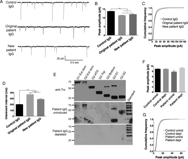
Methods: We developed a fusion protein-based western blotting test for anti-AMPAR encephalitis antibodies. Antibody specificity was also evaluated using immunocytochemistry of HEK293 cells expressing deletion mutants of AMPAR subunits. Purified patient IgG or AMPAR antibody-depleted IgG was applied to live neuronal cultures; amplitude and frequency of miniature excitatory postsynaptic currents (mEPSCs) were measured to evaluate functional effects of antibodies.

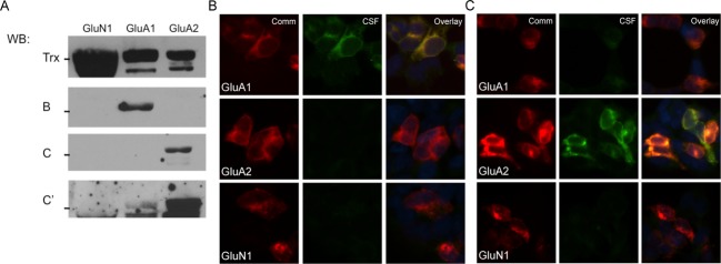
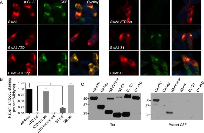
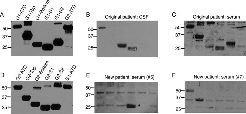
Results: Using both immunocytochemistry and fusion protein western blots, we defined an antigenic region of the receptor in the bottom lobe of the amino terminal domain. Additionally, we used fusion proteins to screen 70 individuals with neurologic symptoms of unknown cause and 44 patients with no neurologic symptoms or symptoms of known neuroimmunological origin for anti-AMPAR antibodies. Fifteen of the 70 individuals had anti-AMPAR antibodies, with broader antigenic reactivity patterns. Using purified IgG from an individual of the original cohort of anti-AMPAR encephalitis patients and a newly discovered patient, we found that application of IgG from either patient cohort caused an AMPAR antibody-dependent decrease in the amplitude and frequency of mEPSCs in cultured neurons.

Interpretation: These results indicate that anti-AMPAR antibodies are widespread and functionally relevant; given the robust response of patients to immunomodulation, this represents a significant treatable patient population.


Figures

Figure 1
Figure 1
Patient CSF recognition of external domain fusion proteins is similar to staining of cells transfected with full subunits. (A) Patient CSF (blots B and C) recognizes a band of the correct size in transfected samples, indicating that native conformation may not be necessary for patient antibody recognition of AMPARs. GluA1, GluA2, and GluN1 all expressed well, with minimal proteolytic cleavage of the desired fusion protein when grown in pLyS BL21 Escherichia coli, as evidenced by blotting for the thioredoxin tag (Trx); even in this cell line, which minimizes protease activity, the GluA3 external domain fusion protein was almost entirely degraded during growth (data not shown). Two representative examples of patient reactivity are shown, as well as a darker exposure of the second patient to demonstrate minor GluA1 reactivity; all previously diagnosed anti-AMPAR encephalitis patients showed reactivity with fusion proteins. (B and C) CSF from two previously diagnosed anti-AMPAR encephalitis patients recognize GluA1 and/or GluA2-transfected HEK293 cells by immunocytochemistry in patterns that correspond to their observed GluA1/GluA2 external domain fusion protein reactivity (CSF from the same patient was used for (B) or (C)-labeled lanes in western blot in (A) and immunocytochemistry). (C) Example of one patient with primary GluA2 reactivity but minor GluA1 reactivity in transfected cells who showed the same differential reactivity with fusion proteins (A, bands C and C′; C, CSF panels). Comm, commercial antibody staining.
Figure 2
Figure 2
Antibodies from patients with anti-AMPAR encephalitis primarily recognize the bottom lobe of the ATD of GluA1 or GluA2. (A) Staining pattern of CSF from a typical anti-AMPAR encephalitis patient on HEK293 cells transfected with GluA2 deletion mutants shows a loss of reactivity with deletion of the bottom lobe of the ATD. (B) Quantification of deletion mutant CSF staining over four patients, three with primary GluA2 reactivity and one with primary GluA1 reactivity, using GluA2 or GluA1 mutants, respectively. (C) Reactivity with receptor domains on western blot. Left, fusion protein expression measured by anti-Thioredoxin antibody (Trx). Right, patient CSF reacts primarily with the ATD and the bottom lobe of the ATD. S1 deletion also decreases antibody binding, but does not appear to bind on western blot, possibly due to the proximity of the S1 domain to the bottom lobe of the ATD. *P < 0.05, ***P < 0.001, one-way ANOVA plus Dunnett's post hoc testing.
Figure 3
Figure 3
Fusion protein reactivity reveals differences in CSF and serum antibody responses and additional patients with anti-AMPAR antibodies in serum. (A and D) Fusion proteins of GluA1 (A) and GluA2 (D) subdomains (anti-Thioredoxin antibody). (B and C) While patient CSF binds the bottom lobe of the GluA1-ATD as well as the ATD and S1 domains, serum from the same patient shows broader reactivity. (E and F) Fusion protein reactivity of two individuals not previously diagnosed with anti-AMPAR encephalitis reveals reactivity with the GluA2-S1 domain (E) and the GluA2-ATD and top lobe (F).
Figure 4
Figure 4
mEPSCs differ in cultured neurons treated with IgG from an individual with no anti-AMPAR reactivity (control IgG), an individual from the original cohort of anti-AMPAR encephalitis with GluA2 bottom lobe ATD reactivity (original patient IgG), and an individual with newly discovered anti-AMPAR antibodies in serum directed against the S1 domain (new patient IgG). (A) Example traces of mEPSCs from neurons treated with IgG from different patients. (B and C) Decreased average peak amplitude of mEPSCs in patient-treated neurons (B) is reflected in a decreased frequency of larger amplitude responses in the cumulative frequency histogram (C). (D) Increased interevent interval in neurons treated with patient material indicates a decrease in event frequency, which is more pronounced with original patient IgG treatment. (E) Incubation of patient IgG with GluA2 fusion proteins specifically depletes anti-AMPAR antibodies (Patient IgG, depleted), while incubation with uninduced bacteria does not (Patient IgG, uninduced; bands in GluA2-ext dom and GluA2-S1 lanes; compare to Fig.3E). Anti-Trx, Thioredoxin loading control. Standard, MagicMark internal exposure standard. (F and G) Patient IgG effect on mEPSC amplitude is dependent on the presence of anti-AMPAR antibodies: preincubation with uninduced bacteria (unind) maintains the patient-specific decrease in mEPSC amplitude (F) and decreased percentage of large events (G), while these effects are lost after AMPAR-specific IgG depletion (depl). n = 6–9 cells, P < 0.0001, one-way ANOVA plus Tukey's post hoc testing.

References

    1. Lancaster E, Dalmau J. Neuronal autoantigens–pathogenesis, associated disorders and antibody testing. Nat Rev Neurol. 2012;8:380–390. - PMC - PubMed
    1. Zuliani L, Graus F, Giometto B, et al. Central nervous system neuronal surface antibody associated syndromes: review and guidelines for recognition. J Neurol Neurosurg Psychiatry. 2012;83:638–645. - PMC - PubMed
    1. Dalmau J, Tuzun E, Wu HY, et al. Paraneoplastic anti-N-methyl-D-aspartate receptor encephalitis associated with ovarian teratoma. Ann Neurol. 2007;61:25–36. - PMC - PubMed
    1. Lai M, Hughes EG, Peng X, et al. AMPA receptor antibodies in limbic encephalitis alter synaptic receptor location. Ann Neurol. 2009;65:424–434. - PMC - PubMed
    1. Lai M, Huijbers MG, Lancaster E, et al. Investigation of LGI1 as the antigen in limbic encephalitis previously attributed to potassium channels: a case series. Lancet Neurol. 2010;9:776–785. - PMC - PubMed

LinkOut - more resources