. 2014 May 22;57(10):3891-911.
doi: 10.1021/jm500220s. Epub 2014 May 1.

2012 Division of medicinal chemistry award address. Trekking the cannabinoid road: a personal perspective

Affiliations

2012 Division of medicinal chemistry award address. Trekking the cannabinoid road: a personal perspective

Alexandros Makriyannis. J Med Chem. .

Abstract

My involvement with the field of cannabinoids spans close to 3 decades and covers a major part of my scientific career. It also reflects the robust progress in this initially largely unexplored area of biology. During this period of time, I have witnessed the growth of modern cannabinoid biology, starting from the discovery of its two receptors and followed by the characterization of its endogenous ligands and the identification of the enzyme systems involved in their biosynthesis and biotransformation. I was fortunate enough to start at the beginning of this new era and participate in a number of the new discoveries. It has been a very exciting journey. With coverage of some key aspects of my work during this period of "modern cannabinoid research," this Award Address, in part historical, intends to give an account of how the field grew, the key discoveries, and the most promising directions for the future.


Figures

Figure 1
Figure 1
Technical Review Proceedings Cover Page
Figure 2
Figure 2
Phyto- and Xeno-Cannabinoids,
Figure 3
Figure 3
Autoradiography of 10 nM [3H]CP55,940 binding in a sagittal section of rat brain. Br St, brainstem; Cer, cerebellum; Col, collculi; CP, caudate-putamen; Cx, cerebral cortex; Ep, entopeduncular nucleus (homolog of GPi); GP, globus pallidus (e, external; I, internal); Hi, hippocampus; Th, thalamus.
Figure 4
Figure 4
Select AM Compounds. AM630 is the first CB2 antagonist., AM411 is the first adamantyl cannabinoid, while AM4054 is a later generation analog. Both are early CB1 receptor agonists. AM2389 is a very potent long-acting CB1 agonist; AM404 is the first early transporter inhibitor, while AM1172 is a second generation transport inhibitor. AM2201 is a very potent aminoalkylindole CB1 agonist.
Figure 5
Figure 5
Endocannabinoid Analogs
Figure 6
Figure 6
Drug and endogenous ligands partition within the cellular membrane where they can: (a) diffuse passively across the bilayer to enter the intracellular space; (b) are tranlocated intracellularly through a yet to be fully identified transport mechanism; (c) interact with the receptor active site through fast lateral diffusion.
Figure 7
Figure 7
(I) Representative solid state 2H-NMR spectra from dipalmitoylphosphatidyl choline (DPPC) model membranes (42°C) containing five cannabinoids each having two deuterium labels at 2 and 4-positions: 2,4-d2-Me-Δ8-THC(A), 2,4-d2-Δ-8-THC(B), 2,4,-d29-THC(C), 2,4-d2-11-OH-9α-HHC(D) and 2,4-d2-11-OH-9β-HHC(E). (II) Orientations of Me-Δ8-THC (top), Δ8-THC and 9-THC (middle), 11-OH-9α-HHC and 11-OH-9β-HHC (bottom) in hydrated DPPC bilayers. The dashed lines represent the direction of the lipid acyl chains.
Figure 8
Figure 8
Electron density profile differences inside the bilayer: Curve B-A is the difference between profiles of dimyristoylphosphatidyl choline (DMPC) + Δ8-THC and DMPC, curve C-A is the difference between those of DMPC + 5’-I-Δ8-THC and DMPC, and curve C-B is the difference between those of DMPC + 5’-I- 8-THC and DMPC + Δ8-THC. The outer pair of the vertical dashed lines indicates the peaks in curves B-A and the inner pair indicates the peaks in C-B. They represent the positions of the center of Δ8-THC and the iodine atom of 5’-I-Δ8-THC in the bilayer, respectively.
Figure 9
Figure 9
A ligand-membrane-receptor model representing the trans-membrane diffusion of CP55940 en route to interacting with the cannabinoid receptor. According to our hypothesis, the ligand preferentially partitions in the membrane bilayer where it assumes a proper orientation and location allowing for a productive collision with the active site.,
Figure 10
Figure 10
2H-solid state NMR experiments identifying the endocannabinoid anandamide assuming an extended conformation in the bilayer with its polar group at the same level as the polar phospholipid head groups. It diffuses laterally to the binding site of the CB1 receptor and interacts with a hydrophobic groove formed by helices 3, 5, and 6. The data were used to model the location of anandamide within the bilayer and its approach to the cannabinoid receptor site through fast lateral diffusion. The C-20 anandamide segment was shown to interact with C6.47 of CB1 Hx6, using covalent ligand labeling experiments and is designated by a yellow star.
Figure 11
Figure 11
On the left, inhibition of [3H]anandamide accumulation by astrocytes by AM404. On the right, effects of AM404 on anandamide-induced inhibition of adenylyl cyclase activity in cortical neurons. AM403 is a control inactive ligand. In all experiments, cells were incubated with the inhibitors for 10 min before the addition of [3H]anandamide for an additional 4 min.
Figure 12
Figure 12
Lipophilic ligands are transported intracellularly by means of extracellular carrier proteins (eg. albumin) and deposited initially at the outer membrane leaflet. Hydrophobic ligands (A, e.g. Me-Δ9-THC) initially occupy a location in the outer leaflet of the membrane below the phospholipid headgroups. Subsequently, a second population of the ligand is found at the center of the bilayer where the two ligand populations are in equilibrium with each other. This suggests a mechanism for the hydrophobic ligands to reach the inner membrane leaflet where they can be taken up by specialized carrier proteins. Amphipathic ligands (B, e.g. Δ9-THC) initially partition at the outer membrane leaflet with their polar groups interacting with the phospholipid headgroups. Through a flip-flop mechanism, the ligands are translocated to the corresponding site at the inner membrane leaflet.
Figure 13
Figure 13. Human Fatty Acid Binding Protein (FABP7)
A. The binding of [3H]-anandamide (AEA) to human brain fatty acid binding protein (FABP7). B. Overlay of 2D 1H-15N Heteronuclear Single Quantum Coherence (HSQC) NMR spectra of free 15N- labeled human FABP7 (blue) and a sample with the addition of anandamide (1:5) (red). C. Docking of AEA into the FABP7 crystal structure reveals three potential H-bond interactions: one between the AEA carbonyl group and the guanidinium group of Arg126, and the other two between the AEA hydroxyl group with the guanidinium group of Arg126.
Figure 14
Figure 14
CB2 Agonists
Figure 15
Figure 15
Covalent Ligands. AM841 is a CB1/CB2 covalent megagonist. AM3677 is an anandamide covalent CB1 receptor agonist; AM9017 is an anandamide covalent CB2 agonist (K. Vadivel and A. Makriyannis, unpublished); AM967 is a photoactivatable CB2 covalent probe; AM1336 is a covalent CB2 receptor antagonist. AM5822 and AM5823 are covalent homo- and heterobifunctional CB1 ligands (Y. C. Leung and A. Makriyannis, unpublished).
Figure 16
Figure 16. Illustration of the CB2 R*/AM841 Binding Site from Modeling Studies
TMHS 1, 4, and 5 have been omitted from this view for simplicity. In the energy-minimized CB2 R*/AM841 complex in which AM841 is covalently attached to C6.47(257), the carbocyclic ring CH2OH of AM841 hydrogen-bonds with S7.39(285) (d = 2.62 Å; O-H-O angle = 175°), while the phenolic hydroxyl of AM841 hydrogen-bonds with S6.58(268) (d = 2.61 Å; O-H-O angle = 176°). Also illustrated here is the formation of a salt bridge between D275 in EC3 and K3.28(109), a residue that is crucial for classical CB binding in the CB1 receptor, but not CB2. In the final, energy-minimized complex illustrated here, K3.28(109) is involved in a salt bridge with D275 of the EC3 loop (d = 2.55 Å; N-H-O angle = 171°) and in a hydrogen bond with N2.63(93) (d = 2.66 Å; N-H-N angle = 168°). D275 also forms a hydrogen bond with S274 in EC3 (d = 2.67 Å; O-H-O angle = 170°) and with S2.60(90) (d = 2.63 Å; O-H-O angle = 160°), a residue that is accessible from within the binding pocket in the CB2 receptor due to the helix distortion produced by S2.54(84).
Figure 17
Figure 17
(a) Chemical structure of AM841. (b) Saturation-binding assay of [3H] CP-55940 radioligand of FLAG-hCB2R-6His in membranes from Sf-21 cells overexpressing this receptor. Membrane incubation with AM841 prior to [3H] CP-55940 binding (▲) reduced the receptor Bmax by ~75% relative to the Bmax in membranes not exposed to AM841 (■). Data are means ±SEM of at least two independent experiments performed in duplicate. (c) Western blot analysis of the purified FLAG-hCB2R-6His preparations with anti-FLAG antibody detection. M, monomer; D, dimer; O, oligomers, (d) MS/MS analysis of Hx6 peptide carrying AM841.
Figure 18
Figure 18
CB1 Antagonists
Figure 19
Figure 19. Example of controlled-deactivation cannabinergic ligand
Compound A [AMG38, (6aR-trans)-3-(1-hexylcyclobutyl)-6a,7,10,10a-tetrahydro-6,6,9-trimethyl-6H-dibenzo[b,d]pyran-1-ol] is a potent CB1 agonist (Ki = 1.5 nM) while compound B is its corresponding analog with similar pharmacophoric groups which also encompasses a key ester group in its side chain that is available for enzymatic cleavage. Through the action of esterases, B yields two fragments (a and b) that are shown to have negligible cannabinergic activity.
Figure 20
Figure 20
Diagrammatic representation of the major hydrolytic inactivation and oxidative biotransformation pathways implicated in anandamide and 2-arachidonyl glycerol catabolism. (٠) Endocannabinoid proteins being studied by AM.
Figure 21
Figure 21
AM3506 was covalently docked to the catalytic Ser241 of rFAAH in the acyl chain binding channel. There is significant hydrogen bonding with the oxyanion hole (formed by the backbone of Ile238, Gly239, Gly240, and Ser241), and also with the backbone carbonyl of Thr488. Hydrogen bonds are denoted by a blue dashed line, and the surface of the binding channels is shown in gray.
Figure 22
Figure 22
AM5206 affords neuronal protection in the hippocampus after KA-induced excitotoxicity in vitro. Organotypic hippocampal slice cultures were used, and a low-power photomicrograph shows their characteristic maintenance of native cellular organization. (A) Nissle staining was also used to assess cellular integrity across treatment groups. CA3 pyramidal zones are shown from slices treated consistently with vehicle (B), those pretreated with vehicle before a 2-h exposure to 60 μM KA (C), and slices pretreated with 10 μM AM5206 before the KA insult (D). After a washout step, the cultured slices were then incubated with vehicle or AM5206 for 24 h before the tissue was fixed, sectioned, and stained. The KA insult resulted in neuronal loss and obvious pyknotic changes that were reduced by the FAAH inhibitor. DG, dentate gyrus; so, stratum oriens; sp, stratum pyramidale; sr, stratum radiatum. Scale bar: a, 400 μM, b-d, 45 μM.
Figure 23
Figure 23
AM5206 affords seizure and neuronal protection after KA induced excitotoxicity in vivo. Seizures were induced by i.p. injection of 9.5 mg/kg KA (n=12 rats), and following the KA administration animals were immediately injected with either vehicle or 8 mg/kg AM5206. Vehicle-treated control rats (veh, n=11) did not receive KA or AM5206. Seizures were scored by blinded raters over a 4-h period (a) and mean scores ± SEM are shown (ANOVA, P<0.0001). At 48 h post injection, hippocampal tissue was rapidly dissected, homogenized, and equal protein aliquots assessed by immunoblot for BDPN, GluR1, synapsin II (syn II), and actin (b). Mean integrated optical densities for GluR1 (c) are shown (±SEM; ANOVA, P<0.001). Post hoc tests compared to KA+ vehicle data, single asterisk P<0.05; triple asterisk, P<0.001.
Figure 24
Figure 24
FAAH and MGL Inhibitors
Figure 25
Figure 25. Hydrogen bonding network in the active site of hMGL
A. Downfield 1H NMR resonances corresponding to the H-bonds in the active site definitively assigned by comparing spectra of wt hMGL and different mutants. B. Active site hydrogen bonding network in hMGL identified using site-directed mutagenesis with total loss of activity observed with D239A, H269A and S122 mutants. The results shed light into the molecular details of hMGL catalysis. C. hMGL inhibitor AM10212 interaction at the active site. This compound was first synthesized at Johnson and Johnson.
Figure 26
Figure 26
MD simulation of hMGL interaction with membrane phospholipid bilayer. (a)Structure of AM6580. Snapshots of hMGL based on HXMS experimental derived data with a phospholipid bilayer membrane of the same composition as in experimental nanodiscs (POPC: POPG, 3:2 molar ratio) after 10 ns of MD simulation. The enzyme is depicted: (b) unliganded as apo-hMGL and (c) occupied with carbamylating inhibitor, AM6580, in the active site. Helix α4 in the lid domain and nearby helix alpha 6 are depicted in red, and AM6580 (c) is highlighted and depicted in green.
Figure 27
Figure 27
3D-cube from the HNCA spectrum of hMGL (900 MHz spectrometer with cryo-probe, 2 days experiment. Triple resonance HNCA and HN(CO)CA spectra were recorded at 310 K on a Bruker Avance 900 MHz NMR spectrometer for the assignment of backbone resonances. All spectra were processed using the Topspin software (Bruker Biospin). Together, these experiments reveal the CA chemical shift for each amino acid residue in hMGL, and provide information linking adjacent residues in the sequence.
Figure 28
Figure 28. Example of Long Range Interactions discovered in the hMGL enzyme
W289 is located in helix H8 at the C-terminal of hMGL separated by a distance of ~18A between W289 and the binding site. Substitution of W289 with Leu leads to total loss of enzymatic activity. (PDB id: 3jw8),
Figure 29
Figure 29
Reduction of KA-induced seizure severity by AM6701(FAAH, MGL inhibitor) and AM6702 (FAAH inhibitor). Seizures were initiated in rats with an intraperitoneal injection of 9.8 mg/kg KA. Immediately following the KA administration, animals were injected with vehicle (n=16) or 5 mg/kg AM6701 or AM6702 (n=6-8). No-insult control rats received 2 vehicle injections (n=13). Seizure scores were tabulated by blinding raters for a 4-h period following the injections, and the mean scores ±SEM are shown. Analysis of variance p<0.0001; post hoc test compared to KA only group: ***p<0.0001.
Figure 30
Figure 30
(A) Putative mechanism of irreversible inhibition of hNAAA by AM 6701 via thiocarbamylation of Cy126. (B) Representation of the active site of hNAAA after treatment of AM6701. Homology model illustrates thiocarbamylation of catalytic nucleophile Cys126 after treatment with AM6701.

Similar articles

Cited by

References

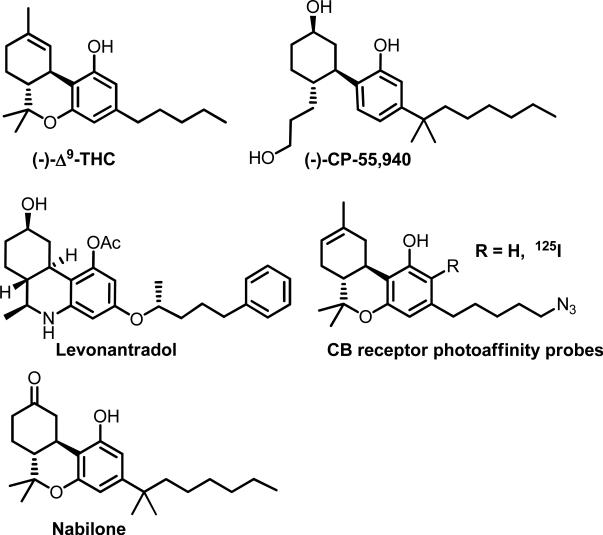
    1. Charalambous A, Yan G, Houston DB, Howlett AC, Compton DR, Martin BR, Makriyannis A. 5′-Azido-delta 8-THC: a novel photoaffinity label for the cannabinoid receptor. J Med Chem. 1992;35:3076–3079. - PubMed
    1. Burstein SH, Audette CA, Charalambous A, Doyle SA, Guo Y, Hunter SA, Makriyannis A. Detection of cannabinoid receptors by photoaffinity labelling. Biochem Biophys Res Commun. 1991;176:492–497. - PubMed
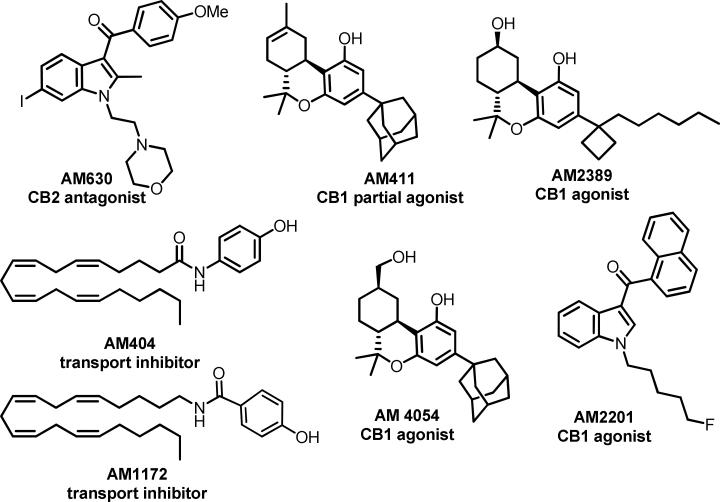
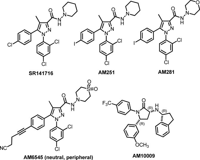
    1. Pertwee R, Griffin G, Fernando S, Li X, Hill A, Makriyannis A. AM630, a competitive cannabinoid receptor antagonist. Life Sci. 1995;56:1949–1955. - PubMed
    1. Guo J, Williams MK, Makriyannis A. Therapeutic Opportunities through the Modulation of Endocannabinoid Transport. Pharmaceut. Reg. Affairs. 2013;3 In press, doi:10.4172/2167-7689.1000e4123. - PMC - PubMed
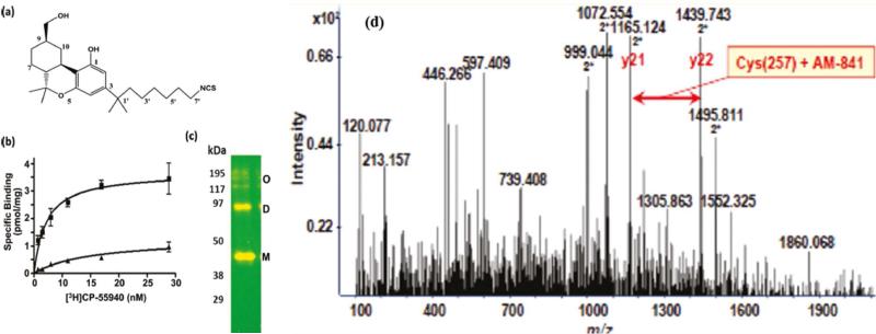
    1. Picone RP, Khanolkar AD, Xu W, Ayotte LA, Thakur GA, Hurst DP, Abood ME, Reggio PH, Fournier DJ, Makriyannis A. (−)-7′-Isothiocyanato-11-hydroxy-1′,1′-dimethylheptylhexahydrocannabinol (AM841), a high-affinity electrophilic ligand, interacts covalently with a cysteine in helix six and activates the CB1 cannabinoid receptor. Mol Pharmacol. 2005;68:1623–1635. - PubMed

Publication types

MeSH terms

LinkOut - more resources