. 2014 Jun;87(1038):20140118.
doi: 10.1259/bjr.20140118. Epub 2014 Apr 8.

Epipericardial fat necrosis: an underdiagnosed condition

Affiliations

Epipericardial fat necrosis: an underdiagnosed condition

K de S Giassi et al. Br J Radiol. 2014 Jun.

Abstract

Objective: Epipericardial fat necrosis (EFN) is an uncommon benign and self-limited condition that leads patients to the emergency department (ED) owing to the onset of acute pleuritic chest pain. The aim of this study was to describe the cases of this disease in our institution and to illustrate the associated clinical and radiological findings.

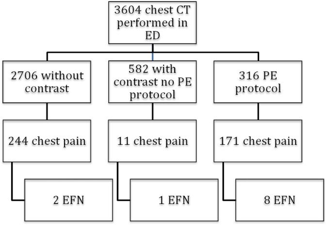
Methods: We reviewed 3604 chest scans referred by the ED from November 2011 to July 2013. Patients diagnosed with epipericardial necrosis had their medical records and original tomography reports analysed.

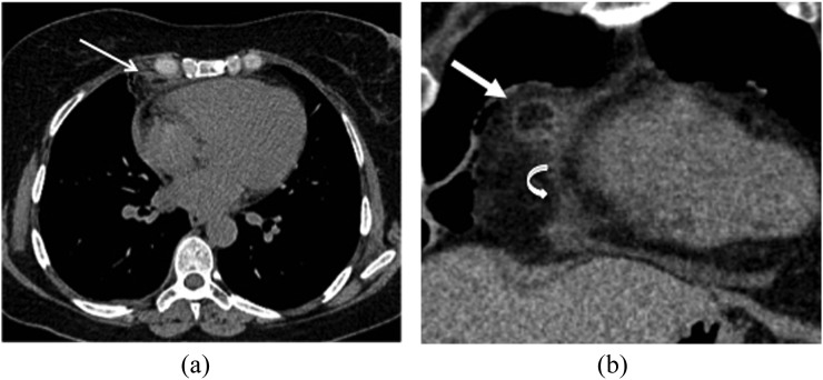
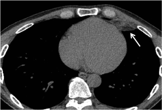
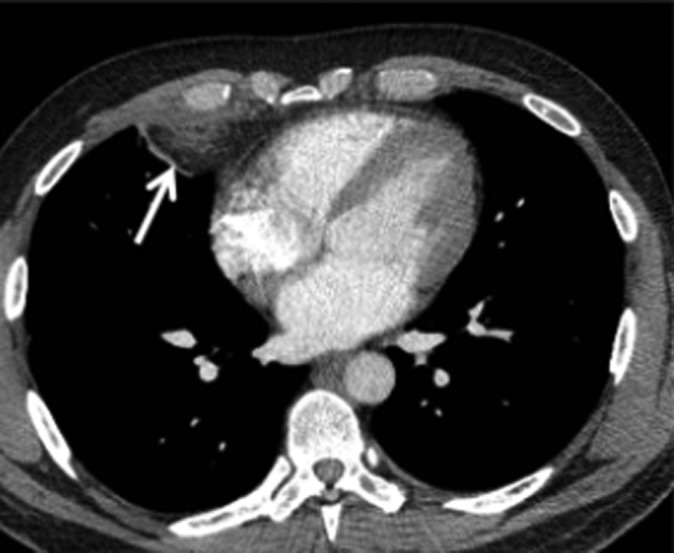
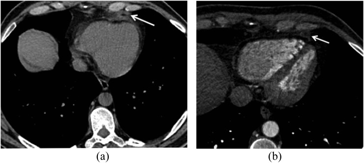
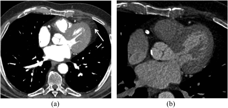
Results: Chest pain was the primary complaint in 426 patients; 11 of them had definitive EFN findings characterized by a round soft-tissue attenuation lesion with a varying degree of strands. All patients presented with pleuritic chest pain on the same side as the lesion. Pericardial thickening, pleural effusion and mild atelectasis were the associated tomography findings. Cardiac enzyme and D-dimer tests performed during the episode were normal in all cases. 27% of the cases only were correctly diagnosed with EFN at the time of presentation.

Conclusion: EFN is a benign inflammatory condition frequently overlooked in the ED by physicians and radiologists but is an important factor in the differential diagnosis of patients with acute chest pain.

Advances in knowledge: The article adds clinically and radiologically useful information about the condition and displays the importance of making the correct diagnosis to avoid unnecessary examinations.


Figures

Figure 1.
Figure 1.
A 47-year-old female with 5-day right pleuritic chest pain. (a) The axial chest CT scan displays a round soft-tissue attenuation lesion with fatty centre and peripheral strands inside the right epipericardial fat (arrow), which is strongly suggestive of epipericardial fat necrosis (EFN). (b) Coronal reformation of the round soft-tissue attenuation lesion suggestive of EFN (straight arrow). There is notable pericardial thickening (curved arrow).
Figure 2.
Figure 2.
A 24-year-old male with 3-day left pleuritic chest pain and epipericardial fat necrosis findings. The axial view of an enhanced chest CT scan displays a small round soft-tissue attenuation lesion with mild stranding (arrow) close to the diaphragmatic pleural surface in the epipericardial fat.
Figure 3.
Figure 3.
A 40-year-old male with 2-day history of right pleuritic chest pain. An axial chest CT scan displays a small, round lesion with a fatty centre surrounded by dense stranding (arrow), which is another finding strongly suggestive of epipericardial fat necrosis.
Figure 4.
Figure 4.
Diagram of the protocols and the patients diagnosed with epipericardial fat necrosis (EFN) in each protocol group. ED, emergency department; PE, pulmonary embolism.
Figure 5.
Figure 5.
A 44-year-old male with 3-day left pleuritic chest pain. (a) The axial view of an unenhanced chest CT scan displays a round soft-tissue attenuation lesion with strands (arrow) in the epipericardial fat and focal pericardial thickening. (b) Axial view of the chest CT control performed 4 weeks later. The lesion has almost completely resolved, with mild residual focal stranding in the epipericardial fat (arrow).
Figure 6.
Figure 6.
An 80-year-old male with 9-h left pleuritic chest pain. (a) An enhanced chest CT scan displays a soft-tissue attenuation lesion and stranding in the left epipericardial fat (arrow). (b) The axial chest CT follow-up scan performed 7 weeks later demonstrates complete resolution of the findings.

References

    1. Hernandez D, Galimany J, Pernas JC, Llauger J. Case 170: pericardial fat necrosis. Radiology 2011; 259: 919–22. doi: 10.1148/radiol.10090786 - DOI - PubMed
    1. Jackson RC, Clagett OT, McDonald JR. Pericardial fat necrosis: report of three cases. J Thorac Surg 1957; 33: 723–9. - PubMed
    1. Pineda V, Caceres J, Andreu J, Vilar J, Domingo ML. Epipericardial fat necrosis: radiologic diagnosis and follow-up. AJR Am J Roentgenol 2005; 185: 1234–6. doi: 10.2214/AJR.04.1310 - DOI - PubMed
    1. Baig A, Campbell B, Russell M, Singh J, Borra S. Epicardial fat necrosis: an uncommon etiology of chest pain. Cardiol J 2011; 19: 424–8. - PubMed
    1. Lee HH, Ryu DS, Jung SS, Jung SM, Choi SJ, Shin DH. MRI findings of pericardial fat necrosis: case report. Korean J Radiol 2011; 12: 390–4. doi: 10.3348/kjr.2011.12.3.390 - DOI - PMC - PubMed

Substances