

Case Reports
doi: 10.1111/jon.12117.
Epub 2014 Apr 7.
Levodopa-responsive hemiparkinsonism secondary to cystic expansion from a coiled cerebral aneurysm
Affiliations
- PMID: 24707971
- PMCID: PMC4418521
- DOI: 10.1111/jon.12117
Item in Clipboard
Case Reports
Levodopa-responsive hemiparkinsonism secondary to cystic expansion from a coiled cerebral aneurysm
J Neuroimaging.
2015 Mar-Apr.
Abstract
An 80-year-old woman with longstanding hemifacial spasm had a 1 cm × 1.5 cm internal carotid artery terminus aneurysm treated with endovascularly delivered bare metal coils. Follow-up imaging revealed an expansile perianeurysmal cyst that coincided with development of contralateral dopa-responsive hemiparkinsonism. This is the first report of perianeurysmal cyst expansion causing levodopa-responsive hemiparkinsonism.
Keywords: Levodopa; aneurysm; cyst; hemifacial spasm; parkinsonism.
Copyright © 2014 by the American Society of Neuroimaging.
Figures
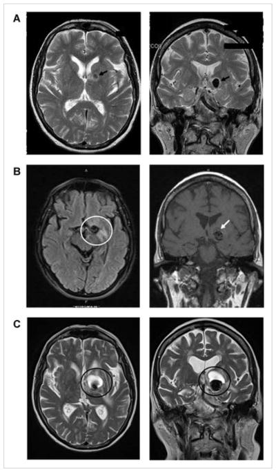
Axial (left column) and coronal (right column) T2 weighted (A, C) and fluid attenuated inversion recovery (FLAIR) MRI images (B) demonstrating aneurysm evolution: (A) prior to intervention, a 12-mm left internal carotid artery terminus aneurysm (black arrows) anterior and superior to the left cerebral peduncle; (B) 14 months after endovascular coil embolization with parenchymal T2 signal hyperintensity around the left callosal and pericallosal regions of the coiled aneurysm site (white circle) and some low signal at the aneurysm apex (white arrow), above the coil mass (represented as area of signal loss) on the coronal view. Low signal is inconsistent with blood product and likely represents developing cyst (T2 images not obtained); (C) 15 months after repeat endovascular coil embolization with enlarged aneurysm and superior expansion of a cystic mass compressing internal capsule, pallidum, and some caudate (dark circles).
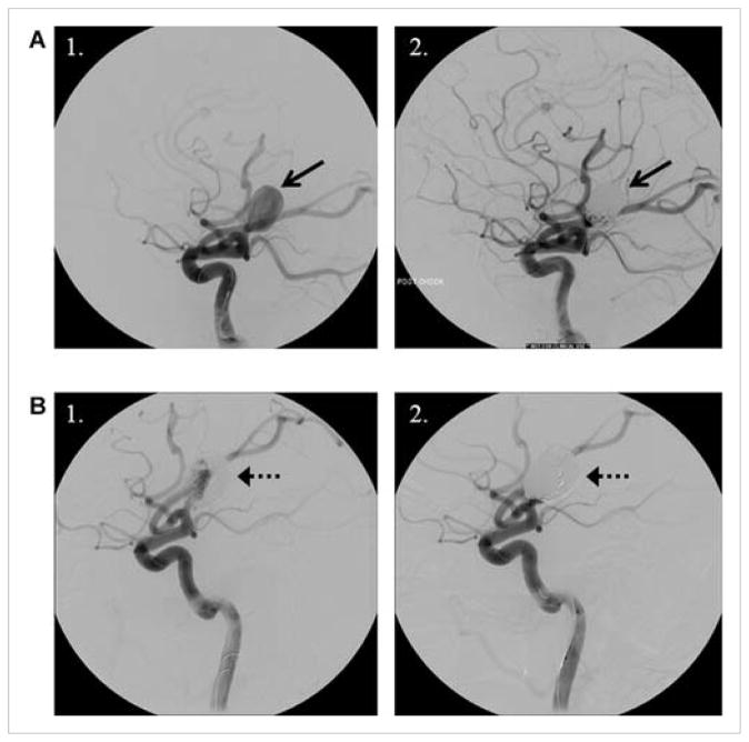
Digital subtraction lateral projection cerebral angiography of the left common carotid artery (A) before placement of coils (1) and after successful initial coil embolization (2), and (B) before retreatment showing compaction of coils at base (1) and successful repeat coil embolization with dense packing at the neck (2). Neck remodeling was performed with a balloon across the neck during placement of coils. Microcatheter is visible in the internal carotid artery in all images. Arrows reference the aneurysm in both image sets.
References
-
- Friedman JA, McIver JI, Collignon FP, et al. Development of a pontine cyst after endovascular coil occlusion of a basilar artery trunk aneurysm: case report. Neurosurgery. 2003;52(3):694–699. discussion 698–699. - PubMed
-
- Konig M, Bakke SJ, Scheie D, et al. Reactive expansive intracerebral process as a complication of endovascular coil treatment of an unruptured intracranial aneurysm: case report. Neurosurgery. 2011;68(5):E1468–E1473. discussion E1473–1474. - PubMed
-
- Marcoux J, Roy D, Bojanowski MW. Acquired arachnoid cyst after a coil-ruptured aneurysm. Case illustration. J Neurosurg. 2002;97(3):722. - PubMed
-
- Meyers PM, Lavine SD, Fitzsimmons BF, et al. Chemical meningitis after cerebral aneurysm treatment using two second-generation aneurysm coils: report of two cases. Neurosurgery. 2004;55(5):1222–1227. - PubMed
Publication types
MeSH terms
Substances
Grants and funding
LinkOut - more resources
Full Text Sources
Other Literature Sources
Medical
