Expression of thyrotropin receptor, thyroglobulin, sodium-iodide symporter, and thyroperoxidase by fibrocytes depends on AIRE
- PMID: 24708100
- PMCID: PMC4079309
- DOI: 10.1210/jc.2013-4271
Expression of thyrotropin receptor, thyroglobulin, sodium-iodide symporter, and thyroperoxidase by fibrocytes depends on AIRE
Abstract
Context: CD34(+) fibrocytes, bone marrow-derived progenitor cells, infiltrate orbital connective tissue in thyroid-associated ophthalmopathy, a manifestation of Graves' disease. In the orbit, they become CD34(+) fibroblasts and coexist with native CD34(-) fibroblasts. Fibrocytes have been shown to express TSH receptor and thyroglobulin.
Objective: The objective of the study was to determine whether a broader repertoire of thyroid protein expression can be detected in fibrocytes and whether a common factor is responsible.
Design/setting/participants: Fibrocytes and fibroblasts were collected and analyzed from healthy individuals and those with Graves' disease in an academic clinical practice.
Main outcome measures: Real-time PCR, Western blot analysis, gene promoter analysis, cell transfections, and flow cytometric cell sorting were performed.
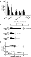
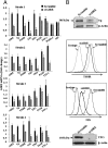
Results: We detect two additional thyroid proteins expressed by fibrocytes, namely sodium-iodide symporter and thyroperoxidase. The autoimmune regulator (AIRE) protein appears necessary for this expression. AIRE expression in fibrocytes results from an active AIRE gene promoter and stable AIRE mRNA. Knocking down AIRE with a targeting small interfering RNA reduces the expression of these thyroid proteins in fibrocytes as well as the transcription factors paired box-8 and thyroid transcription factor-1. When compared with an unaffected first-degree relative, levels of these proteins are substantially reduced in fibrocytes from an individual with an inactivating AIRE mutation. Levels of AIRE and the thyroid proteins are lower in orbital fibroblasts from patients with thyroid-associated ophthalmopathy than in fibrocytes. However, when mixed fibroblast populations are sorted into pure CD34(+) and CD34(-) subsets, the levels of these proteins are dramatically increased selectively in CD34(+) fibroblasts.
Conclusions: Fibrocytes express four proteins, the aggregate expression of which was previously thought to be restricted to thyroid epithelium. These proteins represent the necessary molecular biosynthetic machinery necessary for thyroid hormone production. Our findings implicate AIRE in the promiscuous expression of thyroid proteins in fibrocytes.
Figures


Similar articles
-
Human fibrocytes coexpress thyroglobulin and thyrotropin receptor.Proc Natl Acad Sci U S A. 2012 May 8;109(19):7427-32. doi: 10.1073/pnas.1202064109. Epub 2012 Apr 19. Proc Natl Acad Sci U S A. 2012. PMID: 22517745 Free PMC article.
-
Slit2 Modulates the Inflammatory Phenotype of Orbit-Infiltrating Fibrocytes in Graves' Disease.J Immunol. 2018 Jun 15;200(12):3942-3949. doi: 10.4049/jimmunol.1800259. Epub 2018 May 11. J Immunol. 2018. PMID: 29752312 Free PMC article.
-
TSH receptor and thyroid-specific gene expression in human skin.J Invest Dermatol. 2010 Jan;130(1):93-101. doi: 10.1038/jid.2009.180. J Invest Dermatol. 2010. PMID: 19641516
-
TSH-receptor-expressing fibrocytes and thyroid-associated ophthalmopathy.Nat Rev Endocrinol. 2015 Mar;11(3):171-81. doi: 10.1038/nrendo.2014.226. Epub 2015 Jan 6. Nat Rev Endocrinol. 2015. PMID: 25560705 Free PMC article. Review.
-
Iodide handling disorders (NIS, TPO, TG, IYD).Best Pract Res Clin Endocrinol Metab. 2017 Mar;31(2):195-212. doi: 10.1016/j.beem.2017.03.006. Epub 2017 Apr 4. Best Pract Res Clin Endocrinol Metab. 2017. PMID: 28648508 Review.
Cited by
-
Teprotumumab as a Novel Therapy for Thyroid-Associated Ophthalmopathy.Front Endocrinol (Lausanne). 2020 Dec 17;11:610337. doi: 10.3389/fendo.2020.610337. eCollection 2020. Front Endocrinol (Lausanne). 2020. PMID: 33391187 Free PMC article. Review.
-
Teprotumumab in the management of thyroid eye disease mechanistic insights and adverse reactions: a comprehensive review.Front Endocrinol (Lausanne). 2025 May 27;16:1480195. doi: 10.3389/fendo.2025.1480195. eCollection 2025. Front Endocrinol (Lausanne). 2025. PMID: 40496563 Free PMC article. Review.
-
Teprotumumab Divergently Alters Fibrocyte Gene Expression: Implications for Thyroid-associated Ophthalmopathy.J Clin Endocrinol Metab. 2022 Sep 28;107(10):e4037-e4047. doi: 10.1210/clinem/dgac415. J Clin Endocrinol Metab. 2022. PMID: 35809263 Free PMC article.
-
Therapeutic IGF-I receptor inhibition alters fibrocyte immune phenotype in thyroid-associated ophthalmopathy.Proc Natl Acad Sci U S A. 2021 Dec 28;118(52):e2114244118. doi: 10.1073/pnas.2114244118. Proc Natl Acad Sci U S A. 2021. PMID: 34949642 Free PMC article.
-
Primary hypothyroidism with exuberant dermatological manifestations.An Bras Dermatol. 2020 Nov-Dec;95(6):721-723. doi: 10.1016/j.abd.2019.07.010. Epub 2020 Mar 19. An Bras Dermatol. 2020. PMID: 32482552 Free PMC article.
References
-
- Wang J, Jiao H, Stewart TL, Shankowsky HA, Scott PG, Tredget EE. Improvement in postburn hypertrophic scar after treatment with IFN-α2b is associated with decreased fibrocytes. J Interferon Cytokine Res. 2007;27:921–930 - PubMed
Publication types
MeSH terms
Substances
Grants and funding
LinkOut - more resources
Full Text Sources
Other Literature Sources

