. 2014 Jul;155(7):2524-33.
doi: 10.1210/en.2013-1485. Epub 2014 Apr 7.

Angiotensin II-induced protein kinase D activates the ATF/CREB family of transcription factors and promotes StAR mRNA expression

Affiliations

Angiotensin II-induced protein kinase D activates the ATF/CREB family of transcription factors and promotes StAR mRNA expression

Lawrence O Olala et al. Endocrinology. 2014 Jul.

Abstract

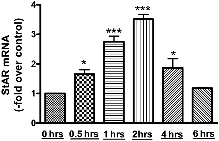
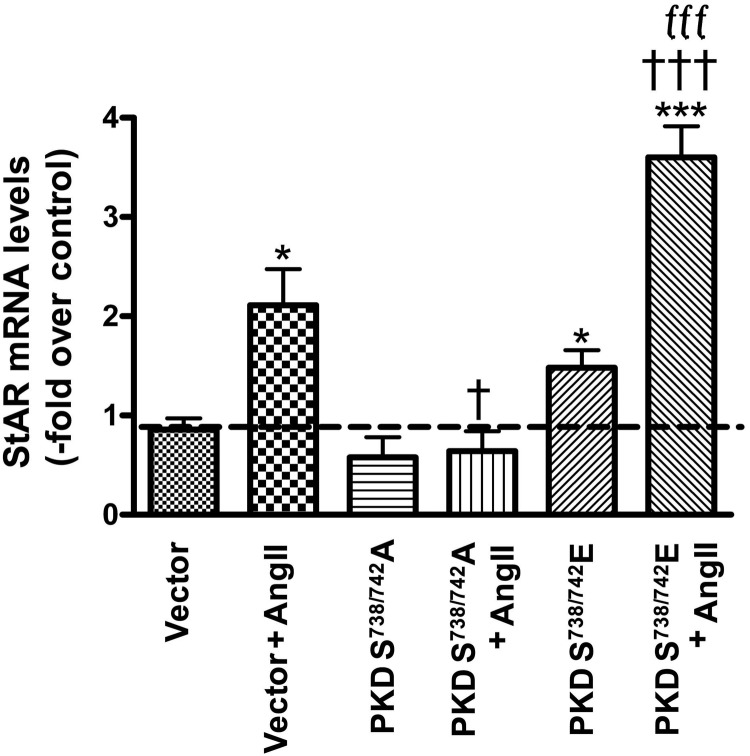
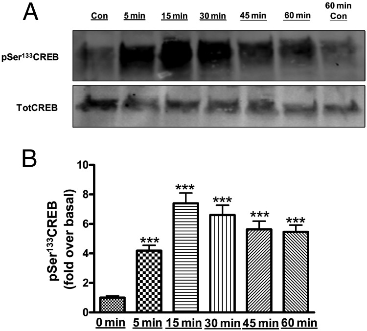
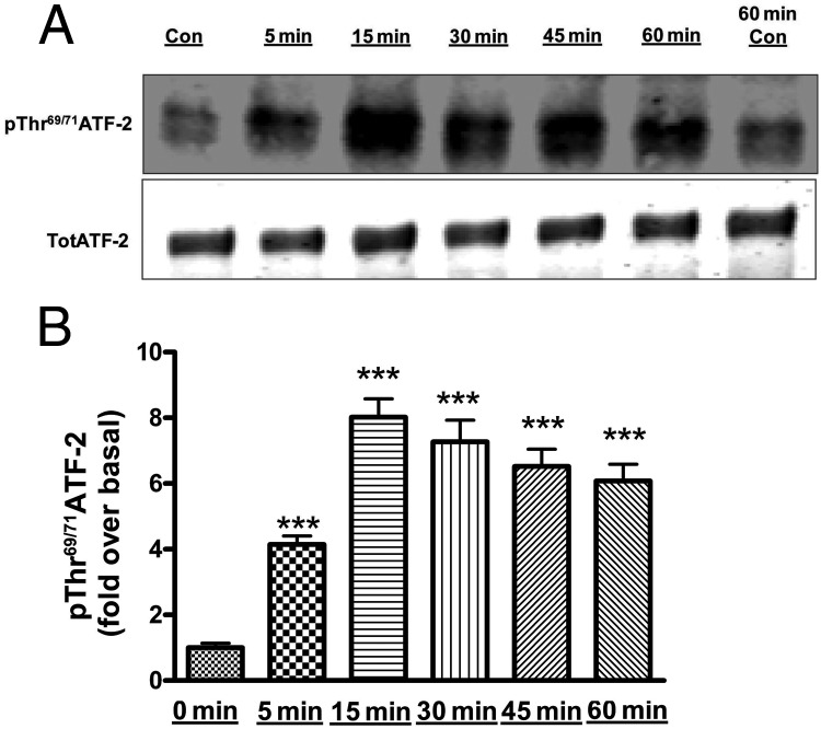
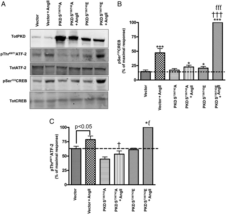
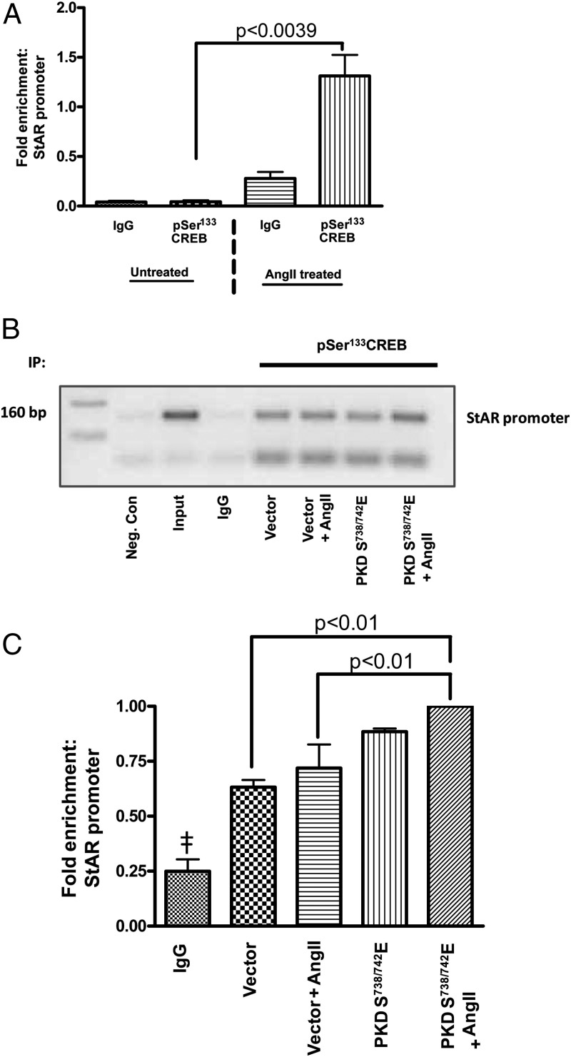
Aldosterone synthesis is initiated upon the transport of cholesterol from the outer to the inner mitochondrial membrane, where the cholesterol is hydrolyzed to pregnenolone. This process is the rate-limiting step in acute aldosterone production and is mediated by the steroidogenic acute regulatory (StAR) protein. We have previously shown that angiotensin II (AngII) activation of the serine/threonine protein kinase D (PKD) promotes acute aldosterone production in bovine adrenal glomerulosa cells, but the mechanism remains unclear. Thus, the purpose of this study was to determine the downstream signaling effectors of AngII-stimulated PKD activity. Our results demonstrate that overexpression of the constitutively active serine-to-glutamate PKD mutant enhances, whereas the dominant-negative serine-to-alanine PKD mutant inhibits, AngII-induced StAR mRNA expression relative to the vector control. PKD has been shown to phosphorylate members of the activating transcription factor (ATF)/cAMP response element binding protein (CREB) family of leucine zipper transcription factors, which have been shown previously to bind the StAR proximal promoter and induce StAR mRNA expression. In primary glomerulosa cells, AngII induces ATF-2 and CREB phosphorylation in a time-dependent manner. Furthermore, overexpression of the constitutively active PKD mutant enhances the AngII-elicited phosphorylation of ATF-2 and CREB, and the dominant-negative mutant inhibits this response. Furthermore, the constitutively active PKD mutant increases the binding of phosphorylated CREB to the StAR promoter. Thus, these data provide insight into the previously reported role of PKD in AngII-induced acute aldosterone production, providing a mechanism by which PKD may be mediating steroidogenesis in primary bovine adrenal glomerulosa cells.


Figures

Figure 1.
Figure 1.
AngII induced StAR mRNA expression in primary bovine adrenal glomerulosa cells in a time-dependent manner. After a 30-minute preincubation with eqKRB+, primary cultures of bovine adrenal glomerulosa cells were treated for 30 minutes or 1, 2, 4, or 6 hours with 10nM AngII. Controls without AngII treatment were performed at times 0 and 6 hours. RNA was isolated and used for qRT-PCR with data normalized to GAPDH and shown as the fold change compared with basal (control 0 min). Values represent means ± SEM from 4 experiments performed in duplicate; *, P < .05 vs control; ***, P < .001 vs control.
Figure 2.
Figure 2.
The constitutively active Ser738/742-to-glutamate PKD mutant promoted, and the dominant-negative Ser738/742-to-alanine PKD mutant inhibited, StAR mRNA expression. Cultured primary bovine glomerulosa cells were incubated for 4 hours with adenovirus expressing pAdtrackCMV (empty vector), or the dominant-negative Ser738/742-to-alanine (PKDS738/742A) or constitutively active Ser738/742-to-glutamate (PKDS738/742E) PKD mutants. On the second day of culture, cells were treated with or without 10nM AngII for 2 hours, and RNA was isolated and used for qRT-PCR with data normalized to GAPDH. The ΔΔCt method was used to analyze the data, and values were expressed as the fold change compared with basal (vector without AngII stimulation) as described in Materials and Methods. Values represent means ± SEM from 4 experiments performed in duplicate; *, P < .05; ***, P < .001 vs control (vector); †, P < .05; †††, P < .001 vs vector + AngII; fff, P < .001 vs PKDS738/742A + AngII.
Figure 3.
Figure 3.
AngII induced phosphorylation (activation) of CREB in primary bovine adrenal glomerulosa cells in a time-dependent manner. After a 30-minute preincubation with eqKRB+, primary cultures of bovine adrenal glomerulosa cells were treated for 5, 15, 30, 45, or 60 minutes with 10nM AngII. Controls (Con; no AngII treatment) were performed at times 0 and 60 minutes. Samples were analyzed by Western blotting to determine phospho-Ser133 CREB levels. A, Representative blot. B, Band intensities from multiple experiments were quantified and normalized to total CREB (TotCREB). Values represent means ± SEM of 6 samples from 3 separate experiments and are expressed as fold over control (the average of control times 0 and 60 min); ***, P < .001 vs control.
Figure 4.
Figure 4.
AngII induced phosphorylation (activation) of ATF-2 in primary bovine adrenal glomerulosa cells in a time-dependent manner. After a 30-minute preincubation with eqKRB+, primary cultures of bovine adrenal glomerulosa cells were treated for 5, 15, 30, 45, and 60 minutes with 10nM AngII. Controls (Con; no AngII treatment) were performed at times 0 and 60 minutes. Samples were analyzed by Western blotting to determine phospho-Thr69/71 ATF-2 levels. A, Representative blot. B, Band intensities from multiple experiments were quantified and normalized to total ATF-2 (TotATF-2). Values represent means ± SEM of 6 samples from 3 separate experiments and are expressed as fold over control (the average of control times 0 and 60 min); ***, P < .001 vs control.
Figure 5.
Figure 5.
The constitutively active Ser738/742-to-glutamate PKD mutant promoted, and the dominant-negative Ser738/742-to-alanine PKD mutant inhibited, AngII-induced CREB and ATF-2 phosphorylation. Cultured primary bovine glomerulosa cells were incubated for 4 hours with adenovirus expressing pAdtrackCMV (empty vector), or the Ser738/742-to-alanine (PKDS738/742A) or Ser738/742-to-glutamate (PKDS738/742E) PKD mutants. On the second day of culture, media were replaced with serum-free media for an additional 16–20 hours before treatment with or without AngII (10nM) for 1 hour. A, Representative blot. B and C, Band intensities from multiple experiments were quantified and normalized to their respective total proteins (TotCREB or TotATF-2), CREB (B) and ATF-2 (C). Analysis was performed on transformed data, and values were expressed relative to the maximal response (PKDS738/742E + AngII) as described in Materials and Methods. Values represent the means ± SEM from 4 experiments performed in duplicate; *, P < .05 vs control (vector); ***, P < .001 vs control (vector); †, P < .05 vs vector + AngII; †††, P < .001 vs vector + AngII; f, P < .05 vs PKDS738/742A + AngII; fff, P < .001 vs PKDS738/742A + AngII.
Figure 6.
Figure 6.
Constitutively active Ser738/742-to-glutamate PKD increased phospho-CREB association with the StAR promoter. Cultured primary bovine glomerulosa cells were treated with or without AngII on day 2 before analysis by ChIP. Alternatively, the cells were incubated for 4 hours with adenovirus expressing pAdtrackCMV (empty vector) or the Ser738/742-to-glutamate PKD mutant (PKDS738/742E). On the second day of culture, media were replaced with serum-free media for an additional 16–20 hours before treatment with or without AngII (10nM) for 1 hour. The ChIP assay was performed as described in Materials and Methods. Cross-linked sheared chromatin was immunoprecipitated with either IgG or antiphospho-CREB antibody. We performed PCR analysis on the recovered chromatin using the −160-bp region of the bovine proximal StAR promoter. A, qRT-PCR analysis from 3 experiments showing the fold enrichment upon AngII treatment relative to the control cells (no AngII treatment) and the IgG immunoprecipitation (IP) control. B, Representative gel using semiquantitative RT-PCR to show the association of phospho-CREB with the proximal StAR promoter. A negative control (Neg. Con) containing water instead of DNA is also shown. C, qRT-PCR analysis from 3 experiments demonstrates the fold enrichment of the StAR promoter relative to the IgG control. Values represent the means ± SEM; ‡, P < .05 vs (vector, vector + AngII, PKDS738/742E, PKDS738/742E + AngII).

Similar articles

Cited by

References

    1. Müller J. Aldosterone: the minority hormone of the adrenal cortex. Steroids. 1995;60(1):2–9 - PubMed
    1. Quinn SJ, Williams GH. Regulation of aldosterone secretion. Annu Rev Physiol. 1988;50:409–426 - PubMed
    1. Calhoun DA. Aldosterone and cardiovascular disease: smoke and fire. Circulation. 2006;114:2572–2574 - PubMed
    1. Delcayre C, Swynghedauw B. Molecular mechanisms of myocardial remodeling. The role of aldosterone. J Mol Cell Cardiol. 2002;34(12):1577–1584 - PubMed
    1. Rocha R, Stier CT, Jr, Kifor I, et al. Aldosterone: a mediator of myocardial necrosis and renal arteriopathy. Endocrinology. 2000;141(10):3871–3878 - PubMed

Publication types

MeSH terms