. 2014 Aug;155(8):2892-9.
doi: 10.1210/en.2013-2175. Epub 2014 Apr 7.

Evidence that estrogen hastens epiphyseal fusion and cessation of longitudinal bone growth by irreversibly depleting the number of resting zone progenitor cells in female rabbits

Affiliations

Evidence that estrogen hastens epiphyseal fusion and cessation of longitudinal bone growth by irreversibly depleting the number of resting zone progenitor cells in female rabbits

Ola Nilsson et al. Endocrinology. 2014 Aug.

Abstract

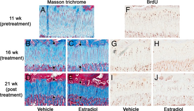
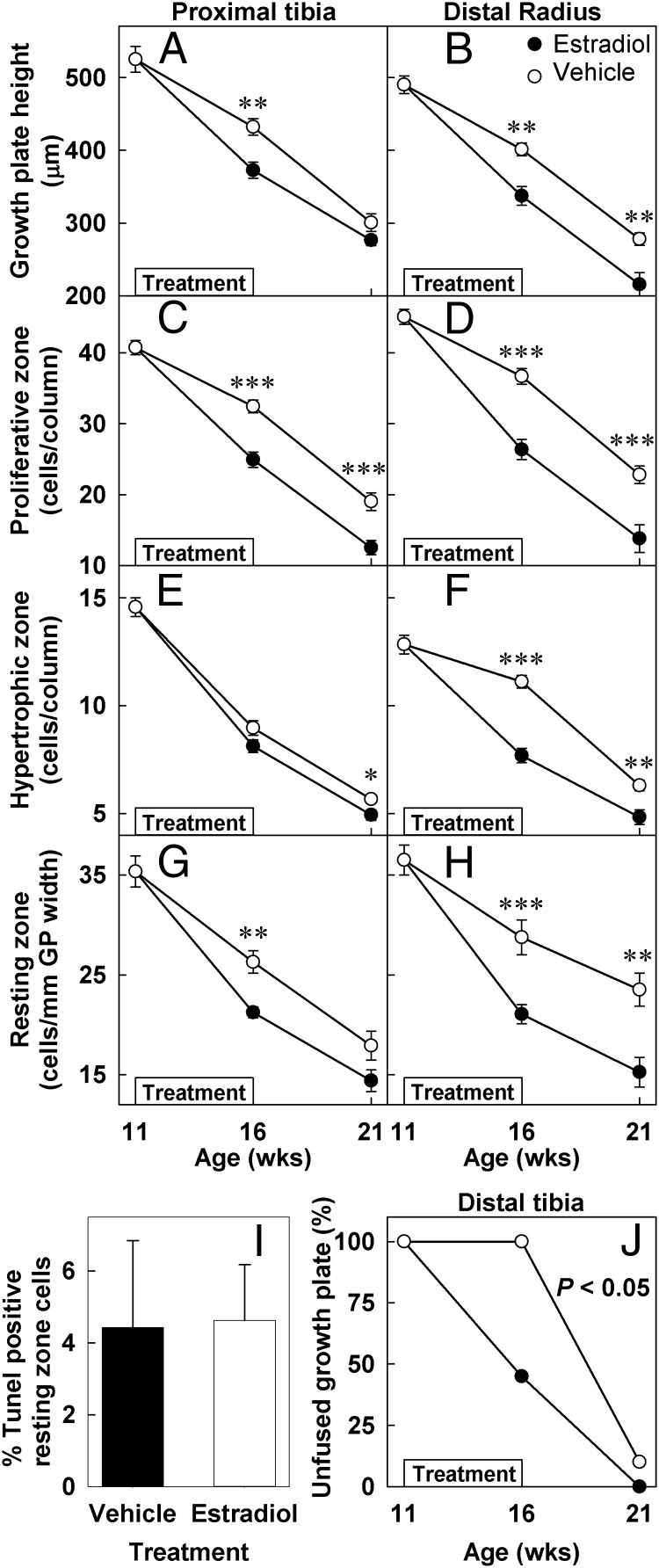
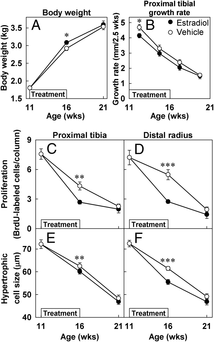
With age, growth plate cartilage undergoes programmed senescence, eventually causing cessation of bone elongation and epiphyseal fusion. Estrogen accelerates this developmental process. We hypothesized that senescence occurs because progenitor cells in the resting zone are depleted in number and that estrogen acts by accelerating this depletion. To test this hypothesis, juvenile ovariectomized rabbits received injections of estradiol cypionate or vehicle for 5 weeks, and then were left untreated for an additional 5 weeks. Exposure to estrogen accelerated the normal decline in growth plate height and in the number of proliferative and hypertrophic chondrocytes. Five weeks after discontinuation of estrogen treatment, these structural parameters remained advanced, indicating an irreversible advancement in structural senescence. Similarly, transient estrogen exposure hastened epiphyseal fusion. Estrogen also caused a more rapid decline in functional parameters of growth plate senescence, including growth rate, proliferation rate, and hypertrophic cell size. However, in contrast to the structural parameters, once the estrogen treatment was discontinued, the growth rate, chondrocyte proliferation rate, and hypertrophic cell size all normalized, suggesting that estrogen has a reversible, suppressive effect on growth plate function. In addition, estrogen accelerated the normal loss of resting zone chondrocytes with age. This decrease in resting zone cell number did not appear to be due to apoptosis. However, it was maintained after the estrogen treatment stopped, suggesting that it represents irreversible depletion. The findings are consistent with the hypothesis that estrogen causes irreversible depletion of progenitor cells in the resting zone, thus irreversibly accelerating structural senescence and hastening epiphyseal fusion. In addition, estrogen reversibly suppresses growth plate function.


Figures

Figure 1.
Figure 1.
PT growth plate morphology and proliferation before (11 weeks), at the end of treatment (16 weeks), and 5 weeks after the estrogen treatment was stopped (21 weeks). Representative sections of PT growth plates from 11-(A and F), 16- (B, C, G, and H), and 21-week-old (D, E, I, and J) control (A, B, D, F. G, and I) and estrogen-treated (C, E, H, and J) animals stained with Masson Trichrome (A–E) or by BrdU immunohistochemistry (F–J). Estrogen treatment (16-week time point) affected the structure of the growth plate, decreasing the overall height as well as the number of cells in each zone. Interestingly, 5 weeks after the estrogen treatment was stopped (21-week time point) these structural changes remained advanced. Estrogen treatment also decreased the number of BrdU-labeled cells in estrogen treated animals (H) compared with controls (G). However, 5 weeks after the estrogen treatment was stopped (21 weeks) the number of BrdU-labeled cells in estrogen- treated animals (J) was similar to that of controls (I). Large arrows indicate the height of the growth plate, whereas small arrows indicate BrdU-labeled cells.
Figure 2.
Figure 2.
Growth plate structure and epiphyseal fusion. Ovariectomized rabbits were treated with estradiol cypionate (closed symbols) or vehicle (open symbols) for 5 weeks starting at 11 weeks of age (denoted by boxes within each graph). Quantitative histology (mean ± SEM) was performed on Masson Trichrome-stained sections of the PT and distal radial growth plates by an observer blinded to treatment and age. In vehicle-treated animals, all structural parameters declined significantly with age (P < .001 by ANOVA) in both PT (A, C, E, and G) and distal radial (B, D, F, and H) growth plates. Estrogen treatment (16-week time point) accelerated the decline in growth plate height (A and B) and in the number of proliferative (C and D) hypertropic (E and F), and resting zone chondrocytes (G and H). Five weeks after the treatment was stopped (21-week time point), all structural growth plate parameters including the number of resting-zone chondrocytes remained advanced in previously estrogen-treated animals compared with controls. The percent of TUNEL (terminal deoxynucleotide transferase mediated dUTP nick end labeling)-positive resting zone chondrocytes was assessed in estrogen- and vehicle-treated animals (16-week time point; I). Estrogen treatment significantly hastened epiphyseal fusion in distal tibia (J). *, P < .05; **, P < .01; ***, P < .001 estradiol vs vehicle.
Figure 3.
Figure 3.
Growth plate function and body weight gain. Ovariectomized rabbits were treated with estradiol cypionate (closed symbols) or vehicle (open symbols) for 5 weeks starting at 11 weeks of age (denoted by boxes within each graph). Tibial growth rate was assessed from serial radiographs obtained every 2.5 weeks, and the calculated growth rates were plotted at the midpoint between the 2 time points. Proliferation rate was defined as the number of BrdU-labeled cells per cell column, assessed by immunohistochemistry. Hypertrophic cell size was defined as the height of the terminal hypertrophic cells, measured on Masson Trichrome-stained sections. Measurements (mean ± SEM) were made by an observer blinded to treatment and age. Estrogen-treated animals gained more weight during treatment, but their weights normalized after the treatment was stopped. In vehicle-treated animals, PT growth rate, proliferation rate, and hypertrophic cell size all declined with age. This decline was accelerated by estrogen (16-week time point) but, after estrogen treatment ended, these functional parameters normalized (21-week time point). *, P < .05; **, P < .01; ***, P < .001.

References

    1. Abad V, Meyers JL, Weise M, et al. The role of the resting zone in growth plate chondrogenesis. Endocrinology. 2002;143(5):1851–1857 - PubMed
    1. Hunziker EB. Mechanism of longitudinal bone growth and its regulation by growth plate chondrocytes. Microsc Res Tech. 1994;28(6):505–519 - PubMed
    1. Kronenberg HM. Developmental regulation of the growth plate. Nature. 2003;423(6937):332–336 - PubMed
    1. Walker KV, Kember NF. Cell kinetics of growth cartilage in the rat tibia. I. Measurements in young male rats. Cell Tissue Kinet. 1972;5(5):401–408 - PubMed
    1. Baron J, Klein KO, Yanovski JA, et al. Induction of growth plate cartilage ossification by basic fibroblast growth factor. Endocrinology. 1994;135(6):2790–2793 - PubMed

Publication types